


 text in
  English (pdf)
 Article in xml format
 Article references
 Send this article by e-mail
 Cited by SciELO
  Cited by Google
  Similars in
SciELO
Similars in
SciELO  Similars in Google
 Permalink
Congenital defects of the posterior arch of the atlas have an estimated incidence of 0.7 to 3% 1. Most patients are asymptomatic and incidentally diagnosed on cervical spine x-rays, and these defects may be mistaken for fractures or proximal dislocations of the cervical spine 2. (Figure 1). Embryologically, there are three ossification centers in the atlas during the seventh week of gestation: the central anterior one produces the anterior tubercle, and the two central lateral ones form the lateral masses and posterior arch which includes the posterior tubercle. Two percent of the population has a fourth ossification center which forms the posterior tubercle; fusion occurs sometime between 3 and 10 years of age 3. Currarino's classification 4 mentions five types of anomalies (Figure 2), with type A occurring in 95% of cases; the presented case is categorized as type E.
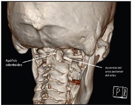
Figure 1 Computed tomography image with 3D reconstruction of the posterior cervical spine, showing total absence of the posterior arch of the atlas, with a direct view of the odontoid process.
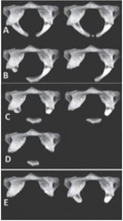
Figure 2 Currarino's classification (4) showing the different types of congenital anomalies of the posterior arch of the atlas. Type A: lack of midline fusion of the posterior arches; B: unilateral cleft due to an absent hemiarch; C: absence of both hemiarches; D: total absence of the posterior arch with a persistent posterior tubercle; E: total absence of the posterior arch and absence of the posterior tubercle.

