SciELO - Scientific Electronic Library Online

 
vol.47 número3Anemia and a kidney massChoque y Shock Choque and Shock índice de autoresíndice de assuntospesquisa de artigos
Home Pagelista alfabética de periódicos  

Serviços Personalizados

Journal

Artigo

Indicadores

Links relacionados

  • Em processo de indexaçãoCitado por Google
  • Não possue artigos similaresSimilares em SciELO
  • Em processo de indexaçãoSimilares em Google

Compartilhar


Acta Medica Colombiana

versão impressa ISSN 0120-2448

Acta Med Colomb vol.47 no.3 Bogotá jul./set. 2022  Epub 28-Mar-2023

https://doi.org/10.36104/amc.2022.2360 

Images in internal medicine

Absence of the posterior arch of the atlas

JORGE IGNACIO MAGAÑA-REYESa  * 

LUIS GERARDO DOMÍNGUEZ-CARRILLOb 

a Especialista en Radiología e Imagenología. Gestalt Imagen, León, Guanajuato; León (México).

b Especialista en Medicina de Rehabilitación. Catedrático Facultad de Medicina de León, Universidad de Guanajuato. León (México).


Congenital defects of the posterior arch of the atlas have an estimated incidence of 0.7 to 3% 1. Most patients are asymptomatic and incidentally diagnosed on cervical spine x-rays, and these defects may be mistaken for fractures or proximal dislocations of the cervical spine 2. (Figure 1). Embryologically, there are three ossification centers in the atlas during the seventh week of gestation: the central anterior one produces the anterior tubercle, and the two central lateral ones form the lateral masses and posterior arch which includes the posterior tubercle. Two percent of the population has a fourth ossification center which forms the posterior tubercle; fusion occurs sometime between 3 and 10 years of age 3. Currarino's classification 4 mentions five types of anomalies (Figure 2), with type A occurring in 95% of cases; the presented case is categorized as type E.
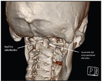
Figure 1 Computed tomography image with 3D reconstruction of the posterior cervical spine, showing total absence of the posterior arch of the atlas, with a direct view of the odontoid process. 
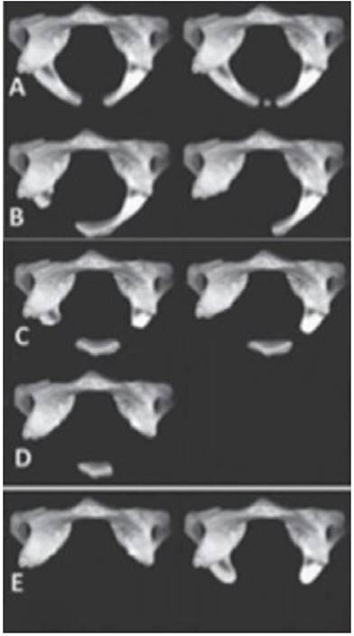
Figure 2 Currarino's classification (4) showing the different types of congenital anomalies of the posterior arch of the atlas. Type A: lack of midline fusion of the posterior arches; B: unilateral cleft due to an absent hemiarch; C: absence of both hemiarches; D: total absence of the posterior arch with a persistent posterior tubercle; E: total absence of the posterior arch and absence of the posterior tubercle. 

References

1. Sabuncuoglu H, Ozdogan S, Karadag D, Kaynak ET. Congenital hypoplasia of the posterior arch of the atlas: case report and extensive review of the literature. Turk Neurosurg. 2011; 21: 97-103. [ Links ]

2. Ouyang ZY, Qiu MJ, Zhao Z, Wu XB, Tong LS. Congenital anomaly of the posterior arch of the atlas: a rare risk factor for posterior circulation stroke. J Neurointerv Surg. 2017; 9 (7): e27. doi: 10.1136/neurintsurg-2016-012731. [ Links ]

3. Junewick JJ, Chin MS, Meesa IR, Ghori S, Boynton SJ, Luttenton CR. Ossification patterns of the atlas vertebra. AJR Am J Roentgenol. 2011; 197: 1229-1234. [ Links ]

4. Currarino G, Rollins N, Diehl JT. Congenital defects of the posterior arch of the atlas: a report of seven cases including an affected mother and son. AJNR Am J Neuroradiol. 1994; 15: 249-254. [ Links ]

Received: October 07, 2021; Accepted: November 03, 2021

* Correspondencia: Dr. Luis Gerardo Domínguez Carrillo. León (México). E-Mail: lgdominguez@hotmail.com

Creative Commons License This is an open-access article distributed under the terms of the Creative Commons Attribution License