SciELO - Scientific Electronic Library Online

 
vol.47 número4Acute arterial thrombosis in SARS-CoV-2 infectionVogt-Koyanagi-Harada disease índice de autoresíndice de assuntospesquisa de artigos
Home Pagelista alfabética de periódicos  

Serviços Personalizados

Journal

Artigo

Indicadores

Links relacionados

  • Em processo de indexaçãoCitado por Google
  • Não possue artigos similaresSimilares em SciELO
  • Em processo de indexaçãoSimilares em Google

Compartilhar


Acta Medica Colombiana

versão impressa ISSN 0120-2448

Acta Med Colomb vol.47 no.4 Bogotá jul./dez. 2022  Epub 29-Maio-2023

https://doi.org/10.36104/amc.2022.2371 

Images in internal medicine

Polycystic kidney disease: more than the kidneys

JOSÉ SANTIAGO CORTÉS-GUZMÁNa  * 

LUIS FERNANDO DURÁN GUTIÉRREZb 

a Residente de Medicina Interna. Facultad de Salud, Universidad Surcolombiana, Hospital Universitario Hernando Moncaleano Perdomo, Neiva (Colombia).

b Jefe del Servicio de Medicina Interna. Hospital Universitario Hern nando Moncaleano Perdomo. Neiva (Colombia).


A 66-year-old man with a history of heart failure (HF), hypertension and chronic kidney disease was admitted to the emergency room with symptoms of acute decompensated HF. A transthoracic echocardiogram showed severe aortic regurgitation. Coronary angiography showed dilation of the ascending aorta (43 mm) and the aortic root. A computed tomography with contrast showed multiple > 5 mm cystic lesions in the liver and kidneys (Figure 1A and B); there was a single cyst in the head of the pancreas (Figure 1C). The patient had normal liver enzymes and elevated CA 19-9. He was diagnosed with autosomal dominant polycystic kidney disease (ADPKD). Brain aneurysms were ruled out with angiography. The patient underwent ascending aorta and aortic root replacement and was discharged with improved symptoms. Autosomal dominant polycystic kidney disease can affect the liver and pancreas, and may be associated with aortic or cerebral artery aneurysms 1,2.
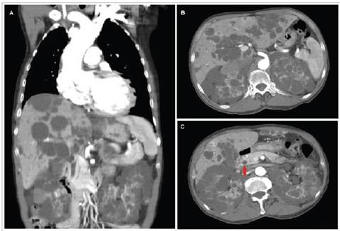
Figure 1 Abdominal tomography with and without contrast A. Coronal section showing dilation of the aortic root and ascending aorta, and cysts in the liver and kidneys. B. Transverse section showing multiple cystic lesions in the parenchyma of the liver and both kidneys. C. Transverse section showing cysts in the head of the pancreas marked by the red arrow and multiple cystic lesions in the liver and kidney parenchyma. 

References

1. Cornec-Le Gall E, Alam A, Perrone RD. Autosomal dominant polycystic kidney disease. Lancet. 2019;393(10174):919-935. doi:10.1016/S0140-6736(18)32782-X [ Links ]

2. Bergmann C, Guay-Woodford LM, Harris PC, Horie S, Peters DJM, Torres VE. Polycystic kidney disease. Nat Rev Dis Prim. 2018;4(1):50. doi:10.1038/s41572-018-0047-y [ Links ]

Received: November 01, 2021; Accepted: February 03, 2022

* Correspondencia: Dr. José Santiago Cortés-Guzmán. Neiva (Colombia). E-Mail: jsancg@gmail.com

Creative Commons License This is an open-access article distributed under the terms of the Creative Commons Attribution License