


 texto em
  Inglês (pdf)
 Artigo em XML
 Referências do artigo
 Enviar este artigo por email
 Citado por SciELO
  Citado por Google
  Similares em
SciELO
Similares em
SciELO  Similares em Google
 Permalink
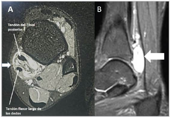
Figure 1 T2-weighted magnetic resonance images of the right ankle in the axial (A) and sagittal (B) planes showing a neoplasm attached to the posterior tibial tendon sheath (white arrows) corresponding to a giant cell tumor of the tendon sheath.
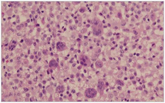
Figure 2 A histopathology photograph showing histiocytic and multinucleated giant cells mixed with scant collagen fibers (H&E 10x).
A giant cell tumor of the tendon sheath (GCTTS) is a rare and solitary soft tissue lesion which arises in the tendon sheath around the ankle and toes. Most cases occur in the hand, and approximately 3-10% of these tumors occur in the ankle and foot. Most patients are young adults. Clinically, the patients report slow, painless growth of a solitary permanent mass over an average of one to two years. There may be a history of trauma, and neurological symptoms are uncommon 1.
Soft tissue edema may be seen on plain x-rays, and the GCTTS may invade the bone and cause visible cystic lesions in 10% of cases. Computed tomography shows the extent of the tumor, and an MRI is useful for determining the extension 2.
The differential diagnosis includes synovial sarcoma, chondromyxoid fibroma, enchondroma, chondrosarcoma and pigmented villonodular synovitis. Histologically, the lesion resembles pigmented villondular synovitis. There is no hemosiderin, and there are macrophages, foam cells and scattered multinucleated giant cells. P63 expression has been found in GCT. Treatment consists of complete and thorough excision of the lesion. Recurrence is reported to be 40% 3.

