


 English (pdf)
 Article in xml format
 Article references
 Send this article by e-mail
 Cited by SciELO
  Cited by Google
  Similars in
SciELO
Similars in
SciELO  Similars in Google
 Permalink
INTRODUCTION
Mastitis caused by intramammary infection (IMI) is one of the most important illnesses in specialized dairy herds worldwide due to the effects on mammary tissue, the decrease of milk production, the poor quality of raw milk, the increase of culling rate ofanimals, and the additional expenses for treatments and milk that is lost because of the antibiotic treatment (Taponen et al. 2016; Krishnamoorthy et al. 2016). Globally, the focus of control programs is for major pathogens like Staphylococcus aureus and Streptococcus agalactiae, but it is important to analyze also minor pathogens like coagulase-negative Staphylococcus species, which are one of the most prevalent groups that cause subclinical mastitis in dairy cows (Vanderhaeghen et al. 2014; Tarazona et al. 2019).
Staphylococci are a diverse group of gram-positive cocci, which can infect the mammary gland. For intramammary infection diagnosis, Staphylococci are classified according to its ability to coagulate plasma in coagulase-positive or coagulase-negative, the microbiological test used to make this determination is the rabbit plasma coagulase test (Andrade et al. 2012). Coagulase-negative staphylococcus (CNS) can be found in teat canals, teat skin and teats ducts. They are classified as minor pathogens causing mastitis, and they rarely differ when IMI is diagnosed, it is also possible that they can cause food-borne disease due to pathogenic factors (De Freitas et al. 2013; Levinson et al. 2016; Dufour et al. 2012).
The CNS regularly isolated in subclinical mastitis are Staphylococcus epidermidis, S. chromogenes, S. sciuri, S. simulans, S. haemolyticus, S. hominis, S. xylosus, S. equorum, S. capitis and S. auricularis (Andrade et al. 2012; Andrade et al. 2018). These bacteria are sometimes referred to environmental staphs, and they are the most frequent organisms isolated from milk samples obtained in herds that have controlled the major pathogens (Krishnamoorthy et al. 2016; Dufour et al. 2012). They are also related with a mild increase of somatic cells count (SCC) in bulk milk in a herd, and with an increase in IL-6 in milk and serum, in concentrations up to 20 times more than in clinically healthy cows (Thorgberg etal. 2009; Bochniarz et al. 2017), and in acute-phase inter-alpha-trypsin inhibitor heavy chain 4. However, there is no consensus regarding the effects of IMI with NEC on milk production, and its physicochemical characteristics (Tomazi et al. 2015).
Some cows with IMI caused by CNS species like S. chromogenes, S. simulans, and S. xylosus had a SCC similar to that observed in IMI caused by S. aureus, others like S. auricularis had minimal impact in SCC (Tomazi et al. 2015; Supre et al. 2011). In Colombia, the studies focused on the previous topic are few (Andrade et al. 2014; Tarazona et al. 2019). Therefore, due to the absence of data related with the prevalence of CNS in Colombia and due to the variability in SCC in IMI caused by CNS, the objective of this study was to determine the prevalence of CNS in a dairy herd in Boyaca and also quantify the effects of every species of CNS in the SCC.
MATERIALS AND METHODS
Population and sampling
The research was carried out in the municipality of Paipa. A longitudinal, prospective, descriptive study was developed. 1 herd was included in the study, this one had a total of 72 animals in production, of which 40 Holstein cows between 2 and 4 calvings, and between 60 and 90 days in milk (DIM), were selected. These cows corresponded to 55% of the animal presents in the herd. The average bulk tank milk somatic cell count (SCC) in the last month was 232.000 cells/ml, and 845 l of milk was produced per day on average. They grazed on kikuyu (Cenchrus clandestinus), and they received water ad libitum. The milking system was mechanic with 1 operator.
This research was approved by the Research Ethics Committee of the Universidad Pedagógica y Tecnológica de Colombia (UPTC), sede Tunja (Approval letter, 22 April 2019).
Sampling and transport
Monthly, milk samples were taken from every teat, between July and December 2019. The samples were taken following the instructions from the National Mastitis Council (2017). All mammary glands were evaluated and no one showed apparent sings like pain or inflammation such as reddish tones in the mammary gland, tenderness, adhesions, and hard areas. Later California mastitis tests (CMT) were performed in all teats. The CMT results were scored and interpreted as either (Godden et al. 2017) 0 (negative or no thickening of mixture), trace (slight thickening), 1 (+, or low thickening but no tendency to form a gel) 2 (++, or immediate thickening, with a slight gel formation), or 3 (+++, or gel formed, and the surface of the mixture becomes elevated) inflammatory response based on the viscosity of the gel formed by mixing reagent with milk (Tarazona et al. 2019; Mendoza et al. 2017; Tolosa et al. 2013).
Milk samples were taken from every teat that showed 0, trace, and 1 results. They were stored in sterile plastic bags, the procedure described by the National Mastitis Council (NMC) was followed (NMC, 2004). The collected samples were kept and transported under refrigeration until they arrived at the laboratory for analysis (Tarazona et al. 2019). All samples were taken by a veterinarian and transported under refrigeration to the milk quality analysis and mastitis control laboratory (MQAMC). They also were transported to the Veterinary Microbiology Laboratory (VML) of the Universidad Pedagógica y Tecnológica de Colombia in Tunja.
Bacteriological tests and species identification
A routine diagnosis was carried out, also the isolation of non-common microorganisms following the methodology proposed in the Laboratory Handbook on Bovine Mastitis, by the NMC (NMC, 2017). 1 ml of milk sample were inoculated on sheep blood agar. After that, the samples were incubated during 24 hours in an aerobic medium at 37°C, gram staining (positive), hemolysis patron, catalase reaction (positive), and coagulase tests (negative) (Soler et al. 2019; Thorgberg et al. 2009). Milk samples that showed growth of 3 or more species of pathogens were considered contaminated, and they were excluded (Dufour et al. 2012; Koop et al. 2012).
Genetic characterization of CNS
The identification of each CNS species was carried out in a private external laboratory through the identification of the rpoB gene, through real-time PCR. However, it was not possible to access the primer sequences.
Colony Forming Units and SCC
SCC was performed using Fossomatic 4000™ equipment from the FOSS Company®, and Colony Forming Units/ml (CFU/ml) was performed using a Bacto-Scan™ FC+ (Vasquez et al. 2012).
Definition of positive case
A positive case (for every mammary quarter) was considered infected by CNS when growth of > 500 Colony Forming Units/ ml (CFU/ml) of a particular organism and only one colony type on the plate were isolated (Dieser et al. 2014; Raspanti et al. 2016).
Statistical analysis
All the data were grouped in an Excel document for windows 2010. Later, they were processed through the statistical program Statgraphics centurion XVI®. Average Geometric measurement SCC was calculated using monthly dates for every infected teat. he correlation between CNS species causing a positive case and average geometric count SCC were compared using variance analysis of Kruskal-Wallis (Anova) with comparison for peers post-hoc (P < 0,05)
RESULTS
Microbiological characterization
Due to the fact that each teat was evaluated separately, a total of 960 samples of milk were taken. he results of the microbiological analyses were: 1) 144 contaminated samples (15%); 2) 120 samples with growing of others type of pathogens principally Streptococcus spp. and Corynebacterium spp. (12,5%) and 3) 77 samples without growing (8%) (figure 1).
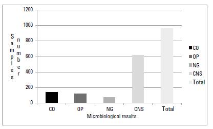
FIGURE 1 Results of microbiological characterization of milk samples. CO: Contaminated; OP: Others Pathogens; NG: No growth; CNS: Coagulase Negative Staphylococcus
The 619 remaining samples (64,47%) were positive for almost 1 strain of CNS. 11 species of CNS were identified (table 1). he duration of infection ranged between 1 and 6 months. Species like S. epidermidis and S. chromogenes were found in several samples in 1 month with high numbers of colonies, while species like S. hyicus, S. auricularis and S. equorum were found in samples in all 6 months with few colonies (table 1).
TABLE 1 Percentage of isolations for every species of CNS and geometric mean of SCC. The letters indicate membership in a statistical group, different letters indicate the statistical difference between groups
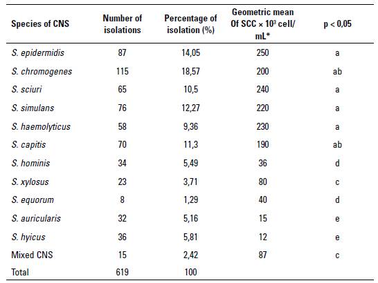
Statistical differences (p < 0,05) were found in the median geometric mean of SCC between species of CNS. The major results of SCC were found for six species of CNS, which were S. epidermidis, S. chromogenes, S. simulans, S. sciuri, S. haemolyticus and S. capitis. For the remaining CNS species, values less than 100 x 103 cell/ml were found, and the mixed infection (S. auricularis and S. equorum) had a result of 87 x 103 cell/ml (table 1).
DISCUSSION
Many factors are associated with an increase of SCC. Even in the absence of bacterial growth, this increase leads usually to traumatic damages (lacerations, cuts, injuries), that get worst due to chemical irritants or external conditions, causing that the teats or the udder, in general, get affected, and increasing the somatic cell count. his could explain the absence of growth of bacterium colonies in the 77 samples (Andrade et al. 2018; Tarazona et al. 2019).
CNS are minor pathogens that caused mastitis. hey are important in the dairy industry worldwide, however, their importance had not been previously evaluated in Colombia. In this study a total of 619 samples (64,47%) were positive to almost 1 CNS species by cultured as well as by pulse gel electrophoresis. These results were similar to those reported by (Hosseinzadeh & Dastmalchi 2014), who determined in Iran that 68,35% of isolations in mastitis milk samples corresponded to CNS; nonetheless, these results were higher compared to the results ofTaponen et al. (2016), who in Finnish farms determined IMI due to CNS in the 13,35% of the samples.
Studies in CNS causing mastitis in dairy cows show a wide variation in the prevalence of distinct species. Hosseinzadeh & Dastmalchi (2014) showed that the CNS species more prevalent in their study were S. haemolyticus (40,7%) and S. chromogenes (15,7%), results that were higher than the one reported here. However, Bochniarz et al. (2017) reported that S. xylosus, S chromogenes, S. haemolyticus, S. simulans and S. sciuri were the most prevalent species in subclinical mastitis milk samples, all these species were found in the present study. Another study in the United States carried out by Gillespie et al. (2009) showed that S. chromogenes and S. epidermidis were the most prevalent species in dairy herds.
Jenkins et al. (2019) in several dairy herds in the United States showed that the most prevalent species of CNS present in that country were S. chromogenes followed by S. haemolyticus, S. simulans, S. epidermidis, S. hominis, S. auricularis, S. sciuri, S. devriesei, S. capitis, S. cohnii and S. warneri, results that were similar to those reported here.
In 89 Canadian herds, Fry et al. (2014) reported that 46% of the samples were positive for one of the 20 species of CNS. hese results are lower compared to the results showed here. From those isolations, S. chromogenes, S. simulans, S. haemolyticus, S. epidermidis and S. xylosus were the most prevalent, results that were similar to those reported here. his similarity showed that CNS had parallel behavior in Colombia compared to North American countries.
However, results by De Freitas et al. (2013) in Brazil determined that S. hyicus, S. warneri and S. epidermidis were the most prevalent species in dairy herds in Sao Paulo, results that were different from those described here, principally related to S. warneri. Similarly, Costa et al. (2008) studied IMI caused by CNS in Brazilian herds located in the states of São Paulo and Pernambuco. They reported that S. warneri, S. hyicus, S. chromogenes, and S. epidermidis were the most prevalent.
In Argentina, Raspanti et al. (2016) determined the prevalence of CNS in dairy herds, showing that S. chromogenes, S. haemolyticus, S. warneri, S. xylosus, S. simulans and S. epidermidis were the principal pathogens present in Argentinian dairy cows, results similar to those found here.
All these studies showed that regardless of the country the species that can infect the mammary gland were similar, and that this similarity can be due to similar management practices between herds evaluated. Also, in several countries management control can be used to combat the pathogens to achieve similar results.
Various factors are associated with pathogenicity, sustainability and spreading of CNS in cows. Factors such as hemolytic and proteolytic activity, higher tolerance to post milking teat disinfection, and the ability to evade immune mechanisms might provide colonization advantages for some species like S. chromogenes and S. haemolyticus in the mammary gland (Bochniarz & Wawron 2012; Hosseinzadeh & Dastmalchi 2014; Piessens et al. 2012; Quirk et al. 2012).
With respect to SCC, the statistical analyses showed that the species S. epidermidis, S. chromogenes, S. sciuri, S. simulans and S. haemolyticus have not statistically differences between the average geometric count of SCC determined for each species, which means that the increases in SCC are similar when any of these pathogen are present in mammary glands. However, not all species generated a similar SCC average (table 1).
The SCC results found here for S. epidermidis, S. chromogenes, S. sciuri, S. simulans and S. haemolyticus were higher than the obtained by Thorgberg et al. (2009), who determined values of SCC for milk samples of persistent or nor persistent subclinical mastitis with the same pathogens, 223 x 103 cells/ml and 189 x 103 cells/ml, respectively. But our results were lower compared to the results of Dufour et al. (2012), who found for S. chromogenes a SCC of277 x 103 cells/ml; for S. haemolyticus, 662 x 103 cells/ml; and for S. simulans, 525 x 103 cells/ml. These differences may be due principally to factors associated with cow immunity, and the quantity of CFU chosen to determine a positive case (1000 CFU/mL), while in this study it was 500 CFU/mL.
It has been shown that S. epidermidis, S. chromogenes and S. simulans generally induced a mild to strong inflammatory reaction as measured by CMT (CMT scores 3-5), whereas persistent IMI with others CNS species, including S. xylosus and S. haemolyticus, mostly had a low CMT score (1-2) (Vanderhaeghen et al. 2014). Our findings were different to those proposed by Vanderhaeghen et al. (2014) due to the pathogens mentioned above in our study, which show CMT scores between trace and 1, and these were related to those mentioned by Taponen et al. (2006), who said that these pathogens can cause subclinical and clinical mastitis in similar proportions.
Sampimom et al. (2009) found lower results with respect to SCC related with S. chromogenes, S. epidermidis and S. capitis of 192, 132, and 187 x 103 cells/ml, respectively. However, for S. xylosus the results were 400 x 103 cells/ml, values that were higher compared to our results. hese results were similar to those mentioned by Supre et al. (2011), who mentioned that S. simulans and S. xylosus can had a substantial effect on milk SCC, an effect comparable to that of S. aureus.
In their study, Fry et al. (2014) obtained similar results related to the principal pathogenic species (S. chromogenes, S. simulans, S. epidermidis, S. xylosus, S. capitis and S. haemolyticus). hey did not find statistical differences between SCC in milk samples of dairy herds in the USA, and they mentioned that these pathogens are primarily ofenvironmental origin, and therefore these species may be acting as opportunistic mastitis pathogens.
An increased prevalence of CNS IMI is associated with several cow and herd level factors, but not with any specific milking or housing system (Taponen et al. 2016). It has been demonstrated that the principally source of CNS was the environment (Piessens et al. 2011), Nagase et al. (2002) demonstrated that CNS like S. warneri, S. hominis and S. capitis can be found in dairymen, which means that these can be a potential source for colonization of these species in bovine udder tissue, as these species are commonly found living on the skin and the mucous membranes of humans. Also, Thorgberg et al. (2009), who used the same technique as in the present study, found S. epidermidis in milk, and the milker's skin, which indicated that this pathogen may be transmitted from the milkers to cows.
CONCLUSIONS
here were determined 11 species of CNS in IMI in cows from Boyaca, Colombia, and the most prevalent species were S. chromogenes, S. simulans, S. epidermidis, S. xylosus, S. capitis and S. haemolyticus. All of this species have an SCC lower than 250 x 103 cells/ml. he results found here are similar to results in different parts of the world, which confirms that they are pathogens that must be constantly evaluated because they can go unnoticed in routine controls, especially in those farms where major pathogens are not a serious problem. The results determined in this study demonstrate that the CNS generates a slight increase in somatic cells.

