


 English (pdf)
 Article in xml format
 Article references
 Send this article by e-mail
 Cited by SciELO
  Cited by Google
  Similars in
SciELO
Similars in
SciELO  Similars in Google
 Permalink
INTRODUCTION
Anatomically, the perineum is the area of the body that caudally covers the pelvis, and surrounds the anal and urogenital canals (Conci 2017). Named as pelvic diaphragm, this characterizes the main structure of the perineum, being composed of the coccygeus muscles, levator ani, external and internal sphincter (König 2011; Fossum 2014). Perineal hernia is the result of weakness and/ or rupture of the muscles and fasciae that make up the pelvic diaphragm, allowing the caudal displacement of abdominal or pelvic organs to the perineum. (Aronson 2017). Its exact cause is still unknown and poorly understood, but it has been proposed to be related to male hormones, exertion and congenital or acquired muscle weakness or atrophy (Fossum 2014), as well as constipation, hyperplastic prostate diseases, recurrent rectopathies, and chronic tenesmus (Bojrab 1981; Silva et al. 2019). In 20% to 50% of cases, the hernia is bilateral (Barreau, 2008) and the dilatation is usually ventrolateral to the anus. (Bellenger and Cafield 2007; Brito 2020). The objective of this work is to report a case of perineal hernia in a male dog and to emphasize the clinical-surgical management performed for this type of condition.
MATERIALS AND METHODS
A 7-year-old male Yorkshire breed dog was admitted at the FullPet Veterinary Clinic in Guarulhos-SP, weighing 4.5 kg. In the anamnesis, the tutor reported that the patient had dyschesia, swelling next to the anus, normorexia, normodipsia and normuria. On physical examination, an aspect of a bag was palpated in the left perineal region with accumulation of feces, mucous membranes were normocolored, non-invasive blood pressure: 110/80 mmHg, respiratory rate: 25 rpm heart rate: 104 bpm and temperature: 38.2 °C.
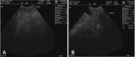
After performing the abdominal ultrasound, a slightly enlarged spleen and an enlarged prostate were identified, with dimensions of approximately 3.29 cm longitudinal axis x 2.54 cm transverse axis (figure 1).
RESULTS
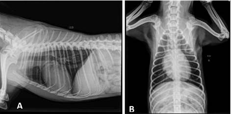
According to the history and clinical findings, the diagnosis of perineal hernia was established, which may be secondary to prostatic alteration, evident on ultrasound. The suggestion was to perform an orchiectomy surgery, with the aim of possible treatment for the reduction of the prostate and surgical correction of perineal hernia. After accepting the surgical procedure, the owner brought the animal for preoperative examinations. On physical examination, the mucous membranes were of normal color, adequate hydration, no pain on abdominal palpation, NDN lymph nodes (nothing noteworthy), left unilateral perineal hernia. The pre-surgical complementary tests performed were blood count, urea, creatinine, ALT, AST, GGT, AF, electrocardiogram and chest x-ray. (LL/ VD) (figure 2). The owner was informed about the possibility of recurrence that after the surgical procedure.
Source: Fullpet Veterinary Clinic.
FIGURE 2 Thoracic x-ray. (A) Chest X-ray (LL), without alterations. (B) Chest X-ray (RV), without alterations.
The animal underwent surgery and intramuscularly acepromazine was administered at a dose of 0.05 mg/kg, combined with tramadol 4 mg/kg as pre-anesthetic medication. Propofol was used at a dosage of 4 mg/kg intravenously and inhalation maintenance with isoflurane for anesthetic induction. The surgical technique used was herniorrhaphy sutures with nylon thread to correct the hernia. An incision was made at the caudal edge of the obturator internus muscle, which was elevated from the ischial surface using a periosteal elevator. Muscle elevation cannot proceed beyond the rim of the obturator foramen to avoid injuring the corresponding vessels and nerves. The tendon of the muscle was incised in its condensed portion, where it begins its lateral passage along the body of the ischium, taking care not to injure the sciatic nerve. The muscle was dorsally elevated and transposed over the hernia. The initial suture was performed between the external anal sphincter muscle, sacrotuberous ligament and the gluteal fascia as dorsally as possible to obtain a surface which could be sutured to the apex of the obturator internus muscle. The caudolateral edge of the muscle flap was sutured to the caudomedial apex of the caudal gluteal artery and the sciatic nerve. The caudomedial edge of the muscle flap was sutured to the external anal sphincter muscle. It is important to point out that all suture threads were positioned as described and later sawed, because in this way it was possible to introduce the threads properly. Then, the suture of the subcutaneous and skin was performed.
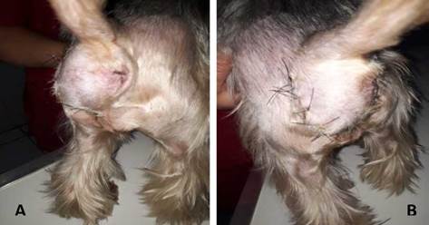
After the procedure, the animal recovered well (figure 3) and was released home.
Source: Fullpet Veterinary Clinic.
FIGURE 3 Post surgical hernia perineal region. (A) Right LD side view; (B) left lateral view (LE).
Nine days after the procedure, the owner returned with the animal and reported normorexia, normodipsia, normuria and normochesia. On physical examination, normal colored mucous membranes, adequate hydration, no pain on abdominal palpation, lymph nodes (DNN), temperature 38.3° C were observed. The surgical wound presented good healing, requesting a return after 7 days to remove the stitches.
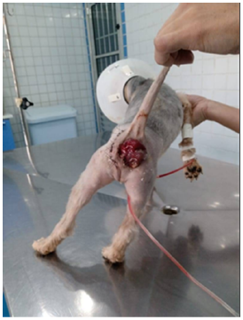
The patient came for the removal of the stitches, but 9 days after the removal the animal presented recurrence, with a rectal prolapse as illustrated (figure 4). It grew where the herniorrhaphy was performed. Complementary tests were requested to investigate the cause of the hernia: TSH, free T4, total T4, T3, fructosamine and glycated hemoglobin. The results of the exams were within the reference values, so the animal was sent again for surgery, for mesh placement (hernia correction). The patient was again directed to the surgical field for preoperative evaluation. The use of biocompatible materials for surgical treatment for the correction of perianal hernia was chosen. The material chosen for the procedure was polypropylene mesh.
Source: Fullpet Veterinary Clinic.
FIGURE 4 Anal prolapse in dog after perineal hernia repair surgery.
For investigating the sample, a surgical procedure of exploratory laparotomy was performed. Presence of splenomegaly was found and enteropexy was performed.
In the postoperative period, the animal had a urethral tube and liquid food to prevent prolapse recurrence. The animal was released after 10 days of surgery with home medications: benzoylmetronidazole 1.5 ml BID + neodexa cream.
Upon return for stitch removal, the tutor reported that the animal was active, with normochesia, normorexia, normodipsia, normuria, and surgical wound showing good healing. Abdominal ultrasound was requested for follow-up and peristalsis and continued intestinal mobility were observed. Two months after the operation, the animal already presented shaped stools, with no difficulty in defecating. After three months, the animal was released with solid food and medical discharge.
DISCUSSION
According to the report, in the anamnesis, the owner reports that the patient has dyschesia, swelling next to the anus, normorexia, normodipsia and normuria. On physical examination, the left perineal region is palpated with accumulation of feces, the mucous membranes are nor-mocolored, non-invasive blood pressure: 110/80 mmHg, respiratory rate: 25 rpm heart rate: 104 bpm and temperature: 38.2° C. As a prevention method used to decrease the chances of recurrence and possibly as a treatment for a reduced prostate, orchiectomy surgery is recommended. This procedure is commonly performed in veterinary practice, which helps to prevent pathologies, and in addition to prevention, it is also a method adopted for possible treatments of pathologies of reproductive origin (Oliveira et al. 2010). Testicular and scrotal neoplasms, orchitis, epididymitis, trauma, or abscesses are primary indications for this procedure. As stated in the literature, prostatic hypertrophy, prostatitis, cryptorchidism, perianal adenoma, and perineal hernia are seen as an indication for orchiectomy, serving as prevention and adjunct treatment of these pathologies (Towle 2012; Crane 2014).
The increased perineal volume in the case of the patient in this study may have an undetermined cause. According to Penaforte Junior et al. (2015) and Assumpção (2016), approximately 97% of cases occur in male dogs and, of these, 95% in non-orchiectomized dogs. This can be explained according to the fragility of the insertions of the levator ani muscle in the male, and the pressure caused by the prostate which, when hypertrophied, causes an extra pressure load on the pelvic diaphragm muscles (Serafini et al. 2011). The actual cause of muscle weakness is still unknown, but some factors have been proposed, such as neurogenic or senile muscle atrophy, enlarged prostate, myopathies, and hormonal changes (Rahal 2005; Machado et al 2020). The pelvic diaphragm represents the main structure of the perineum, which is formed by the levator ani muscles, external anal sphincter, coccygeus, and internal anal sphincter (König 2011; Fossum 2014). Perineal hernia is caused by the separation and weakening of the muscles and fascia that are part of the formation of the pelvic diaphragm; the muscular pelvic diaphragm that supports the rectal wall, which deflects causing pelvic and/or abdominal contents to bulge (Bellenger and Canfield 2007).
Perineal hernia is described as a low-prevalence condition that affects animals over 7 years of age. (Bellenger and Canfield 2007; Assumpção 2016). The profile of the patient described in this report perhaps fits both the age group and the reproductive status because it is an uncastrated animal, according to the broad classification investigated by Fernandes (2019). There are also reports of genetic predispositions in dogs of the German Shepherd, Boxer, Collie, Poodle, Daschund and Pekingese breeds (Assumpção 2016; Silva et al. 2019).
During the evaluation, in the examination of the patient, it is possible to observe the growth in terms of volume in the left lateral region of the anus. After performing the abdominal ultrasound, a slightly enlarged spleen and prostate were identified. The normal sonographic appearance of the prostate in the dog is very variable in shape and texture, depending on age, sexual status (neutered or intact) and history of previous prostate disease. As with shape and texture, the echogenicity of the prostate can vary, being slightly more echogenic than the spleen (Junior 2012; Moresco and Gonçalves, 2019).
The animal mentioned in this report underwent surgery with the following pre-anesthetic medication: acepromazine at a dose of 0.05 mg/kg, combined with intramuscular tramadol 4 mg/kg. For anesthetic induction, propofol with a dosage of 4 mg/kg was used intravenously and inhalation maintenance with isoflurane. Brito (2020) used midazolam, at a dose of 0.5 mg/kg, by intravenous access, propofol, at a dose of 3mg/kg, and ketamine at a dose of 1 mg/kg, they were used for induction, both were used for the intravenous access in the case of a patient with correction of perineal hernia.
Among the techniques used to correct this condition, the use of biocompatible materials has grown in small animal surgery, such as the use of bovine pericardium (Marques et al. 2015), peritônio bovino (Bastos et al. 2005), equine pericardium (Zerwes et al. 2011), natural latex (Paulo et al. 2005), and polypropylene fabric (Rego et al. 2016; Ferraz et al. 2017). In the correction of the hernia of this patient, the surgical technique used was the traditional suture with nylon thread. Surgical techniques frequently used in pelvic diaphragm reconstruction include the traditional suturing method, also known as anatomic replacement, transposition of the obturator internus muscle, with or without section of the muscle tendon; the transposition of the superficial gluteal muscle and the transposition of the obturator internus muscle combined with the transposition of the superficial gluteal muscle.
25 days after undergoing the surgical procedure and post-operative evaluation returns, the patient returned to the clinic with recurrence due to anal prolapse, presented an increase where the herniorrhaphy was performed, which consists of the replacement of the contents of the hernia sac in its normal place. Possible postoperative complications include wound infection, fecal incontinence, tenesmus, rectal prolapse and sciatic nerve paralysis (Ribeiro, 2010; Moreira et al. 2020). Relapses usually occur in animals with more severe preoperative signs and in those with bilateral involvement. Complementary tests were requested to investigate the cause of the hernia: TSH, free T4, total T4, T3, fructosamine and glycated hemoglobin. These tests aim to evaluate the amount of hormones available in the bloodstream for possible use by tissue organs, to assess whether hormone production is within the standards to provide energy for metabolic activities. These tests aim to evaluate the amount of hormones available in the bloodstream for possible use by tissue organs, to assess whether hormone production is within the standards to provide energy for metabolic activities.
The material chosen for the correction procedure was the polypropylene mesh. The use of the mesh is more viable both for its low cost and for the response in the correction of hernias. As it is woven with monofilament thread and interspersed with pores, it allows the infiltration of fibroblasts and also stimulates and allows the production of collagen, which offers a less aggressive inflammatory response. According to Silva et al. 2019, the polypropylene mesh still has an inexpressible level of reactivity, low potential for bacterial adhesion and even when placed in a highly contaminated environment, the mesh accepts full incorporation by granulation tissue. In this way, the material will remain in place for an indefinite period, which guarantees the fundamental and necessary mechanical barrier to prevent the animal from having a hernia recurrence again after this surgical correction.
CONCLUSION
The technique, traditional suture with nylon thread, associated with orchiectomy is a possible method of treatment for the case of perineal hernia reported in a dog, and may be indicated for routine use of this type of problem. The most frequent clinical signs are tenesmus, constipation, and perineal swelling. For an adequate and accurate diagnosis, the physical examination must include rectal palpation, and radiographic and ultrasound examinations may also be necessary, as in this case. In cases of recurrence, complementary methods are used to correct the hernia, such as the use of polypropylene mesh, which proved to be more viable due to its low cost and the response in the correction of hernias, allowing the infiltration of fibroblasts, that stimulates and allows the production of collagen.

