


 English (pdf)
 Article in xml format
 Article references
 Send this article by e-mail
 Cited by SciELO
  Cited by Google
  Similars in
SciELO
Similars in
SciELO  Similars in Google
 Permalink
INTRODUCTION
Phimosis is the inability to move the penis through the preputial orifice, so it remains trapped inside the foreskin. This is caused by inadequate development that produces narrowing or complete closure of the orifice (Kruger et al. 1996; Sarierler and Kara 1998; May and Hauptman 2009; Vadalia et al. 2014). This disease can appear in both dogs and cats, but it is less frequent in felines (Sarierler and Kara 1998; Bright and Mellanby 2004; May and Hauptman 2009; Yoon and Jeong 2013). So far phimosis in cats is not associated with a specific race as a risk factor or carrier of this condition.
However, it has been established that the average age for developing phimosis is between 10.5 and 18 weeks (May and Hauptman 2009; De Vlaming et al 2019). It can be a congenital or acquired disease. When acquired, it is related to traumatic events that unchained edema, inflammatory processes, and in some cases cicatrization (May and Hauptman 2009; Vadalia et al. 2014). Clinical symptoms depend on the narrowness of the preputial orifice. Strangury, pollakiuria, hematuria, inflammation, and constant licking are the main signals. Nevertheless, there can be asymptomatic patients with slight stenosis in the orifice, preputial ulcerations in much narrower orifices, and cases with a total absence of the orifice; death can be a consequence if symptoms remain untreated (Kruger et al. 1996; Yoon and Jeong 2013). Diagnosis is based on physical examination, where stenosis in the preputial orifice, narrowing, or impossibility of exposure of the penis can be appreciated (May and Hauptman 2009; de Vlaming et al. 2019). Concerning treatment, there are reports of corticosteroid ointments and procedures for expansion of the preputial orifice without resection of surrounding tissue, but the main option is preputioplasty (Papazoglou and Kazakos 2002; HedLund 2007). In order to choose the best surgical approach, the following classification has been proposed: phimosis type 1 (cats with stenosis in the preputial orifice and absence of adherences between the penis and the prepuce), and phimosis type 2 (cats with stenosis in the preputial orifice and adherences between the penis and the prepuce). According to this classification, in cats with phimosis type 1, preputioplasty with a circular stapler is indicated, meanwhile in cats with phimosis type 2, preputial urethrostomy (Bright and Mellanby 2004; Yoon and Jeong 2013; de Vlaming et al. 2019).
Exposure of the tip of the penis is the main complication reported in patients subjected to preputioplasty. Normally, this exposure diminishes within post-surgical time, however, there is a possibility of reduced exposure instead of disappearing completely (de Vlaming et al. 2019).
The objective of this report is to contribute information about clinical and surgical decisions and the main complications in cats with phimosis.
EXAMINATION OF THE PATIENT
A common cat, 6 weeks old, is brought to consultation due to hematuria, pollakiuria, periuria, vocalization, and excessive licking before and after micturition. The owner declares she found the cat in the street a week ago and since that moment the patient has shown the same symptoms.
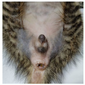
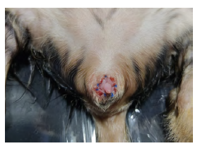
The patient walks as usual, is attentive to external stimuli, and has a calm attitude. The systemic state does not show apparent alterations. At the examination, the penis shows preputial distension, the size of the orifice is reduced, and it is impossible to expose the penis (figure 1). After the physical examination, the diagnosis is confirmed. Surgery is suggested as a therapeutic approach. A complete blood count is carried out and the results are in between the reference marks. A urine sample was obtained through cystocentesis and then sent to an antibiogram and urine culture. There was no bacteria growth in the result.
TREATMENT APPROACH
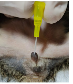
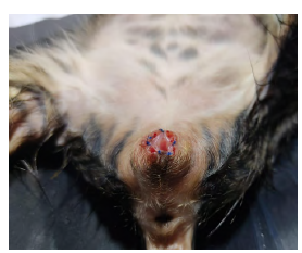
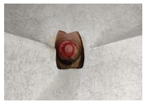
Fentanil was prescribed as pre-medication (5µg/kg, IV), followed by a dose of propofol as an inducing agent (2 mg/kg, IV). During the procedure, an oxygen mask was used, and anesthesia was provided through sevoflurane. A catheter number 24 was introduced through the preputial orifice (figure 2). Using the catheter as a guide, a small incision was done in the preputial orifice going to the abdominous, and from this incision, a circular extraction of preputial tissue was carried out. A simple interrupted suture between the mucosa and the preputial skin was done (figure 3). A couple of hours after the procedure, the patient urinates as usual. Three days after the patient is brought back into consultation. The owner says the patient spends more time than usual in the sandbox and does not urinate. Physical examination evinces a granulated tissue blocking the movement of the penis (figure 4). It is established that from the circular resection of tissue that initially occluded the penis, the cicatrization process produced a new obstruction in the preputial orifice. The patient undergoes surgery once again with the purpose of solving the relapse. Granulated tissue is removed and most of the preputial tissue is resected (figure 5). After the surgery, ampicillin/sulbactam (20 mg/kg, IV, twice a day) and tramadol (2 mg/kg, IV, once a day) were prescribed as a treatment. Right after the surgery, the patient can urinate, and four days after the patient is discharged since there is no sign of clinical symptoms. Two weeks after, the patient is brought for a medical check, where it can be appreciated that the tip of the penis is exposed and there are no signs of the disease (figure 6). After seven months of following the patient's process, there is a minor exposure of the penis and there are no signs of the disease. Telephonic monitoring is carried out for a year, and the owner does not report any signs of the initial pathology, except for some occasional licking.
Source: own elaboration.
FIGURE 2 Feline patient in dorsal decubitus. A catheter number 24 is used as a guide to mark the preputial orifice.
Source: own elaboration.
FIGURE 3 Feline patient in dorsal decubitus. Circular resection of the foreskin and pattern of simple interrupted suture between the mucosa and the skin.

Source: own elaboration.
FIGURE 4 Feline patient in dorsal decubitus. Examination three days after the surgery. Granulated tissue occludes the penis.
Source: own elaboration.
FIGURE 5 Feline patient in dorsal decubitus. Corrective surgery with a generous circular resection. There is more exposure of the penis.
DISCUSSION
Phimosis in cats has been reported in a lower frequency in comparison with phimosis in dogs (Ettinger et al. 2017). Some isolated cases have been reported, and two more studies include no more than ten patients in a lapse of eight years (Bright and Mellanby 2004; May and Hauptman 2009; de Vlaming et al. 2019). The patient of this study was diagnosed with phimosis due to the clinical signs and the physical symptoms found after the explorations of the foreskin. Additional tests were not necessary for the diagnosis, but they were prescribed in order to avoid complications such as crystalluria, UTIs, or renal disease. The age of the patient indicates that it is a case of congenital origin, however, it has been reported that acquired phimosis is the most common result after a traumatism, such as excessive cleaning of the mother or the rest of the litter (De Vlaming et al. 2019). There was no information about the interaction with the mother or the rest of the litter, so it was impossible to establish if the case was congenital or acquired. Since the use of corticosteroid ointments has not cured the disease, according to the published studies, the surgical approach was considered the most suitable option (Bright and Mellanby 2004; De Vlaming et al. 2019). According to the classification proposed by De Vlaming et al. (2019), this is a case of phimosis type 1, where there is a narrow and swollen preputial orifice, with edema and no adherences between the penis and the preputial tissue. Concerning this classification, this kind of patient benefits from preputioplasty with a circular stapler. Due to the diminished preputial orifice, characteristic of patients with phimosis type 1, using a catheter through the urethra is not suitable (Bright and Mellanby 2004; May and Hauptman 2009). For this specific case, since there was a small preputial opening, catheter number 24 was used as a guide for a more precise surgical correction. With the purpose of increasing the preputial orifice diameter a preputioplasty with a circular stapler was performed. After finishing the procedure, the penis showed an appropriate exposure. Within days, an excess of granulated tissue appears and there is a relapse for preputial stenosis. This complication is associated with the fact that during the first surgical approach, just a small resection of preputial tissue was performed and after some days the cicatrization tissue started to block again the preputial orifice, which is why the clinical symptoms appeared again. This process was solved by resecting the granulated tissue with a bigger amount of preputial tissue, leaving as a result a major exposure of the penis. Exposure of the tip of the penis is the number one post-surgery complication. Most of the cases were solved within 9.5 days (Olsen and Salwei 2001; Yoon and Jeong 2013; De Vlaming et al. 2019), meanwhile the patient in this report maintained a small exposure after a seven-month period. The persistence in the exposure of the penis, in the long term, can create complications due to an excess of cleaning, which produces traumatism, localized infections, and Urinary Tract Infection (UTI) symptoms. Monitoring the patient during a year after the surgical procedure evinced occasional preputial licking but no signs of the disease according to the information provided by the owner. Even though the culture was negative, anti-biotherapy was performed empirically due to excessive surgical manipulation. However, it is not recommended to do this before having a positive culture and related clinical symptoms (Weese et al. 2019).
CONCLUSION
Preputioplasty with a circular stapler, a generous extraction of preputial tissue, and a certain exposure of the tip of the penis is the ideal approach for diminishing the apparition of excessive granulated tissue, to avoid post-surgery complications. It is advised to perform the resection of preputial tissue without fear of an excessive exposure of the tip of the penis to avoid scar tissue occluding once again the preputial orifice. The exposure solves itself within a short time, and as the patient keeps growing, the foreskin covers the penis again. It is important to monitor the patients that maintain the exposure for longer periods in order to avoid future complications related to this process. According to this study and the findings in other documents, it is a fact that, up to the present, phimosis is not a frequent disease in cats, which is why it is important to provide information about clinical conditions, surgical procedures, and possible complications.

