


 English (pdf)
 Article in xml format
 Article references
 Send this article by e-mail
 Cited by SciELO
  Cited by Google
  Similars in
SciELO
Similars in
SciELO  Similars in Google
 Permalink
INTRODUCTION
Fascioliasis is a disease caused by Fasciola hepatica, a parasite that belongs to the phylum Platyhelminthes, class Trematoda, order Digenea, and family Fasciolidae (Valderrama 2016) and infects mainly bovine, ovine, and caprine, although it can also affect porcine, donkeys, alpacas, and llamas. Its transmission occurs through the ingestion of contaminated food, and it is caused by trematode worms known as flukes. The infection occurs when drinking water or ingesting aquatic or semi-aquatic plants whose metacercariae are adhered to the stem or the leaves, this explains why the risk of fascioliasis in bovine rises during pluvial precipitation (Merino & Valderrama 2017).
Endemic zones are distributed at a worldwide level; however, the most affected localities can be found in the South American Andes. This parasite affects the liver and lungs but severe morbidity occurs when the worm gets to the main bile ducts (OPS 2021), which causes higher prevalence, mainly in the least developed countries (Carrión-Ascarza et al. 2021) with percentages of infection in Europe, Asia, and Africa of0.1% to 38% (Getahun et al. 2020; Khoramian et al. 2014; Shamsi et al. 2020; Theodoropoulos et al. 2002), and 0.6% to 41.5% in America (Giraldo et al. 2016; Londoño et al. 2020; Nuñez et al. 2017; Palacio et al. 2020; Pinilla et al. 2020; Ramírez-Hubener et al. 2019; Rojas y Cartín 2016; Stoore et al. 2018). Nevertheless, Peru shows the highest rates (38.2% to 59.5%) (Chávez et al. 2012; Hubener et al. 2019; Julon et al. 2020), especially in Inter-Andean Valleys (up to 4.500 meters) (Espinoza et al. 2010).
This disease has been considered a relevant veterinary problem since 1987, but during the last twenty years, it has been recognized as the vector transmission illness with the highest degree of extension (Valderrama 2016) due to the significant productive and financial loss in the stockbreeding industry (Ashrafi 2015). From a financial point of view, fascioliasis is the most important helminth infection in terms of negative impact on animal production, with no less than 50 million USD per year (Espinoza et al. 2010).
In Peru, this disease has been found in 17 out of 24 regions (Stoore et al. 2018), and it is the second parasitic infection, causing a loss of 10.5 million USD per year (Palacio et al. 2020). The negative impact of stockbreeding is related to the number of livers (Valderrama 2016). Viscera seizure is relevant when the innocuousness and quality of the food are affected (Carrión-Ascarza et al. 2021). Such is the case that infected viscera must be discarded, which causes elevated financial loss in terms of animal production, lessening the development of the stockbreeding industry. However, due to the scarce registered information about stockbreeding production in Peru, it is difficult to evaluate the negative financial impact caused by viscera seizure.
Apurimac is a hyper-endemic location for fascioliasis, especially when it comes to bovine and ovine, which produces an important monthly financial decrease due to viscera seizure (Carrión-Ascarza et al. 2021). Therefore, since the breeding of porcine, bovine, ovine, and caprine is a relevant activity in the region, as well as an important source of food for its inhabitants, including viscera consumption, the frequency of fascioliasis in slaughtered animals was determined in slaughterhouses of Andahuaylas and the financial loss due to seizure of livers.
MATERIALS AND METHODS
Type of study
This study was quantitative at a basic level, analytical design, observational type, and cross-sectional. At the same time, a retrospective study corresponding to the period 2011-2016 was carried out.
Location of the study
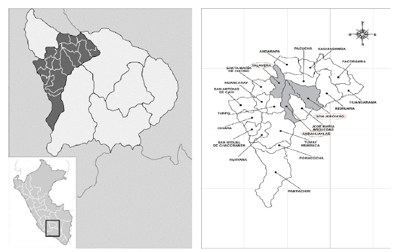
Andahuaylas is a province in the Northeast of the Apurimac region, in the coordinates UTM: 635312 E and 722384 E in the East axis, and 8516644 N and 8391547 N in the North axis, latitude 13°38'55", longitude 73°22'0". Its surface measures 4.034.2 km2 (19.1% of the region) at 2.865 masl. The temperature variates and increases corresponding to the altitude; in the morning it can be around 25°C and at night it falls below 3°C (SENAMHI 2018, INEI 2018). The study was carried out between October and December 2016 in the municipal slaughterhouses of Andahuaylas, San Jeronimo, and Talavera.
Studied animals
In Andahuaylas, the cattle population is divided as follows: 73.608 bovines, 124.589 ovines, 50.786 porcine, 14.914 alpacas, which corresponds to 25.3% bovine, 24.8% ovine 56.3% porcine, and 6.6% alpacas of the complete population of this region (INEI 2012). The studied zone is shown in figure 1.
Authorization for the use of information included in the seizure charts
The retrospective data from the corresponding slaughterhouses have the approval of the National Service of Plant Sanity (Senasa in Spanish) from both the province and the inspectors of Talavera de la Reyna, Andahuaylas, and San Jerónimo (Apurimac Region) (document signed on September 13th, 2018).
Methods of study
Every journey started at 8:00 a.m. with the slaughtering process at slaughterhouses, following all the post-mortem inspection protocols according to the Manual on Good Practices for the Meat Industry (FAO 2007) and the Sanitary Regulations for Slaughtering Animals for Human Consumption (MINAGRI 2012). Thus, the methodology for viscera inspections was carried out: external observation (surface, color), size, and consistency palpation.
Afterward, the liver was placed on a flat surface, where the visceral face and then the base of the caudate lobe were examined to explore the biliary duct, looking for any yellow-greyish collocation inflammation, bleeding, and necrotic areas under the fibrous capsule of the liver and black bile. The veterinarian was the person in charge of the seizure and discard processes.
To establish the origin of the slaughtered animals, an interview with the owners was carried out. Likewise, the age of the animal was determined by observing teeth eruption as well as adult teeth inside the buccal cavity. To estimate the financial loss due to Fasciola hepatica, the livers from all species were weighed immediately after slaughtering and a value for the loss in USD was determined, as shown in table 1.
TABLE 1 Average weight and price of the liver of animals slaughtered in the province of Andahuaylas (2011-2016) (1 USD = 3.22 Peruvian soles)
| Type of Animal | Average weight of the liver | Average price for the kilogram of liver | Average price of the liver |
|---|---|---|---|
| kg | USD | USD | |
| Bovine | 2.75 | 2.76 | 7.59 |
| Porcine | 1.75 | 1.86 | 3.20 |
| Ovine | 0.75 | 2.76 | 2.07 |
| Caprine | 0.75 | 2.76 | 2.07 |
Source: own elaboration.
On the other hand, data corresponding to the period 2011-2016 was recovered. These files were found in poor condition and deteriorated in the municipal warehouses and the archives of the National Service of Agroalimentary Sanity and Quality of Andahuaylas. For this purpose, an epidemiologic card on diseases found in slaughterhouses was used. Then, all the information from the veterinary inspection was digitalized and included in a database (Microsoft Excel 2013) considering the species, sex, and origin of the slaughtered animal. The information was analyzed with the software Epidat 4.2 by using Pearson's Chi-square test to establish differences between proportions, and the t-test to estimate equality of means assuming a confidence level of 95% and confidence intervals of α = 0.05.
RESULTS
Fascioliasis in animals of Andahuaylas (October-December 2016)
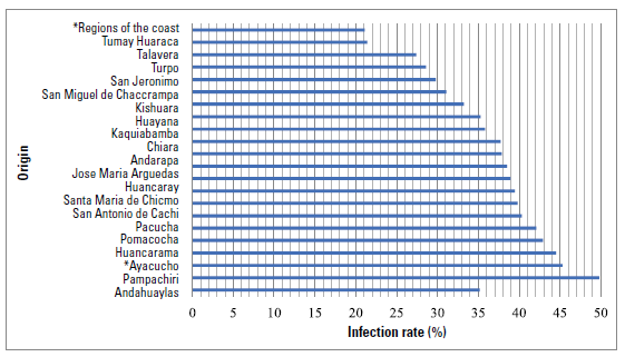
Figure 2 shows the origin of the animals with fascioliasis, with an emphasis on the Pampachiri district with 49%. Likewise, animals from Ayacucho (45.3%) and the Peruvian Coast (21.1%) were put in these slaughterhouses.
Source: own elaboration.
FIGURE 2 Origin of the slaughtered animals and frequency of fascioliasis in slaughterhouses in the province of Andahuaylas, October-December 2016 (p<0.01). *Zones that do not belong to the province of Andahuaylas.
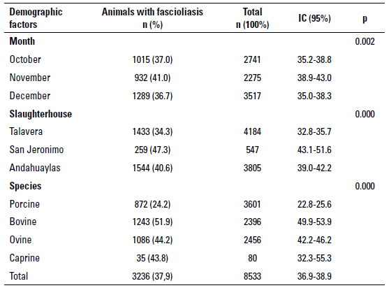
Between October and December 2016, 8.535 animals were slaughtered -porcine (42.2%), bovine (28.1%), ovine (28.8%), and caprine (0.9%)- in the slaughterhouses of Andahuaylas, San Jeronimo, and Talavera. Bovine was the species with the most significant financial loss due to liver seizure as a consequence of fascioliasis (9.434.4 USD), followed by porcine (2.790 USD), ovine (2.248.02 USD), and caprine (72.5 USD). Table 2 shows that fascioliasis had a frequency of 37.9% (IC 95% = 36.9-38.9) during the aforementioned period. November was the month with the most infections (41%) (p < 0.01): San Jeronimo was the Slaughterhouse with the most infections (47.3%) (p < 0.01) and bovine was the most affected species (51.9%) (p < 0.01).
TABLE 2 Fascioliasis in animals slaughtered in the province of Andahuaylas between October and December 2016
Source: own elaboration.
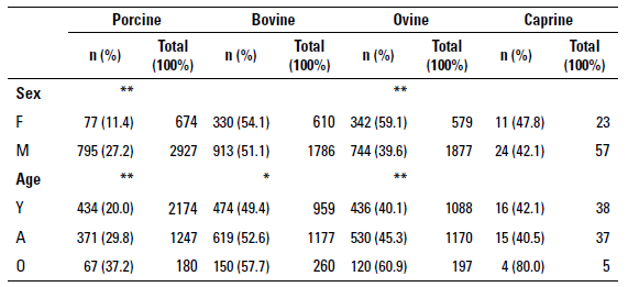
Table 3 shows that male porcine and female ovine developed fascioliasis more frequently than others (27.2% and 59.1% respectively), as well as older porcine, bovine, and ovine: 37.2%, 57.7%, and 60.9%, respectively. On the other hand, table 4 shows there was no significant difference between the average weight of the healthy animal carcasses and the average weight of the cattle carcasses infected with fascioliasis (p > 0.05).
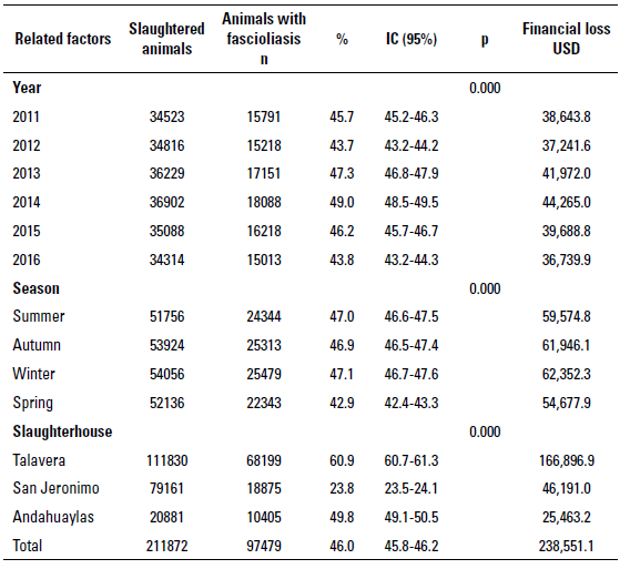
Fascioliasis in animals of Andahuaylas between 2011-2016
During this period, the frequency of fascioliasis was 46% (IC 95% = 45.8-46.2), with a financial loss due to liver seizure of 238.551.1 USD. The most significant percentages of infection (49%) and financial loss occurred during 2014 (44.265 USD) (p < 0.01). Spring was the season with the lowest frequency of fascioliasis (42.9%) and the smallest financial loss (54.677.9 USD) (p < 0.01), as shown in table 5. Even though April was the month with the highest frequency of fascioliasis, the most significant financial loss occurred in March and August (21.033.7 USD) (p < 0.01). On the other hand, Talavera was the slaughterhouse with the highest percentages of infection (60.9%) and financial loss (166.986.9) (p < 0.01). For this period (2011-2016), the average monthly financial loss was 3.313.19 USD, and the average annual loss was 39.758.25 USD.
TABLE 4 Average weight (kg) of healthy and infected with fascioliasis animals' carcasses in slaughterhouses in the province of Andahuaylas between October and December 2016
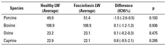
LW=Live weight
Source: own elaboration.
DISCUSSION
Fascioliasis in animals of Andahuaylas (October-December 2016)
Animals coming from the Pampachiri district showed the highest percentage of infection (49.8%), possibly due to its geographic location on the right side of Chicha River (rich in aquatic vegetation), in the highest spot ofAndahuaylas province (3.364 masl), and with mainly cold weather (0°C-11°C in the night) (SENAMHI 2018). It is necessary to consider the fact that, at a higher altitude, the rain season coincides with the summer, when the number of snails of the gender Lymnaea and the risk of infection rise due to the close relationship with the apparition of water, which favors the survival of snails that participate as intermediate hosts (OPS 2021). Thus, extensive breeding enhances the manifestation of the disease, since the animals are in direct contact with the parasite during its contagious stage (Valderrama 2016), disseminating its eggs (Ashrafi 2015).
Bovine was the most affected species during the aforementioned period (51.9%), reaching hyper-endemic levels of infection (> 50%) according to the ranking of Valderrama (2016). This hyper-endemic level in bovine is similar to other reports from different regions of Peru (55.2%-90%) (Carrión-Ascarza et al. 2021; Julon et al. 2020; Mehmood et al. 2017; Valderrama 2016) and other countries of America such as Bolivia, Brazil, Venezuela, Argentina, Mexico, and Colombia (52.4%-96.5%) (Mehmood et al. 2017; Pinilla et al. 2020; Valderrama 2016); Africa: Chad, Sudan, Tanzania, and Uganda (63.8%-91%); Asia: Nepal, Saudi Arabia, Turkey, and Vietnam (51%-69.2%); Europe: Germany, Ireland, Poland, and Wales (53.9%-86%); and Australia in Oceania (86%) (Mehmood et al. 2017).
Unlike other species, bovine would be more vulnerable to getting infected by F. hepatica due to its permanence in less elevated and more humid areas, as well as the search for pastures directly from the soil (Pérez et al. 2010). Ovine, porcine, and caprine probably feed on bush species, which diminishes the probability of exposure to contamination by metacercariae. Due to the resistance of bovine, fascioliasis is rarely a cause of death, but it is more frequent in smaller species (Larroza et al. 2010). In addition, the coexistence with ovine species could be a risk factor, since sheep are more sensitive to infections by Trematoda because they do not develop resistance to new infections, therefore they continuously contribute to contaminating pastures for a long period (Asim et al. 2015), which in turn could be harming for porcine and caprine. Nevertheless, porcine species seem to have the lowest prevalence (24.2%), due to their liver parenchyma and its solid consistency, with a thin membrane composed of thick connective tissue, covered in the mesothelium. Besides, it is divided into polygonal lobes defined by septum of connective tissue, typical of this species (UBA 2022).
Male porcine and female ovine showed the highest frequency offascioliasis (27.2% and 59.1%, respectively). The reason behind this could be the social practice of keeping female porcine and male ovines in much better conditions concerning handling and feeding in comparison to those of the opposite sex living in the pastures (Asim et al. 2015).
Older porcine, bovine, and ovine showed a higher percentage of infection (37.2%, 57.7%, and 60.9%, respectively), as several authors have reported (Carrión-Ascarza et al. 2021; Julon et al. 2020; Valderrama 2016), mentioning that this age group of ruminants is the most affected by Fasciola hepatica in Peru. Older animals tend to show more prevalence for this trematode since they endure a longer period of exposure to the infection. Therefore, elevated levels of infection are not probable in young animals; in addition, mechanisms of cell and humoral immunity protect them against parasites during the first months of life (Asim et al. 2015; Pinilla et al. 2020).
There was no difference between the average weight of healthy animal carcasses and the average weight of fascioliasis infected animals' carcasses, as reported in another study in the province of Abancay. This is explained by the fact that the infectious process, which is progressive and could even last years, depends on multiple factors, such as rusticity, a trait of local animals (Carrión-Ascarza et al. 2021), their origin (Valderrama 2016), and the period of pluvial precipitation, due to the increase in the animals' weight along this season (Merino and Valderrama 2017).
Fascioliasis in animals of Andahuaylas between 2011-2016
During this period, the general frequency of the disease was 46%, which classifies the province ofAndahuaylas as a mesoendemic zone of infection (10%-50%), according to the recommended ranking (Valderrama, 2016). This level ofprevalence, even though in a lower percentage, was like the values reported in the province of Aymaraes (24.6%) as well as in other spots of the country (Valderrama 2016). However, it is lower than what was reported in the province ofAbancay, where hyper-endemic levels of infection (51.3%) were reported (Carrión-Ascarza et al. 2021).
On the other hand, this study showed that the percentage of infection remained between 46.9%-47.1% during summer, autumn, and winter, going considerably down during spring (42.9%), since rain enhances optimal conditions for the development of snails after a drought, mainly when there are periods of heavy precipitations (Valderrama, 2016).
Fasciola hepatica needs humidity in all its stages, which induces variations in the spreading of the disease according to geographic location and season of the year (Ticona et al. 2010). This is why acute epidemics are less frequent in winter (Soca-Pérez et al. 2016). Likewise, droughts and floods have an impact on epidemiology. since droughts disrupt the focal points of snails and floods contribute to their survival and propagation (Valderrama 2016).
Regularly, Fasciola hepatica eggs spread by the feces of infected animals develop in the snails between spring and the beginning of summer, producing cercariae and metacercariae at the end of summer. Therefore, animals that ingest them show symptoms of the disease at the end of autumn and during winter. However, the eggs disseminated by animals, that then infect snails, grow with the mild temperatures of spring, therefore metacercariae appear at the end of spring or the beginning of summer. Then, metacercariae are ingested by animals, causing symptoms during summer and autumn (OPS 2021). The infection originating in winter is inferior to the one in spring, probably because of the high rate of mortality of snails that appear in winter, but especially of the mortality of infected snails (Valderrama 2016).
During drought, snails gather in the stream shores and springs (Soca-Pérez et al. 2016). In this context, animals catch lots of metacercariae, and later, after six or eight weeks they endure the disease in its acute form. During rainy periods, pastures are abundant in metacercariae, but in drought periods, larvae remain in wet and low areas. Cercariae set themselves in the pastures to be eaten by the animals, which explains why there is a high amount of cercariae at the end of summer and autumn, especially when the cattle feed on swampy spots. However, infections appear at the beginning of spring due to the surviving metacercariae (Palacio et al. 2020).
During the period 2011-2016 the average financial damage due to liver seizure was 39.758.25 USD, lower than the numbers reported in Brazil (5.718.813.1 USD) (Quevedo et al. 2018), Mexico (4.239.667 USD) (Rodríguez-Vivas et al. 2017), and Cuba (58.209.1 USD) (Palacio et al. 2020). However, this loss is perceived as highly significant, considering that in the province of Andahuaylas, there is a poverty level of 68.9% and 26.5% of extreme poverty (INEI 2018). Likewise, the average monthly loss was 3.313.19 USD, only similar to what was reported in Abancay (Carrión-Ascarza et al. 2021), Cuba (Palacio et al. 2020), Colombia (Ramírez-Londoño et al. 2020), and Costa Rica (Rojas & Cartín 2016), and other reports showed the minimum monthly loss in Paraguay (Núñez et al. 2017), Cuba (Palacio et al. 2020), Greece (Theodoropoulos et al. 2002), and Iran (Khoramian et al. 2014). This contrast occurs due to different slaughtering and handling techniques performed in every slaughterhouse, as well as to the high percentage of fascioliasis reported (Arias-Pacheco et al. 2020).
Bovine species showed the most significant loss due to liver seizure because of Fasciola hepatica, followed by porcine, ovine, and caprine. This result is like what was found in Abancay (Carrión-Ascarza et al. 2021). Therefore, it is necessary to consider that the financial impact of fascioliasis could be even bigger, since this parasite must be affecting, directly and indirectly, the production and reproduction of animal meat and milk, as well as the development of other diseases, due to the fact the liver is crucial in most of the vital processes of cattle, and connected to the host immunity (Rodríguez-Vivas et al. 2017; Soca-Pérez et al. 2016; Theodoropoulos et al. 2002).

