Servicios Personalizados
Revista
Articulo
Indicadores
-
 Citado por SciELO
 -
 Accesos
Links relacionados
-
 Citado por Google
 -
 Similares en
SciELO
 -
 Similares en Google
Compartir
Colombian Journal of Anestesiology
versión impresa ISSN 0120-3347
Rev. colomb. anestesiol. v.39 n.3 Bogotá jul./oct. 2011
https://doi.org/10.5554/rca.v39i3.247
Reporte de Caso
Anesthetic Management of Congenital Epulis
Ana Sepúlveda Blanco*, Soledad Bellas Cotán**, Ramón Reina González***, Antonio Ontanilla López****
* Médica residente de III año en anestesiología y reanimación, Hospital Infantil Virgen del Rocío, Sevilla, España. Correspondencia: C/Alfonso de Cossio 5 8C CP 41004, Sevilla, España. Correo electrónico: anitasepul@hotmail.com
** Médica. Residente Anestesiología III año. Universidad CES. Medellín. Colombia.
*** Médico adjunto, especialista en anestesiología y reanimación, Hospital Infantil Virgen del Rocío, Sevilla, España.
**** Director, Unidad de Gestión Hospital Infantil Virgen del Rocío, Sevilla, España.
Recibido: febrero 28 de 2011. Enviado para modificaciones: abril 14 de 2011. Aceptado: mayo 18 de 2011.
Summary
Introduction. epulis of the newborn is a granular cell tumor arising in the mucosa of the dental ridge. It presents as a pedunculated soft tissue mass that can be lobular or multinodular. It is more common in females than in males (8:1) perhaps due to hormonal factors. It may be accompanied by other congenital malformations. Anesthetic management is based on a potentially difficult intubation and the risk of bleeding.
Objectives. To present the case of a newborn with congenital epulis and to review this pathology and its anesthetic management.
Methods and Results. Clinical case presentation. Conclusions. Several types of anesthesia have been described depending, among other factors, on tumor size and on the professionals involved in excising the lesion. In our case, and given the characteristics of the tumor, we chose inhalation sedation with O2 / air / sevoflurane, lateral decubitus position and local infiltration at the base of implantation. Good collaboration between the surgeon and the anesthetist is critical for success.
Key Words: Anesthesia, granular cell tumor, congenital abnormalities, laryngoscopy, gingival neoplasms. (Source: MeSH, NLM).
Clinical case
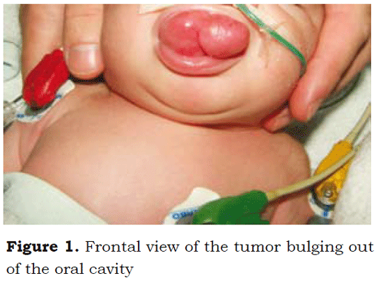
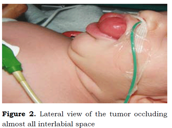
Forty-eight-hour old newborn weighing 3.2 kg, diagnosed with congenital epulis, referred for priority surgical excision. The pre-anesthetic exam was normal for the age of the patient. The examination revealed a large tumor protruding outside the oral cavity and filling almost the entire space between the lips, with discrete sideto- side mobility (Figure 1). Upon palpation, the mass had a gummy consistency and was attached to the upper gingival ridge by means of a pedicle 0.5 cm in length and 3-5 mm thick, with a broader implantation base (Figure 2).
Anesthetic management seemed complex due to a potentially difficult intubation because the size of the tumor prevented adequate direct laryngoscopy. Additionally, there was a potential risk of bleeding, characteristic of these tumors. Of the various options described in the literature for the management of these patients, a local anesthetic infiltration at the implantation base was selected, together with the use of a vasoconstrictor (1 % lidocaine + epinephrine), lateral decubitus positioning and facial mask sedation with a 50 % O2/ air mix plus 3 % sevofluorane, under spontaneous breathing.
The tumor was excised successfully (Figure 3). After reinforcing hemostasis with the electro scalpel and gauze compression, the intervention was completed with the patient fully awake, and transfer back to the Neonatology Unit.
Discussion
Congenital epulis was first described by Neumann in 1871. It is a lesion arising from the alveolar crest in neonates, typically localized on the maxillary crest, particularly in the area of the incisor and canine teeth. It may vary in size, from just a few millimeters up to 9 cm in diameter. The main differential diagnoses include Bohn's nodules, Epstein's pearls, mucocele, eruption cyst, infantile melanotic neuroectodermal tumor, and Abrikossoff's tumor (1). Because of is location, this lesion may impair breathing or feeding. After surgical removal, there is no relapse or dental abnormality, and there are very few reports of spontaneous involution. Histologically, it is a granular cell tumor (2) rarely associated with other major congenital abnormalities, except for polydactily and neurofibromatosis (3).
Multiple theories have been proposed to explain its histogenesis, and one of the most widely accepted is the possible influence of ovarian hormones on the fetus, considering that the incidence is higher among female fetuses compared to males (8:1). Consequently, the proposal for prenatal diagnosis is to conduct immunohistochemistry tests for estrogen influence and progesterone receptors (4,5). However, that theory is still under investigation and most congenital epulis are postnatal findings.
There is no consensus regarding the anesthetic management of these patients (6,7) who are a true challenge for the anesthetist because of the risk of bleeding as well as the obvious airway compromise.
Canavan Holliday and Lawson propose orotracheal intubation with the patient on spontaneous ventilation, using a mix of halothane and oxygen under direct laryngoscopy for induction, with the help of an assistant whose role is to mobilize the epulis gently towards the oral commissure. Merrett and Crawford describe the excision of small epulis masses under local anesthesia. Swami et al. prefer general anesthesia and orotracheal intubation in order to secure the airway, given the possibility of bleeding and aspiration. In our case, the infiltration with the local anesthetic, the role of the vasopressor in preventing intraoperative bleeding, and rapid excision, contributed to a less aggressive anesthetic management. Also, critical for the success of the operation were the participation of experienced nursing staff and a good communication between the surgeon and the anesthetist.
Conclusions
Epulis represents a true challenge for the anesthetist, in particular as concerns airway management. Given the absence of guidelines, the different options must be carefully assessed depending on the case and the expertise of the staff involved.
REFERENCES
1. Kizlansky V, Saint Genez D, Casas G, et al. Épulis congénito. Dermatol Pediatr Latinoam. 2009;7:38- 41.
2. Senoo H, Iida S, Kishino M, et al. Solitary congenital granular cell lesion of tongue. Oral Surg Oral Med Oral Pathol Oral Radiol Endod. 2007;104:e45-8.
3. Canavan-Holliday KS, Lawson RA. Anaesthetic management of the newborn with multiple congenital epulides. Br J Anaesth. 2004;93:742-4.
4. Subramaniam R, Shah R, Kapur V. Congenital epulis. J Postgrad Med. 1993;39:36.
5. Reinshagen K, Wessel LM, Roth H, et al. Congenital epulis: A rare diagnosis in pediatric surgery. Eur J Pediatr Surg. 2002;12:124-6.
6. Kusukawa J, Kuhara S, Koga C, et al. Congenital granular cell tumor (congenital epulis) in the fetus: a case report. J Oral Maxillofac Surg. 1997;55:1356-9.
7. Merret SJ, Crawford PJM. Congenital epulis of the newborn: a case report. Int J Paediatr Dent. 2003;13:127-9.
1. Kizlansky V, Saint Genez D, Casas G, et al. Épulis congénito. Dermatol Pediatr Latinoam. 2009;7:38- 41. [ Links ]
2. Senoo H, Iida S, Kishino M, et al. Solitary congenital granular cell lesion of tongue. Oral Surg Oral Med Oral Pathol Oral Radiol Endod. 2007;104:e45-8. [ Links ]
3. Canavan-Holliday KS, Lawson RA. Anaesthetic management of the newborn with multiple congenital epulides. Br J Anaesth. 2004;93:742-4. [ Links ]
4. Subramaniam R, Shah R, Kapur V. Congenital epulis. J Postgrad Med. 1993;39:36. [ Links ]
5. Reinshagen K, Wessel LM, Roth H, et al. Congenital epulis: A rare diagnosis in pediatric surgery. Eur J Pediatr Surg. 2002;12:124-6. [ Links ]
6. Kusukawa J, Kuhara S, Koga C, et al. Congenital granular cell tumor (congenital epulis) in the fetus: a case report. J Oral Maxillofac Surg. 1997;55:1356-9. [ Links ]
7. Merret SJ, Crawford PJM. Congenital epulis of the newborn: a case report. Int J Paediatr Dent. 2003;13:127-9. [ Links ]


 texto en


