Servicios Personalizados
Revista
Articulo
Indicadores
-
 Citado por SciELO
 -
 Accesos
Links relacionados
-
 Citado por Google
 -
 Similares en
SciELO
 -
 Similares en Google
Compartir
Colombian Journal of Anestesiology
versión impresa ISSN 0120-3347
Rev. colomb. anestesiol. v.39 n.4 Bogotá oct./dic. 2011
https://doi.org/10.5554/rca.v39i4.235
Reporte de Caso
Anesthetic Management and Radiological Findings in Craniopagus Conjoined Twins on Nuclear Magnetic Resonante
Roberto Rivera Díaz*, José Luis Ascencio Lancheros**, Valentina Cifuentes Hoyos**
* Anestesiólogo. Docente de Anestesia y Dolor, Universidad CES, Instituto Colombiano del Dolor. Correspondencia: Carrera 48 No. 19A-40 Unidad 1205 Torre Médica Ciudad del Río, Medellín, Colombia. Correo electrónico: robertorivera@incodol.com
** Neuroradiólogo. Instituto Neurológico de Antioquia. Medellín, Colombia.
*** Medica general. Docente Facultad de Medicina Universidad CES. Medellín, Colombia.
Recibido: mayo 29 de 2011. Enviado para modificaciones: agosto 22 de 2011. Aceptado: septiembre 3 de 2011.
Summary
The prevalence of conjoined twins is extremely low, but whenever a case occurs, it is of the utmost importance to have a multidisciplinary team consisting of an anesthesiologist, a radiologist, a pediatrician, and specialists in various surgical areas, depending on the type of conjoined twins. Likewise, it is critical to consider all the physiological, pharmacodynamic and anatomical implications, including cross circulation. The goal is to plan appropriately in order to enable the best possible result. This report is intended to present a case of 3 day-old craniopagus conjoined female twins scheduled for nuclear magnetic resonance (MRI).
Keywords: Twins, conjoined, craniopagus, cross circulation, magnetic resonance spectroscopy, anesthesia, airway management. (Source: MeSH, NLM).
Introduction
The prevalence of the craniopagus type of conjoined twins is 1 out of every 2.5 million live births. Only 6.2 % of conjoined twins are joined at the head (craniopagus), representing the least common type (1).
There are many anesthetic implications and indications for the management of conjoined twins, considering that they may need anesthesia or sedation before definitive surgery for diagnostic procedures such as nuclear magnetic resonance (2), computed tomography, echocardiography, cerebral angiography, or for other surgical procedures (application of silicone expanders to promote progressive skin stretching and help with flap reconstruction during the separation procedure) (3).
The most significant anesthetic challenges in these patients include airway management, positioning to avoid neurological lesions, trauma due to airway compression or obstruction, associated pathologies, and the percentage of cross circulation (a factor of significant pharmacodynamic and pharmacokynetic importance with hemodynamic implications, and which is more pronounced in this type of conjoined twins) (4).
Descripción del caso
Three day-old female craniopagus conjoined twins scheduled at the Neurological Institute of Antioquia for plain magnetic resonance imaging (MRI) of the head, the chest and the abdomen, using contrast for vein resonance. Approximate duration of the scan: 3 hours.
Anesthetic Management
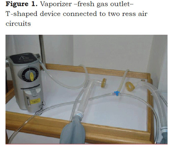
Once the informed consent was signed by the parent, an anesthesiologist and an anesthesia resident initiated basic monitoring (oxymetry, respiratory band, cardioscope) using MRI-compatible monitoring and anesthesia equipment. An oxygen source connected to a sevofluorane vaporizer was used. A three-way stopcock or a T-shaped device was placed at the outlet of the fresh gas for connection to air ress circuits (Figure 1).
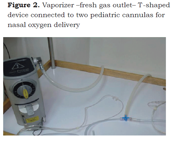
The anesthesiologist and the resident initiated induction simultaneously with at a fresh gas flow rate of 4 liters per minute and 4% sevofluorane. Once the two babies were unconscious the two ress air circuits were exchanged at the level of the three-way stopcock for two pediatric nasal cannulas (Figure 2), the oxygen flow rate was brought down to 2 liters and the sevofluorane concentration was maintained at 2.5 % during the rest of the procedure.
After checking the position of the babies, small pillows were used to avoid abnormal neck extension. During the 3 hours of the study, the patients remained adequately sedated and immobilized, and there were no adverse events such as desaturation or a drop in respiratory rate. The halogenated agent was interrupted at the end of the procedure and the girls woke up after 90 seconds.
Radiological Findings
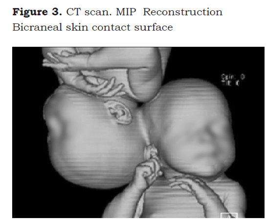
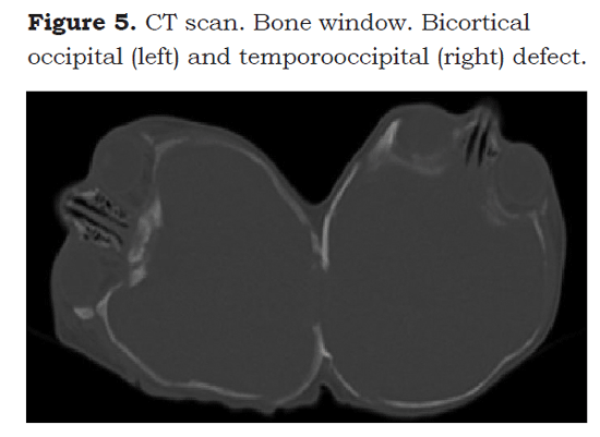
A CT scan was performed using a 6-channel multidectector scanner in order to obtain the anatomical data for the bone and soft tissue windows. Multi-planar reconstructions (MPR) and maximum intensity projections (MIP) were also obtained. A bicortical occipital bone defect was identified in one of the twins and a temporo-occipital defect was identified in the other twin (Figures 3, 4 y 5).
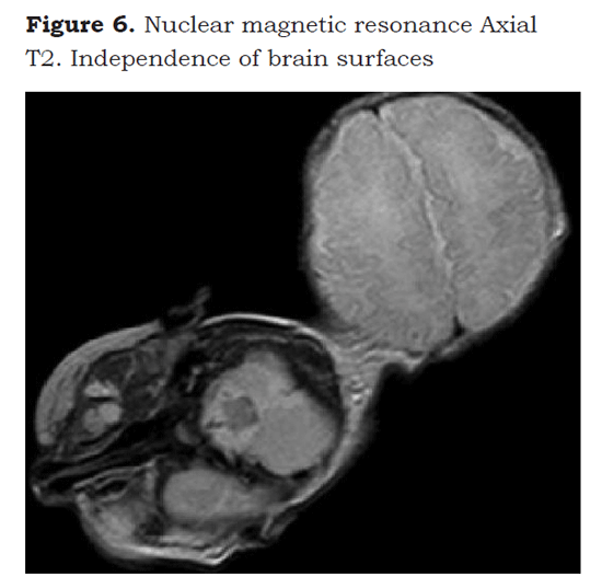
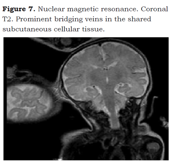
Using a 1.5 Tesla superconductor magnet, a high-resolution MRI was performed, showing integrity and independence of the cerebral cortices, and absence of cortical developmental malformations in the supratentorial and the infratentorial compartments. In the shared subcutaneous cellular tissue, the presence of several prominent shunted veins was identified (Figures 6 y 7).
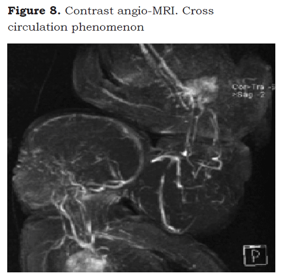
In the angio-MRI sequence performed in the arterial and contrast venous phases, the presence of shunted arterial polygons or of dural venous sinuses was ruled out in both patients. It is worth noting that the contrast injection (gadolinium) revealed cross circulation because when one of the twins was injected, the circulatory system of her sister was immediately visualized (Figure 8).
Discussion
Since they were initially reported in the literature, conjoined twins have created curiosity, and that is even more so with the rare cases of craniopagus twins. Conjoined twins are genetically identical and, consequently, they are of the same sex (more often female than male, in a 4:1 proportion). No association has been established between this occurrence and race, maternal age or parity, or inheritance.
Relatively few craniopagus twins survive the perinatal period; approximately 40 % are born and an additional 33 % die during the neonatal period, usually as a result of congenital abnormalities of other organs. On the other hand, it is estimated than more than 90 % of craniopagus twins die before 10 years of age (5).
Craniopagus conjoined twins are defined on the basis of the cranial fusion only, and the definition excludes fusions involving the foramen magnum, the skull base, the vertebrae or the face itself. The chest and abdomen are separate, and each twin has its own umbilicus and umbilical cord.
O’Connell (6) was the first to develop a comprehensive classification for this type of conjoined twins, distinguishing at first two categories: partial and total. In partial craniopagus twins only a limited area is affected, with an intact skull or only minimal cranial defects; total craniopagus twins are defined as sharing an extensive surface area with a large connection between the cranial cavities.
Another classification that contributes importantly to the interpretation of this fusion is that by Bucholz et al. (7), who further classified craniopagus conjoined twins into 4 types: frontal, parietal, temporoparietal and occipital. O’Connell also went on to make an additional subdivision of parietal craniopagus based on the degree of rotation of one of the heads in relation to the other. Thus, in type 1 both twins face in the same direction; in type 2, they face in opposite direction at a rotation angle greater than 140°; and in type 3, there is an intermediate rotation angle of the longitudinal axis of one head in relation to the other (8).
In craniopagus twins there can also be fusion of some regions of the cerebral cortex, and neurovascular abnormalities, both venous as well as arterial; moreover, they may share bone and soft tissue. The finding of a shared dural venous sinus is of the highest significance because it increases the risk associated with surgical separation. Most failed separations have resulted in hemorrhagic complications, due mainly to the lack of expertise of the surgeon and the presence of dural venus sinus variants; hence the importance of careful assessment and preoperative planning. For this reason, new imaging techniques have been used to reconstruct the fusion, and it is of vital importance to remember that the anatomy is unique in every case of craniopagus conjoined twins (5).
Craniopagus conjoined twins have other surgical risks requiring cardiac, pulmonary and renal function workup in order to ensure an optimal condition before taking them to surgery, because the surgical procedure may involve additional stress factors such as neuroanesthesia, blood loss, cerebral edema and cerebral perfusion during the procedure.
Several risk factors need to be considered when it comes to the surgical separation of craniopagus conjoined twins: 1) The degree of shared scalp; 2) The degree of shared cranium; 3) The extent of share dura; 4) The extent of fused cortex; 5) The extent of shared arterial connections and cross circulation; 6) The extent of common venous sinuses; 7) The presence or absence of independent deep venous drainage; and 8) The presence or absence of a common or separate ventricular system, or of hydrocephalus. All these factors have a direct influence on the size and timing of the surgical strategy (5).
It has been mentioned that, before separation surgery, the twins may need anesthesia for different types of procedures. The technique will depend on many variables including the length and invasiveness of the procedure, the analgesic requirements, the positioning, and the need or not for endotracheal intubation. In this particular case report, the indication for anesthesia was determined by the type of study: nuclear magnetic resonance lasting approximately three hours and requiring immobilization in order to ensure good quality images.
The review of the literature showed that the techniques employed are based on intravenous drugs such as fentanyl, ketamine and propofol. In our research group we used a sedation technique with sevofluorane as single agent at low concentrations to ensure unconsciousness. The procedure did not involve a pain stimulus and, consequently, did not require the use of other drugs or anesthetic concentrations of the halogenated agent. The procedure was completed in its entirety with adequate hemodynamic stability and no difficulties.
It is recommended to delay the separation surgery until the twins have gained sufficient weight and are able to better tolerate the blood loss. Case reports mention more than 50 % mortality when the separation surgery is performed during the first four months of age. There are similar cases where several silicone expander implantation surgeries were performed over the previous months before definitive separation in order to gain extra scalp to facilitate the closure of the defect. This strategy could be very useful for the type of conjoined twins presented in this case report (9).
Conclusions
Because the prevalence of conjoined twins is extremely low, all the literature on the subject is based on case reports. Successful separations have been achieved thanks to a multidisciplinary team approach, relevant workup and good planning. Reports refer to sevofluorane as an alternative for anesthesia induction and maintenance. The latter has been accomplished using balanced techniques with intravenous drugs (10).
The team in charge of this case report that sedation with sevofluorane may be an adequate option in these cases, particularly for diagnostic procedures where the goal is to ensure immobility and a minimal or absent painful stimulus in order to facilitate the study. Moreover, the researchers confirm the finding described in the literature of the cross circulation phenomenon that is so pronounced in craniopagus conjoined twins.
REFERENCES
1. Huang WQ, Fang JY, Xiao LC, et al. Anesthetic management for separation of craniopagus twins. Acta Anaesthesiol Scand. 2004;48:919-21.
2. Parameswari A, Vakamudi M, Raghupathy V, et al. Anaesthetic management of total craniopagus twins for magnetic resonance imaging and cerebral angiography. Br J Anaesth. 2010;105:368-70.
3. Girshin M, Broderick C, Patel D, et al. Anesthetic management of staged separation of craniopagus conjoined twins. Paediatr Anaesth. 2006;16:347-51.
4. Thomas JM. Anaesthesia for conjoined twins. Childs Nerv Syst. 2004;20:538-46.
5. Browd SR, Goodrich JT, Walker ML. Craniopagus twins. J Neurosurg Pediatrics. 2008;1:1-20.
6. O’Connell JE. Craniopagus twins: surgical anatomy and embryology and their implications. J Neurol Neurosurg Psychiatry. 1976;39:1-22.
7. Bucholz RD, Yoon KW, Shively RE. Temporoparietal craniopagus. Case report and review of the literature. J Neurosurg. 1987;66:72-9.
8. Stone JL, Goodrich JT. The craniopagus malformation: classification and implications for surgical separation. Brain. 2006;129:1084-95.
9. Khan ZH, Tabatabai SA, Saberi H. Anesthetic and surgical experience in a case of total vertical craniopagus. Surg Neurol. 1999;52:62-7.
10. Pietrini D, Valenti M, Pusateri A, et al. Perioperative management of face-to-face craniopagus twins separation. Pediatric Anesthesia. 2005;15:519-24.
1. Huang WQ, Fang JY, Xiao LC, et al. Anesthetic management for separation of craniopagus twins. Acta Anaesthesiol Scand. 2004;48:919-21. [ Links ]
2. Parameswari A, Vakamudi M, Raghupathy V, et al. Anaesthetic management of total craniopagus twins for magnetic resonance imaging and cerebral angiography. Br J Anaesth. 2010;105:368-70. [ Links ]
3. Girshin M, Broderick C, Patel D, et al. Anesthetic management of staged separation of craniopagus conjoined twins. Paediatr Anaesth. 2006;16:347-51. [ Links ]
4. Thomas JM. Anaesthesia for conjoined twins. Childs Nerv Syst. 2004;20:538-46. [ Links ]
5. Browd SR, Goodrich JT, Walker ML. Craniopagus twins. J Neurosurg Pediatrics. 2008;1:1-20. [ Links ]
6. O'Connell JE. Craniopagus twins: surgical anatomy and embryology and their implications. J Neurol Neurosurg Psychiatry. 1976;39:1-22. [ Links ]
7. Bucholz RD, Yoon KW, Shively RE. Temporoparietal craniopagus. Case report and review of the literature. J Neurosurg. 1987;66:72-9. [ Links ]
8. Stone JL, Goodrich JT. The craniopagus malformation: classification and implications for surgical separation. Brain. 2006;129:1084-95. [ Links ]
9. Khan ZH, Tabatabai SA, Saberi H. Anesthetic and surgical experience in a case of total vertical craniopagus. Surg Neurol. 1999;52:62-7. [ Links ]
10. Pietrini D, Valenti M, Pusateri A, et al. Perioperative management of face-to-face craniopagus twins separation. Pediatric Anesthesia. 2005;15:519-24. [ Links ]


 texto en

