


Services on Demand
Journal
Article
 text in
  English (pdf)
 Article in xml format
 Article references
 Send this article by e-mail
Indicators
-
 Cited by SciELO
 -
 Access statistics
Related links
-
 Cited by Google
 -
 Similars in
SciELO
Similars in
SciELO -
 Similars in Google
Share

 Permalink
Colombian Journal of Anestesiology
Print version ISSN 0120-3347
Rev. colomb. anestesiol. vol.40 no.2 Bogotá Apr./June 2012
https://doi.org/10.1016/S0120-3347(12)70030-8
http://dx.doi.org/10.1016/S0120-3347(12)70030-8
Case report
Tracheotomy Cannula as a Foreign Body in Pediatric Airway Management
Cánula de traqueostomía como cuerpo extraño en vía aérea pediátrica
Víctor Daniel Giraldo Cobo*
MD, Anesthesiologist, Hospital Universitario del Valle, Cali, Colombia
Corresponding author: calle 5 # 36-08, Cali, Colombia. E-mail: victorvictordan@yahoo.com (V.D. Giraldo Cobo).
ARTICLE INFO
Article history: Received: December 20, 2011 Accepted: February 14, 2012
ABSTRACT
Proper airway management is a challenge for every anesthesiologist. During a shift in the emergency room, a difficult airway often appears both in cases of trauma and medical pathology. The tracheotomy cannula is a device suited to ensure airway opening of patients with obstruction or lesion of the upper respiratory tract. In this medical case, a 7 year old female child is admitted at the emergency service with an episode of respiratory distress caused by rupture of a silver tracheotomy cannula, previously implanted due to a submandibular tumoral lesion. This is a rare case with little investigation in international medical literature The case was approached with interdisciplinary management including Pediatric surgery and Pulmonology with satisfactory outcome.
Keywords: Tracheotomy Airway management Anesthesia Lymphangioma
© 2011 Sociedad Colombiana de Anestesiología y Reanimación. Published by Elsevier.
All rights reserved.
RESUMEN
El manejo adecuado de la vía aérea es un reto para el médico anestesiólogo. Durante un turno de urgencias es frecuente, tanto en traumatismos como en patologías médicas, la presentación de casos clínicos con vía aérea difícil. La cánula de traqueostomía es un dispositivo diseñado para permeabilizar el tracto respiratorio de pacientes con obstrucciones o lesiones en la vía aérea superior. En este caso clínico, se presenta a una niña de 7 años de edad que ingresó al servicio de urgencias con episodio de dificultad respiratoria por rotura de una cánula de plata de traqueostomía que se le había colocado por lesión tumoral submandibular, caso de poca presentación y con pocos reportes en la literatura médica internacional. Fue manejado en forma interdisciplinaria con el grupo de cirugía pediátrica y neumología pediátrica, con buen resultado clínico.
Palabras clave: Traqueostomía Manejo de la vía aérea Anestesia Linfangioma
© 2011 Sociedad Colombiana de Anestesiología y Reanimación. Publicado por Elsevier.
Todos los derechos reservados.
Case report
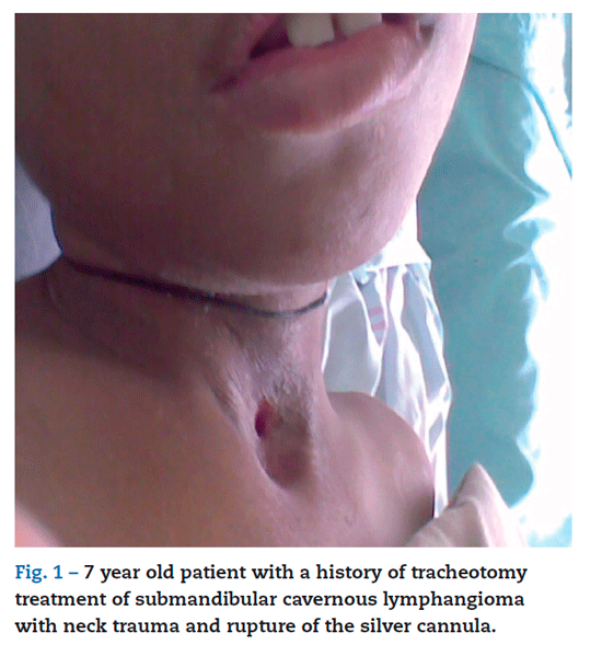
This is the case of a seven year old female patient with a history of a submandibular cavernous lymphangioma type lesion1 extended to the supraglottic area, undergoing outpatient care management for several years with a tracheotomy cannula.2
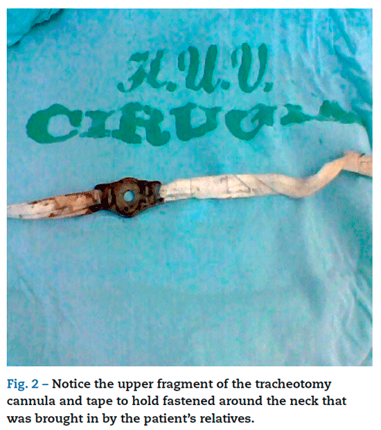
The child had neck trauma caused by a fall while playing outside, which resulted in rupture and fragmenting of the silver tracheotomy cannula3 and subsequent airway obstruction by the a piece of the cannula. Her family arrives at the Hospital Universitario del Valle Pediatric Emergency service in Cali with the patient4,5 due to an episode of mild respiratory distress and showed the physicians the upper fragment of the cannula (figs. 1 and 2).
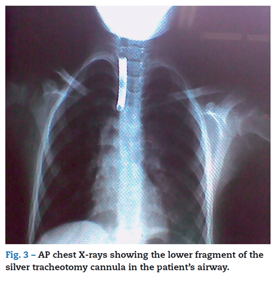
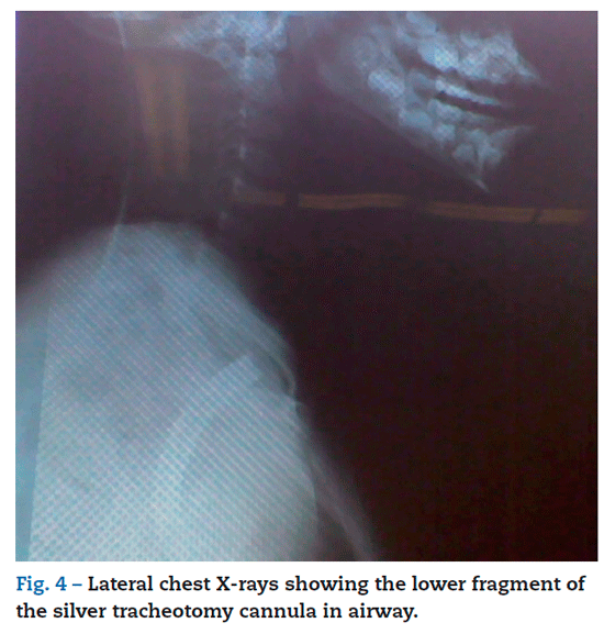
Pediatric Surgery assessment requested emergency admittance into the operating room for foreign body extraction from the airway.6-8. Anterior-posterior and lateral chest X-rays are carried out with portable X-ray equipment (figs. 3 and 4) showing the lower fragment of the cannula in the patient's airway.10,11
Medical history
Pathologic record. A submandibular mass is found at birth, which progressively increased its size and compromised the airway at a supraglottic level, causing respiratory distress, seizures, gastrointestinal reflux, duodenitis and recurrent pneumonia.
Surgical history. At age 11 months, a biopsy sample of the mass is taken, an open tracheotomy and placement of a silver cannula are carried out.
Biopsy findings. Cavernous lymphangioma.1
Pharmacological management. Valproic acid suspension 5 ml every 12 hours.
Anesthetic management
Once the case was reported, we reviewed the medical record and carried out physical examination and assessment of the X-rays9 jointly with the Pediatric Surgery and Pulmonology services. Clinical, surgical and anesthetic management hypotheses are established12-15 considering different alternatives during the procedure of extraction.16-19
Surgical procedure plan
We are equipped with a flexible bronchoscope,20 a rigid bronchoscope and a pediatric surgery equipment for performing a foreign body extraction from the airway in a pediatric patient.21
Basic monitoring, cardioscopy in EKG lead II, pulse oximetry, non-invasive blood pressure measurement with pediatric bracelet and capnography16,17 were carried out. The patient was placed in supine and Semi-fowler position.22 The patient weighed 20 kg and was previously oxygenated
using a no. 2 pediatric mask. We used ranitidine22 and metoclopramide for aspiration prophylaxis. We gave 1 mg of midazolam and begun intravenous anesthesia with 50 mg of sodium thiopental and continued maintenance with 3-4% sevofluorane, achieving spontaneous respiration
during the entire procedure. An oropharyngeal Guedel airway cannula was placed. Local infiltration at tracheotomy orifice was performed by the pediatric surgeon using 20 mg of 1% lidocaine. Next, the surgeon carried out an incision surrounding the ostium, expanded the orifice and extracted the lower piece of the silver cannula through the
tracheotomy opening with a Kelly surgical clamp. There were no complications during the procedure (fig. 5).

We continue to secure the airway with a no. 4 pediatric endotracheal tube through the orifice of the tracheotomy and the patient adapted to the manual ventilation system, sustaining spontaneous respiration. Considering the event of neck trauma during the the patient's fall and the history of the supraglottic mass, Pediatric Pulmonology proceeds to a diagnostic flexible bronchoscopy. Findings reported a displaced, pale, lobed mass in hypopharynx compatible with a cavernous lymphangioma type lesion obstructing the airway in supraglottic and parapharyngeal areas; and observation of vocal chords was difficult.
We decided to continue airway management with a plastic tracheotomy cannula. The endotracheal tube was then removed and a no. 5 plastic tracheotomy cannula through the opening of the tracheotomy. The patient tolerated the procedure well. A 30 mg/kg dose of dipyrone is given as analgesia together with 0.6 mg of dexamethasone. Once the patient regained consciousness, she initially remained in the post-anesthetic care room. She was awake, hemodynamically stable, and in her family's company. She was later transferred to clinical post-operative monitoring and management of respiratory therapy in the Anna Frank Pediatric Surgery ward at Hospital Universitario del Valle, Cali with a positive outcome. Before discharge, the plastic cannula was replaced by a Ruschelit Biesalski.
Discussion
Interdisciplinary management is crucial for patients with airway obstruction by a foreign body. This strategy provides alternatives during the initial approach and case management from anesthetic and surgical standpoints,16,17 properly assessing different risk and benefit variables.
The patient arrived at the emergency service after neck trauma. Aspiration prophylaxis was fulfilled with ranitidine22 and metoclopramide. In this case's management of airway trauma no muscular relaxants were used during anesthesia, therefore preserving physiological mechanisms of spontaneous ventilation in order to prevent collapsing of structures of the damaged airway. Also during anesthesia, proper and balanced inhalation and intravenous anesthetic techniques were ensured. For this, the algorithm for difficult airway management and ASA guides must be taken into account12,13.
Nevertheless, anesthetic management through controlled ventilation with use of muscle relaxants for extraction of foreign bodies in the aerodigestive tract is also an option. It shows lower incidences of lung atelectasis, cardiac arrhytmias and myocardic depression; associated to lower doses of inhaled and intravenous agents.23
It is necessary to provide the operation room with diagnostic and medical equipment as well as surgical tools for emergency airway management; including sternotomy equipment for distal approach of the intrathoracic trachea when securing the airway in extremely urgent cases.24
Once the medical, surgical and anesthetic managements are established and therapeutic alternatives are clear, the priority procedure must be carried out immediately. Additionally, a place in the pediatric intensive care unit must be requested for post-operative management.
Morbidity and complications that may occur during the extraction of foreign bodies from the aerodigestive tract must be considered25. The patient's family must be notified and asked for authorization to carry out the procedure with informed consent.
The silver tracheostomy cannula is a device suited for airway management of pediatric and adult patients with severe obstructive lesions. Its use was favored in the past for outpatient care, though currently it is progressively decreasing due to advances in new tracheotomy cannulae3. The weakening of the metallurgic alloy caused by oral secretions eventually leads to deterioration of these devices. It is difficult to determine in this case whether if the cannula was structurally damaged, or if the metal or the welding contributed to its breaking associated to trauma.
Although common in emergency services, obstruction by foreign bodies in the aerodigestive tract such as this is unusual: pediatric trauma resulting in airway obstruction by the tracheotomy cannula due to a tumoral lesion management.
Funding
Author's own resources.
Conflict of interests
None declared.
Acknowledgements
To the staff in the operating room of the Hospital Universitario del Valle, Cali, for their collaboration and work, and the doctors who participated in this case, Dr Guillermo Sarmiento, Paediatric surgeon, and Dr Alvaro Sánchez, Paediatric Chest Physician (RIP).
REFERENCES
1. Regezi-Sciubba. Patología bucal. Correlaciones clinicopatológicas. 3.a ed. McGraw-Hill Interamericana; 2000.
2. López MA, Aragón MF. Conceptos actuales en traqueostomía. Acta Otorrinolaringol Cir Cabez Cuello. 2002;304.
3. Tracheostomy tubes and related appliances. Respiratory Care. 2005;50.
4. Guildner CW, Williams D, Subitch T. Airway obstructed by foreign material: the Heimlich maneuver. JACEP. 1976;5:675-7.
5. Redding JS. The choking controversy: critique of evidence on the Heimlich maneuver. Crit Care Med. 1979;7:475-9.
6. Echandia CA. Aspiración de cuerpo extraño. Colombia Med. 1995;26:21-6.
7. Méndez M, Molina I, Bonilla E. Aspiración y deglución de cuerpos extraños: revisión de 1034 casos. Rev Colomb Anestesiol. 1993;21:21-5.
8. Trujillo ML, Villamizar JE. Cuerpos extraños en vía aerodigestiva en los niños. Experiencia de siete años, Hospital Universitario Erasmo Meoz. Rev Med Unab. 2008;11:195-200.
9. Weber AL. Radiologic evaluation of the trachea. Chest Surg Clin North Am. 1996;6:637-73.
10. Ospina J. Cuerpos extraños en el tracto aerodigestivo infantil. El papel del otorrinolaringólogo pediatra. Acta Otorrinolaringol Cir Cabez Cuello. 2005;33:36-46.
11. Digoy P. Diagnosis and manegement of upper aerodigestive tract foreign bodies. Otolaryngol Clin North Am. 2008;41: 485-96.
12. Practice Guidelines for management of the difficult airway an update report by the American Society of Anesthesiologist Task Force on Management of the Difficult Airway. Anesthesiology. 2003;98:1269-77.
13. Coombes X, Le Roux B, Suen P, Dumerat M, Motamed C. Unanticipated difficult airway in anesthetized patients: prospective validation of a management algorithm. Anesthesiology. 2004;100:1146-50.
14. Iohom G, Ronayne M, Cunningham AJ. Prediction of difficult tracheal intubation. Eur J Anesthesiol. 2003;20:31-6.
15. Mallampati SR, Gatt SP, Gugino LD. A clinical sign to predict difficult tracheal intubation: A prospective study. Can J Anesth. 1985;32:429.
16. Benumof JL. Anesthesia for Thoracic Surgery. 2.a ed. Philadelphia: WB Saunders; 1995.
17. Arango E, Hernández V, Rey A, Niño C, Raffan F. Manejo perioperatorio del paciente para resección de la tráquea. Acta Otorrinolaringol Cir Cabez Cuello. 2002;30.
18. Vergara JC. Anatomía quirúrgica de la laringe y la tráquea. Acta Otorrinolaringol Cir Cabez Cuello. 2002;30.
19. Techniques for performing tracheostomy. Respiratory Care. 2005;50.
20. Flores S, García R, Núñez C. Extracción de cuerpos extraños de la vía aérea en niños mediante broncoscopia flexible. Rev Ins Nal Enf Resp Mex. 2005;18:103-8.
21. Fernández JH, Villareal E, Cepeda JR. Broncoscopia y traqueostomía, simultáneas, para la extracción de cuerpo extraño. Rev Mex Pediatr. 1994;61:29-30.
22. Gómez LM, Jaramillo J, Osorio J, Reyes G, Salcedo E. Guía de práctica clínica: manejo de la vía aérea del paciente pediátrico con estómago lleno II parte. Rev Colomb Anestesiol. 2007;35:183-94.
23. Jacob R, Cote C, Thirlwell J. Entendiendo la anestesia pediátrica. 2.a ed. BI Publications; 2010.
24. Vargas A, Noriega J. Manejo de vía aérea en resección de tráquea por toracotomía. Rev Colomb Anestesiol. 2006;34: 191-5.
25. Baquero, Guijarro E. Cuerpo extraño en esófago causal de fístula traqueoesofágica. Reporte de un caso. Salud Uninorte. 2002;16:45-52.
1. Regezi-Sciubba. Patología bucal. Correlaciones clinicopatológicas. 3.a ed. McGraw-Hill Interamericana; 2000. [ Links ]
2. López MA, Aragón MF. Conceptos actuales en traqueostomía. Acta Otorrinolaringol Cir Cabez Cuello. 2002;304. [ Links ]
3. Tracheostomy tubes and related appliances. Respiratory Care. 2005;50. [ Links ]
4. Guildner CW, Williams D, Subitch T. Airway obstructed by foreign material: the Heimlich maneuver. JACEP. 1976;5:675-7. [ Links ]
5. Redding JS. The choking controversy: critique of evidence on the Heimlich maneuver. Crit Care Med. 1979;7:475-9. [ Links ]
6. Echandia CA. Aspiración de cuerpo extraño. Colombia Med. 1995;26:21-6. [ Links ]
7. Méndez M, Molina I, Bonilla E. Aspiración y deglución de cuerpos extraños: revisión de 1034 casos. Rev Colomb Anestesiol. 1993;21:21-5. [ Links ]
8. Trujillo ML, Villamizar JE. Cuerpos extraños en vía aerodigestiva en los niños. Experiencia de siete años, Hospital Universitario Erasmo Meoz. Rev Med Unab. 2008;11:195-200. [ Links ]
9. Weber AL. Radiologic evaluation of the trachea. Chest Surg Clin North Am. 1996;6:637-73. [ Links ]
10. Ospina J. Cuerpos extraños en el tracto aerodigestivo infantil. El papel del otorrinolaringólogo pediatra. Acta Otorrinolaringol Cir Cabez Cuello. 2005;33:36-46. [ Links ]
11. Digoy P. Diagnosis and manegement of upper aerodigestive tract foreign bodies. Otolaryngol Clin North Am. 2008;41: 485-96. [ Links ]
12. Practice Guidelines for management of the difficult airway an update report by the American Society of Anesthesiologist Task Force on Management of the Difficult Airway. Anesthesiology. 2003;98:1269-77. [ Links ]
13. Coombes X, Le Roux B, Suen P, Dumerat M, Motamed C. Unanticipated difficult airway in anesthetized patients: prospective validation of a management algorithm. Anesthesiology. 2004;100:1146-50. [ Links ]
14. Iohom G, Ronayne M, Cunningham AJ. Prediction of difficult tracheal intubation. Eur J Anesthesiol. 2003;20:31-6. [ Links ]
15. Mallampati SR, Gatt SP, Gugino LD. A clinical sign to predict difficult tracheal intubation: A prospective study. Can J Anesth. 1985;32:429. [ Links ]
16. Benumof JL. Anesthesia for Thoracic Surgery. 2.a ed. Philadelphia: WB Saunders; 1995. [ Links ]
17. Arango E, Hernández V, Rey A, Niño C, Raffan F. Manejo perioperatorio del paciente para resección de la tráquea. Acta Otorrinolaringol Cir Cabez Cuello. 2002;30. [ Links ]
18. Vergara JC. Anatomía quirúrgica de la laringe y la tráquea. Acta Otorrinolaringol Cir Cabez Cuello. 2002;30. [ Links ]
19. Techniques for performing tracheostomy. Respiratory Care. 2005;50. [ Links ]
20. Flores S, García R, Núñez C. Extracción de cuerpos extraños de la vía aérea en niños mediante broncoscopia flexible. Rev Ins Nal Enf Resp Mex. 2005;18:103-8. [ Links ]
21. Fernández JH, Villareal E, Cepeda JR. Broncoscopia y traqueostomía, simultáneas, para la extracción de cuerpo extraño. Rev Mex Pediatr. 1994;61:29-30. [ Links ]
22. Gómez LM, Jaramillo J, Osorio J, Reyes G, Salcedo E. Guía de práctica clínica: manejo de la vía aérea del paciente pediátrico con estómago lleno II parte. Rev Colomb Anestesiol. 2007;35:183-94. [ Links ]
23. Jacob R, Cote C, Thirlwell J. Entendiendo la anestesia pediátrica. 2.a ed. BI Publications; 2010. [ Links ]
24. Vargas A, Noriega J. Manejo de vía aérea en resección de tráquea por toracotomía. Rev Colomb Anestesiol. 2006;34: 191-5. [ Links ]
25. Baquero, Guijarro E. Cuerpo extraño en esófago causal de fístula traqueoesofágica. Reporte de un caso. Salud Uninorte. 2002;16:45-52. [ Links ]
