


Servicios Personalizados
Revista
Articulo
 texto en
  Inglés (pdf)
 Articulo en XML
 Referencias del artículo
 Enviar articulo por email
Indicadores
-
 Citado por SciELO
 -
 Accesos
Links relacionados
-
 Citado por Google
 -
 Similares en
SciELO
Similares en
SciELO -
 Similares en Google
Compartir

 Permalink
Colombian Journal of Anestesiology
versión impresa ISSN 0120-3347
Rev. colomb. anestesiol. vol.40 no.4 Bogotá oct./dic. 2012
https://doi.org/10.1016/j.rca.2012.07.006
http://dx.doi.org/10.1016/j.rcae.2012.08.002
Interesting images
Correcting the tetralogy of Fallot: The role of the transesophageal intraoperative echocardiography
Corrección de tetralogía de Fallot: papel del ecocardiograma transesofágico intraoperatorio>
David M. Orozco Vinasco *, Mauricio Abello Sanchez, Javier Osorio Esquivel
Department of Anesthesia, Fundación Clínica Shaio, Bogotá, Colombia article info
* Please cite this article as: Orozco Vinasco DM, et al. Corrección de tetralogía de Fallot: papel del ecocardiograma transesofágico intraoperatorio.
* Corresponding author at: Fundación Clínica Shaio, Diagonal 115A N 70c-75, Oficina de Anestesia, Bogotá, Colombia. E-mail address: david.orozco@shaio.org (D.M. Orozco Vinasco).
© 2012 Published by Elsevier España, S.L. on behalf of Sociedad Colombiana de Anestesiología y Reanimación.
ARTICLE INFO
Article history: Received 8 April 2012 Accepted 17 July 2012 Available
online 19 September 2012
Abstract
The classical approach to evaluate the correction of the tetralogy of Fallot (TOF) consists in calculating the ratio between the right systolic and systemic pressure; however, with the advent of transesophageal echocardiography, new approaches have been developed.
We shall present a case of a 15-year-old patient with TOF who underwent correction and the management was based on the intraoperative echocardiography.
We will also address the role of the echocardiographic parameters in the evaluation of the correction of the TOF.
Keywords: Echocardiography, Tetralogy of Fallot, Perioperative period, Anesthesia
© 2012 Published by Elsevier España, S.L. on behalf of Sociedad Colombiana de Anestesiología y Reanimación.
Resumen
La forma clásica de evaluación de la corrección de la tetralogía de Fallot (TDF) consiste en el cálculo de la relación entre las presiones sistòlica ventricular derecha y sistèmica. Sin embargo, el advenimiento del ecocardiograma transesofágico ha desarrollado nuevos enfoques al respecto.
Presentaremos el caso de un paciente de 15 años con TDF el cual fue llevado a corrección, y cuyo manejo se basó en el ecocardiograma intraoperatorio.
También discutiremos el papel de los parámetros ecocardiográficos en la evaluación de la corrección de la TDF.
Palabras clave: Ecocardiografía Tetralogía de Fallot Periodo perioperatorio Anestesia
© 2012 Publicado por Elsevier España, S.L. en nombre de Sociedad Colombiana de Anestesiología y Reanimación.
Clinical case
Transesophageal echocardiography has become an invaluable tool to the cardiovascular anesthesiologist because it provides detailed information about the anatomy and cardiac function. It has become the standard of care in valve replacement surgery, aortic and coronary artery surgery.1
Its use has also been validated in congenital heart surgery, and has shown to be useful not just for evaluating the corrections, but also as a guideline in hemodynamic therapy.2
The TOF is characterized by the presence of a ventricular septal defect (VSD), valvular or sub-valvular obstruction of the right ventricular outflow tract (RVOT), dextroposition of the aorta with overriding, and right ventricle hypertrophy. The broad spectrum of this malformation, the dynamic physiological changes, including the effects of the anesthetic agents, makes it a really challenging lesion. A case is presented where the surgical approach was based on the intraoperative trans-esophageal echocardiography.
A 15-year-old patient was referred to us for the correction of a TOF. The patient was scheduled for closure of the VSD with patch and expansion of the RVOTF.
Following an uneventful anesthetic induction and initiation of mechanical ventilation, a transesophageal echocar-diographic probe was placed; as part of complete study, we evaluated the following characteristics, specifically prior to the correction and once again after the cardiopulmonary bypass3:
Determination of the peak and mean gradient between the right ventricle and the pulmonary artery.
Color Doppler examination of the inter-ventricular septum to outline the defect and assess the patch closure.
Right Ventricular Ejection Fraction (RVEF) in a mid-esophageal 4-chamber view.
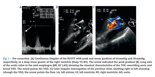
Our echocardiographic examination showed the classical findings of a TOF: extensive ventricular septal communication with right to left shunt, severe hypertrophy of the right
ventricle with mild RVEF depression calculated at 35%, RV outlet tract obstruction with peak and mean gradients of 66 and 29mmHg, respectively (Fig. 1).
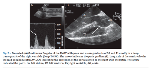
The patient was operated on using a transatrial, transpul-monary approach and 100 min of pump time; following the correction, but prior to decannulation, a control echocardio-gram was done, with the following results: ventricular patch with no residual shunt, left alignment of the aorta, reduced peak and mean RV outlet tract obstruction to 29 and 11 mmHg, mild pulmonary failure, unchanged RVEF (Fig. 2).
The correction was considered adequate,4 and the patient was transferred to the Pediatric Intensive Care Unit (PICU) for two days and was discharged from hospital after six days of being admitted.
In our case report, the right ventricular function and the evaluation of the RVOTF were the two most important echocardiographic measurements in the management of the patient.
The right ventricle is a complex structure that contrary to the left ventricle cannot be described with conventional geometric models and this makes the evaluation of the ventricular function more difficult.
There are several echocardiographic parameters to assess the systolic function of the right ventricle, such as the right Tei index, displacement of the tricuspid ring, fractional area change and volumetric methods; however, none have been validated in the same manner as the ejection fraction for the left ventricle.
Any of these measurements could have been used in our patient; what is really important is the comparison between before and after the correction. The obstruction of the RVOTF determines the severity of the symptoms in this particular heart disease and hence determining the RVOTF gradient during the post-repair period is key to only to prevent a low cardiac output in the immediate postop, but during re-operations in the long term.
The classical approach to estimate the RVOTF gradient is to calculate the ratio between the right ventricle systolicpressure divided into the systemic systolic pressure; if the result is equal or less than 0.75, the correction is considered acceptable.4
The advent of intraoperative transesophageal echocardi-ography has led to the exact determination of the peak and mean RVOTF gradient using the continuous Doppler method according to Bernoulli's principle that establishes the relationship between flow acceleration and the increase in pressure through an orifice. In the case of correcting a TOF, a mean gradient above 54 mmHg is an indication for revision of surgery.4
The instant mean gradient represents a more physiological measurement than the relationship between the systolic pressure of the right ventricle and the systemic pressure, because this relationship will be affected by multiple factors; i.e., in the case of systemic hypotension the ratio increases but does not represent a change in the degree of obstruction of the RVOTF.
To conclude, we would like to highlight the benefits of trans-esophageal echocardiography for evaluating the correction of the TOF.
Funding
Authors' own funds.
Conflict of interest
None declared.
REFERENCIAS1. Practice guidelines for perioperative transesophageal echocardiography. An updated report by the American Society of Anesthesiologists and the Society of Cardiovascular Anesthesiologists task force on transesophageal echocardiography. Anesthesiology. 2010;112: 1-7.
2. Garg R, Murthy K, Rao S, Muralidhar K. Intraoperative transesophageal echocardiography in congenital heart disease. Ann Card Anaesth. 2009;12:173.
3. Joyce J, Hwang E, Wiles H, Bradley SM, Crawford FA. Reliability of intraoperative transesophageal echocardiography during tetralogy of Fallot repair. Echocardiography. 2000;17:319-27.
4. Kaushal S, Radhakrishanan S, Dagar K, Iyer P, Girotra S, Shrivastava S, et al. Significant intraoperative right ventricular outflow gradients after repair for tetralogy of Fallot: to revise or not to revise? Ann Thorac Surg. 1999;68:1705-13.
1. Practice guidelines for perioperative transesophageal echocardiography. An updated report by the American Society of Anesthesiologists and the Society of Cardiovascular Anesthesiologists task force on transesophageal echocardiography. Anesthesiology. 2010;112: 1-7. [ Links ]
2. Garg R, Murthy K, Rao S, Muralidhar K. Intraoperative transesophageal echocardiography in congenital heart disease. Ann Card Anaesth. 2009;12:173. [ Links ]
3. Joyce J, Hwang E, Wiles H, Bradley SM, Crawford FA. Reliability of intraoperative transesophageal echocardiography during tetralogy of Fallot repair. Echocardiography. 2000;17:319-27. [ Links ]
4. Kaushal S, Radhakrishanan S, Dagar K, Iyer P, Girotra S, Shrivastava S, et al. Significant intraoperative right ventricular outflow gradients after repair for tetralogy of Fallot: to revise or not to revise? Ann Thorac Surg. 1999;68:1705-13. [ Links ]
