


Services on Demand
Journal
Article
 text in
  English (pdf)
 Article in xml format
 Article references
 Send this article by e-mail
Indicators
-
 Cited by SciELO
 -
 Access statistics
Related links
-
 Cited by Google
 -
 Similars in
SciELO
Similars in
SciELO -
 Similars in Google
Share

 Permalink
Colombian Journal of Anestesiology
Print version ISSN 0120-3347
Rev. colomb. anestesiol. vol.43 no.4 Bogotá Oct./Dec. 2015
Scientific and Technological Research
Infraclavicular block in paediatric anaesthesia: Concordance between the modified Wilson approach and ultrasound in determining the ideal puncture site*
Abordaje infraclavicular en pediatría: concordancia del abordaje de Wilson modificado y el ultrasonido para la localización del sitio de punción ideal
Andrea Carolina Pérez-Pradillaa,b,**, Ana Angélica Peña-Riverónc, Laura Catalina Chaparro-Gómezb, Lailigh Citlallis Castro-Ortizb, Elizabeth Velázquez-Delgadob, Rosangel Acevedo de la Peñab
a Paediatric Anaesthetist, Instituto Roosevelt de Ortopedia Infantil, Bogotá D.C., Colombia
b Anaesthetist, Higher Specialization Postgraduate Resident, Instituto Nacional de Rehabilitación, Mexico, Federal District, Mexico
c Anaesthetist and Pain Specialist, Anaesthesia Division, Instituto Nacional de Rehabilitación, Mexico, Federal District, Mexico
* Please cite this article as: Pérez-Pradilla AC, Peña-Riverón AA, Chaparro-Gómez LC, Castro-Ortiz LC, Velázquez-Delgado E, Acevedo-de la Peña R. Abordaje infraclavicular en pediatría: concordancia del abordaje de Wilson modificado y el ultrasonido para la localización del sitio de punción ideal. Rev Colomb Anestesiol. 2015;43:269-277.
** Corresponding author at: Calzada México Xochimilco N° 289, Colonia Arenal de Guadalupe, CP 14389, Torre de Ortopedia, División de Anestesiología, México, Distrito Federal, México.
E-mail address: cperezpradilla@gmail.com (A.C. Pérez-Pradilla).
Article info
Article history: Received 26 January 2014 Accepted 1 July 2015 Available online 20 August 2015
Abstract
Introduction: The modified Wilson infraclavicular approach (MWIA) was described in our institution for brachial plexus blocks in paediatric patients. However, concordance studies between this approach and ultrasound for the identification of ideal puncture site have no been reported.
Objective: To determine the concordance between MWIA and ultrasound for localization of the ideal puncture site.
Materials and methods: Descriptive observational study; we included 100 healthy patients between 1 and 16 years of age, with parental consent, over a 5-month period. Continuous variables were described and kappa statistics were used for concordance evaluation. We also conducted a multivariate analysis to confirm the relationship between the measured distances and weight and height.
Results: The distance fromthe skin to the brachial plexus, aswell as the distance between the coracoid process and the brachial plexus, and the distance from the coracoid process to the pleura were all smaller in abduction, with no statistically significant difference. Height and weight are independent factors that determine the distance between the coracoid process and the posterior cord, both in adduction and abduction. The concordance of MWIA vs. ultrasound for determining the ideal puncture site was 47% in both positions.
Conclusions: Concordance between MWIA and ultrasound for the determination of the ideal puncture site is low when it comes to anatomic localization; however, this technique must be evaluated in randomized clinical studies in order to determine its efficacy and usefulness. Height and weight are independent factors that determine the distance between the coracoid process and the posterior cord in adduction and abduction.
Keywords: Anesthesia, Conduction ultrasonography, Brachial plexus, Pediatrics, Nerve block.
Resumen
Introducción: En pacientes pediátricos, el sitio óptimo de inyección para el abordaje infraclavicular sigue siendo sujeto de debate; no hay estudios de concordancia entre el abordaje infraclavicular de Wilson modificado para bloqueo del plexo braquial (AIWM) y el ultrasonido para la localización del sitio ideal de punción.
Objetivo: Determinar la concordancia entre AIWM y ultrasonido para localizar el sitio ideal de punción.
Materiales y métodos: estudio observacional descriptivo; se incluyeron 100 niños sanos entre 1-16 años, en un periodo de 5 meses. Se describieron variables continuas y se evaluó la concordancia mediante la estadística kappa. Se realizó análisis multivariado para confirmar la correlación entre las distancias, el peso y la talla.
Resultados: Se encontró una menor profundidad del plexo braquial y menor distancia entre el plexo braquial y la apófisis coracoides y entre la coracoides y la pleura en abducción, sin significancia estadística. El peso y la talla son factores independientes que determinan la distancia entre la apófisis coracoides y el cordón posterior en abducción y en aducción. La concordancia del sitio ideal de punción entre AIWM y ultrasonido fue 0,47 en ambas posiciones.
Conclusiones: La concordancia entre el AIWM y el ultrasonido para determinar el sitio ideal de punción es sorprendentemente baja. Sin embargo, desde el punto de vista de utilidad y eficacia clínica, estas técnicas deben ser comparadas y evaluadas con estudios clínicos aleatorizados. El peso y la talla determinan de forma independiente la distancia entre el cordón posterior y la apófisis coracoides en aducción y abducción del brazo.
Palabras clave: Anestesia de conducción, Ultrasonografía, Plexo braquial, Pediatría, Bloqueo nervioso.
Introduction
According to studies conducted in the Netherlands, distal radius and wrist fractures account for approximately 25% of all paediatric fractures1 and traumatic injuries involving the hand range between 1800 and 2600 per 100,000 inhabitants per year in the Netherlands and Denmark, respectively.2 There are also congenital disorders involving the upper limb in 0.16-0.18% of live births in the United Kingdom.3
Significant technological and scientific advances in the field of regional anaesthesia over the past decade have led to improvements in our knowledge of the anatomy, and to better surgical approaches, with improved safety profiles. Together with greater availability of resources, this has promoted the use of regional anaesthesia in paediatric patients.4-9 However, the use of regional anaesthesia techniques in children is limited by the size of the structures, the proximity of the neural structures to critical structures (pleura, blood vessels), poorly defined superficial landmarks, a tight safety margin, a higher potential for toxicity related to local anaesthetics, and the need for sedation or general anaesthesia, which might mask any signs of alarm (paraesthesias).10,11
The infraclavicular approach for brachial plexus blocks is indicated for hand, forearm, elbow and arm surgery (the shoulder excluded). Its advantages include a higher probability of achieving axillary and musculocutaneous nerve blocks when compared with the axillary approach; ease of fixation of the continuous analgesia catheters; lower impact on pulmonary function when compared to the supraclavicular and the interscalene approaches; and no arm abduction requirement.12,13 Disadvantages include the possibility of not achieving adequate posterior cord block when single injections are used; also, in the event of vascular puncture due to the depth of the structures, it is difficult to apply compressive manoeuvres.12
There are variations to the infraclavicular block: the cora-coid approach,14,15 the lateral sagittal approach,16 the vertical approach,17 and the modified Raj approach.18,19 The coracoid approach is the most popular in the United States.
The coracoid approach was first described by Whiffler21 and later modified by Wilson who, with the help of nuclear magnetic resonance imaging (NMR), determined that the ideal puncture site in adults was 2 cm medial and 2 cm caudal to the lateral tip of the coracoid process.15
In paediatric patients, the optimal injection site for the infraclavicular approach is still controversial.11 The modified Raj and the vertical approaches are considered "dangerous" because of the proximity of the puncture site to the cervical pleura.22,23 There are no publications at the present time in relation to Whiffler's approach or Wilson's modification in children. However, a modified Wilson technique for paediatric patients was developed at the National Rehabilitation Institute, consisting of using the coracoid process as the anatomical landmark and finding the puncture site medial and caudal to it according to the patient's weight. This modification has not been published before.
In 1999, Kapral described another modification of Whiffler's technique called the "lateral vertical" approach.24 Kapral reported a high rate of success using this technique (1 %) with a broad range of nerve blocks with a broad range of nerve block spectrum (including the musculocutaneous, tho-racodorsal, axillary and medial cutaneous brachial nerves) and absence of complications in adult patients.24 Later, Fleis-chmann and Ponde reported the use of this technique in paediatric patients, modifying the location of the puncture site, this time 0.5 cm caudal to the coracoid process, with similar results as those found by Kapral.25,26 In 2004, Klaastad described the "lateral sagittal" technique in adults;27 however, there are few reports on the use of this technique in paediatric patients.28,29
The first report of an ultrasound-guided brachial plexus block through the infraclavicular approach in paediatric patients was described by Marhofer, who compared this technique with a neurostimulation-guided lateral vertical approach in a randomized clinical trial. He found shorter latency periods and longer block durations in the ultrasound group, with no difference in block quality after 30 min.30
Despite the many advantages of ultrasound-guided techniques, the main limitations are still the cost of the equipment and the need for specialized training. Moreover, it does not totally eliminate the risk of inadvertent intraneural injection.31 On the other hand, safety recommendations in regional anaesthesia include triple monitoring (i.e., neurostimulation, ultrasound and injection pressure measurements).32
The objective of our study was to determine concordance between the modified Wilson infraclavicular approach and the use of ultrasound for the anatomic localization of nerve structures and of the ideal puncture site.
Materials and methods
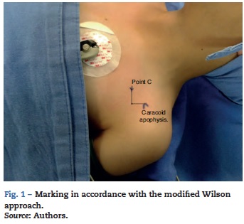
A descriptive observational study was conducted. Patient recruitment was initiated once the approval of the Scientific Research Commission and the Ethics and Research Committee of the National Rehabilitation Institute was obtained. Patients included healthy children 1-16 years of age scheduled for open or closed reduction surgery for fractures of any extremity; patients with anatomical variants obscuring the visualization of the cords in the infraclavicular region were excluded. The parents of the children signed an informed consent and patients 12 or older signed an informed assent. Puncture site (C point) marking was done in accordance with the modified Wilson infraclavicular (MWIA) coracoid approach, as follows: for patients weighing less than 10 kg, 1 cm caudal and 1 cm medial to the coracoid process; for patients weighing 10-20 kg, 1.5 cm caudal and 1.5 cm medial to the coracoid process; for patients weighing more than 20 kg, 2 cm caudal and 2 cm medial to the coracoid process (Fig. 1).
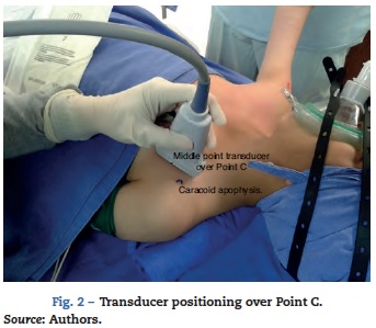
A Sonosite Micromaxx (Bothell, WA, USA) ultrasound machine with a high-frequency linear transducer (13-6 MHz) was used for making measurements of any of the two extremities. The centre point of the transducer was placed on the C point marked as MWIA in a parasagittal orientation (Fig. 2), and the necessary depth and focus adjustments were made in order to ensure visualization of the target structures.
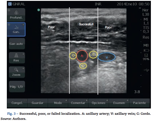
Localization was rated as "successful" when the axillary artery and the brachial plexus cords were visualized in the central third of the screen; "poor" if the axillary artery and the brachial plexus cords were seen on the lateral or medial third of the image; and "failed" when these structures were not visualized (Fig. 3).
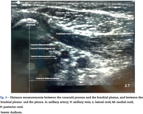
The Vienna classification described by Marhofer et al. was used to evaluate quality of imaging visualization.33 Additionally, distance measurements were made from the most prominent point in the coracoid process to the brachial plexus cords, from the skin to the cords, from the pleura to the coracoid process, and the position of the cords in relation to axillary artery was described (Fig. 4). For image acquisition, the transducer was positioned on an oblique sagittal plane.
Measurements were made with the arm in adduction and 90° of abduction with the patient under sedation or general anaesthesia for the scheduled surgical procedure. It is important to mention that the ultrasound scans were performed by two investigators wit experience in ultrasound-guided infraclavicular blocks.
The sample size was determined on the basis of convenience, requiring 100 patients over a 5-month period. The data were entered in an Excel database, and the STATA 10 software was used for data analysis. A measurement of central tendency was made for continuous variables. Kappa statistics were used to evaluate concordance between nominal variables. A linear regression was also performed in order to confirm the correlation between the distance from the coracoid process to the posterior cord and weight and height.
Results
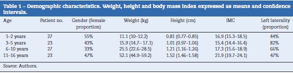
The study enrolled 100 patients. Demographic characteristics are shown by age group (Table 1). The patients were distributed homogenously among age groups. Gender distribution was also homogenous except in the 6-10 group (33% female patients). Children under S tended to have a low BMI (body mass index) (<18).
There is a significant difference in distances measured from the coracoid process to the brachial plexus in adduction and abduction between patients <10kg and those 11-20 kg. However, in patients of more than 20 kg there is no significant difference. Distances from the skin to the cords, the coracoid process to the cords, and from the coracoid process to the pleura were shorter in abduction in all age groups. However, the difference was not statistically significant (Tables 2 and 3).
Localization of the target anatomical structures using ultrasound on "C point" in patients under 10 kgfailed in 8.4% of patients, both in adduction as well as abduction; it was poor in 41% of patients in adduction and in 33.3% in abduction; and it was successful only in 66% of patients in adduction and S8.3% of patients in abduction.
In patients 10-20 kg, localization of the target anatomical structures using ultrasound on "point C" failed in 13% of patients in adduction and 0% in abduction; it was poor in 41% of patients in adduction and in 43% in abduction; and was successful in 46% of patients in adduction and S6% in abduction.
In patients over 20 kg, localization of the target anatomical structures using ultrasound on "point C" failed in 13% of patients in adduction and in 4.1% in abduction; it was poor in 49% in adduction and in 59.1% in abduction; and it was successful in 38% in adduction and in 36% in abduction. No statistically significant differences were found in terms of successful ultrasound identification in adduction or abduction according to the weight of the patients.
In terms of the quality of the visualization, 88% of the measurements were given a score of 2, and the rest were given a score of 3 under the Vienna classification. There were no statistically significant differences between age groups regarding the quality of visualization.
A multivariate analysis was conducted in order to determine the correlation between the distance from the coracoid process to the posterior cord in accordance with weight and height, and a statistically significant correlation was found in adduction (weight: p = 0.015, height: p <0.01) and in abduction (weight and height p < 0.01).
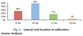
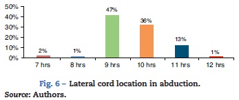
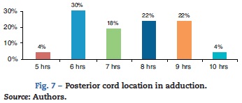
Position of the cord in the brachial plexus in relation to the axillary artery was evaluated in adduction and abduction, using a clock distribution. Considering that no intervention was performed and cords were, therefore, not identified using neurostimulation, cord positions described in previous anatomical studies were used. The lateral cord was defined as the most superficial and closest to the coracoid process; the posterior cord was defined as the most posterior in relation to the artery; and the medial cord was defined as the one most frequently located between the axillary artery and vein.
The lateral cord was found between 7 and 12 o'clock, and the most frequent location was at 10 o'clock in adduction (50%) and 9 o'clock in abduction (47%) (Figs. 5 and 6).
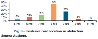
The posterior cord was found between 5 and 11 o'clock. The most frequent location was at 6 o'clock in adduction (30%) and at 8 o'clock in abduction (42%) (Figs. 7 and 8).
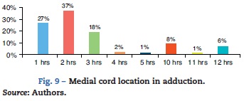
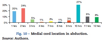
The greatest location variability was found for the medial cord, mainly in abduction. The most frequent location was at 2 o'clock in adduction (37%) and at 10 o'clock in abduction (27%) (Figs. 9 and 10).
Concordance between the modified Wilson approach and ultrasound for determining the ideal puncture site was 0.47 in adduction and abduction.
Discussion
The objective of this study was to determine concordance between the MWIA and ultrasound for the location of the ideal anatomical puncture site, considering the lack of clinical studies regarding this topic in the literature, and the fact that there is only one similar study in adult patients. The study by Kuo et al.,34 assessed the accuracy of Wilson's coracoid approach for the location of the ideal puncture site and compared it with ultrasound in adult patients. It was found that the accuracy rate for the landmark-based approach was 74.4%.34 In our study, concordance between anatomical landmarks (MWIA) and ultrasound for identifying the ideal puncture site was only 0.47, suggesting that the landmark-based approach might not be adequate for predicting the ideal puncture site in paediatric patients. However, due to the usual individual anatomical variability, it is not very probable that a method guided by anatomical landmarks can be successful in 100% of cases without the need for needle repositioning.
It is important to mention that, given the size of paediatric patients, a 1 cm distance difference, perhaps irrelevant in adults, might be quite significant in a child. There are other factors influencing success with the first attempt, including the entry angle of the needle in relation to the skin, which might lead to a failed block even when the exact entry point is determined by anatomical landmarks or by other means. In this regard, ultrasound provides the advantage of direct visualization of needle orientation.
Multiple studies have been conducted in adult populations using nuclear magnetic resonance (NMR) in order to advance the development of landmark-based approaches.15,27,35,36 However, the use of NMR or computed tomography-based studies in healthy children is limited by the need to provide sedation or general anaesthesia, mainly in smaller children who might suffer adverse events associated with anaesthetic agents or the exposure to ionizing radiation.37-40 From this perspective, studies using ultrasound are warranted.
The study by Ruiz et al. assessed the influence of arm abduction on the distance of the axillary artery from the skin, the coracoid process and the pleura, using ultrasound in adult patients. They reported a shorter depth of the axillary artery in abduction, but they did not find any differences in pleural depth in abduction, or regarding the distance between the coracoid process and the axillary artery.41 The study by Greher et al. sought to identify the accuracy of the vertical infraclavicular approach developed by Kilka by means of ultrasound measurements in healthy adults. They found that the overall accuracy of Kilka's vertical approach was not sufficient to predict the optimal puncture site.22
In our study, there was no statistically significant difference in the depth of the brachial plexus, the distance between the cords and the coracoid process, or the distance between the coracoid process and the pleura in abduction. This is consistent with the results from studies performed in adults. Ultrasound may possibly underestimate the depth of the brachial plexus structures due to the compressive effect from the transducer. The measurements obtained suggest that the distance between the coracoid process and the brachial plexus is modified to a greater degree in children under 20 kg. Additionally, it was found that weight and height are independent variables that modify the distance between the coracoid process and the posterior cord. This suggests that a formula based on weight and height should be developed for children under 20 kg in order to calculate the distance between the coracoid process and the brachial plexus when it comes to performing infraclavicular blocks.
As for the location of the brachial plexus cords in relation to the axillary artery, it was found to be highly variable in particular for the medial cord. In an earlier study by Sauter et al. using NMR in adult patients, the authors found the brachial plexus cords between 3 and 11 o'clock.35 A limitation of our study was that the cords were not identified using neurostimulation, hence our inability to assert that they were correctly identified.
Patients were not studied bilaterally, and this may also be a limitation of this study, considering reports of individual right/left asymmetry in 61% of cases.12
In one of its publications, the Paediatric Regional Anaesthesia Network (PRAN) reported a higher tendency towards performing ultrasound-guided peripheral blocks in the upper extremity (82%), leading to the assumption that ultrasound availability may explain the increasing number of peripheral nerve blocks.4 In the most recent study by ADARPEF (L'Association Des Anesthésistes-Réanimateurs Pédia-triques d'Expression Francaise), there was also a higher trend towards the performance of peripheral nerve blocks in pae-diatric patients.42 Despite the advantages of ultrasound,31 availability of these machines is low in many developing countries, hence the continuing use of landmark and neurostimulation-guided approaches.
Conclusions
There is low concordance between the modified Wilson infraclavicular coracoid approach and the use of ultrasound for determining the ideal puncture site; however, no clinical studies have been conducted to assess the success, the need for needle repositioning or the complications of this approach.
Weight and height are independent factors that determine the distance between the coracoid process and the posterior cord in adduction as well as in abduction. The infraclavicular brachial plexus is slightly more superficial and lies closer to the coracoid process in upper limb abduction; there is also variability in the distribution of the cords around the axillary artery in adduction as well as in abduction.
Funding
The authors did not receive sponsorship to undertake this article.
Conflict of interest
The authors have no conflicts of interest to declare.
Ethical disclosures
Protection of human and animal subjects. The authors declare that no experiments were performed on humans or animals for this study.
Confidentiality of data. The authors declare that they have followed the protocols of their work centre on the publication of patient data.
Right to privacy and informed consent. The authors have obtained the written informed consent of the patients or subjects mentioned in the article. The corresponding author is in possession of this document.
References
1. De Putter CE, van Beeck EF, Looman CW, Toet H, Hovius SE, Selles RW. Trends in wrist fractures in children and adolescents, 1997-2009. J Hand Surg Am. 2011;36:1810-5. [ Links ]
2. Shah SS, Rochette LM, Smith GA. Epidemiology of pediatric hand injuries presenting to United States emergency departments, 1990 to 2009. J Trauma Acute Care Surg. 2012;72:1688-94. [ Links ]
3. Bates SJ, Hansen SL, Jones NF. Reconstruction of congenital differences of the hand. Plast Reconstr Surg. 2009;124:128e-43e. [ Links ]
4. Polaner DM, Taenzer AH, Walker BJ, Bosenberg A, Krane EJ, Suresh S, et al. Pediatric Regional Anesthesia Network (PRAN): a multi-institutional study of the use and incidence of complications of pediatric regional anesthesia. Anesth Analg. 2012;115:1353-64. [ Links ]
5. Bosenberg A. Neuraxial blockade and cardiac surgery in children. Paediatr Anaesth. 2003;13:559-60. [ Links ]
6. Bosenberg A. Pediatric regional anesthesia update. Paediatr Anaesth. 2004;14:398-402. [ Links ]
7. Bosenberg AT. Epidural analgesia for major neonatal surgery. Paediatr Anaesth. 1998;8:479-83. [ Links ]
8. Kehlet H. Epidural analgesia and postoperative outcome. Lancet. 2008;372:2109. [ Links ]
9. Kehlet H. Fast-track colorectal surgery. Lancet. 2008;371:791-3. [ Links ]
10. Tsui B, Suresh S. Ultrasound imaging for regional anesthesia in infants, children, and adolescents: a review of current literature and its application in the practice of extremity and trunk blocks. Anesthesiology. 2010;112:473-92. [ Links ]
11. Willschke H, Marhofer P, Machata AM, Lonnqvist PA. Current trends in paediatric regional anaesthesia. Anaesthesia. 2010;65 Suppl. 1:97-104. [ Links ]
12. Neal JM, Gerancher JC, Hebl JR, Ilfeld BM, McCartney JL, Franco CD, et al. Upper extremity regional anesthesia: essentials of our current understanding, 2008. Reg Anesth Pain Med. 2009;34:134-70. [ Links ]
13. Hadzic A. Textbook of regional anesthesia and acute pain management. McGraw Hill; 2010. [ Links ]
14. Salazar CH, Espinosa W. Infraclavicular brachial plexus block: variation in approach and results in 360 cases. Reg Anesth Pain Med. 1999;24:411-6. 15. Wilson JL, Brown DL, Wong GY, Ehman RL, Cahill DR. [ Links ]
15. Infraclavicular brachial plexus block: parasagittal anatomy important to the coracoid technique. Anesth Analg. 1998;87:870-3. [ Links ]
16. Koscielniak-Nielsen ZJ, Rasmussen H, Hesselbjerg L, Gurkan Y, Belhage B. Clinical evaluation of the lateral sagittal infraclavicular block developed by MRI studies. Reg Anesth Pain Med. 2005;30:329-34. [ Links ]
17. Rettig HC, Gielen MJ, Boersma E, Klein J, Groen GJ. Vertical infraclavicular block of the brachial plexus: effects on hemidiaphragmatic movement and ventilatory function. Reg Anesth Pain Med. 2005;30:529-35. [ Links ]
18. Raj PP, Montgomery SJ, Nettles D, Jenkins MT. Infraclavicular brachial plexus block - a new approach. Anesth Analg. 1973;52:897-904. [ Links ]
19. Sims JK. A modification of landmarks for infraclavicular approach to brachial plexus block. Anesth Analg. 1977;56:554-5. [ Links ]
20. Rodriguez J, Barcena M, Alvarez J. Restricted infraclavicular distribution of the local anesthetic solution after infraclavicular brachial plexus block. Reg Anesth Pain Med. 2003;28:33-6. [ Links ]
21. Whiffler K. Coracoid block - a safe and easy technique. Br J Anaesth. 1981;53:845-8. [ Links ]
22. Greher M, Retzl G, Niel P, Kamolz L, Marhofer P, Kapral S. Ultrasonographic assessment of topographic anatomy in volunteers suggests a modification of the infraclavicular vertical brachial plexus block. Br J Anaesth. 2002;88:632-6. [ Links ]
23. Marhofer P. Vertical infraclavicular brachial plexus block in children: a preliminary study. Paediatr Anaesth. 2005;15:530-1. [ Links ]
24. Kapral S, Jandrasits O, Schabernig C, Likar R, Reddy B, Mayer N, et al. Lateral infraclavicular plexus block vs. axillary block for hand and forearm surgery. Acta Anaesthesiol Scand. 1999;43:1047-52. [ Links ]
25. Fleischmann E, Marhofer P, Greher M, Waltl B, Sitzwohl C, Kapral S. Brachial plexus anaesthesia in children: lateral infraclavicular vs axillary approach. Paediatr Anaesth. 2003;13:103-8. [ Links ]
26. Ponde V, Athani B, Thruppal S. Infraclavicular coracoid approach brachial plexus block for radial club hand repair. Paediatr Anaesth. 2007;17:863-6. [ Links ]
27. Klaastad O, Smith HJ, Smedby O, Winther-Larssen EH, Brodal P, Breivik H, et al. A novel infraclavicular brachial plexus block: the lateral and sagittal technique, developed by magnetic resonance imaging studies. Anesth Analg. 2004;98:252-6. [ Links ]
28. Sedeek KA, Goujard E. The lateral sagittal infraclavicular block in children. Anesth Analg. 2007;105:295-7. [ Links ]
29. Gurkan Y, Ozdamar D, Solak M, Toker K. Lateral sagittal infraclavicular block is a clinically effective block in children. Eur J Anaesth. 2008;25:949-51. [ Links ]
30. Marhofer P, Sitzwohl C, Greher M, Kapral S. Ultrasound guidance for infraclavicular brachial plexus anaesthesia in children. Anaesthesia. 2004;59:642-6. [ Links ]
31. Dillane D, Tsui BC. Is there still a place for the use of nerve stimulation? Paediatr Anaesth. 2012;22:102-8. [ Links ]
32. Gadson J. Monitoring and documentation. In: Hadzic A, editor. Hadzic's peripheral nerve blocks and anatomy for ultrasound-guided regional anesthesia. New York: McGraw-Hill; 2012. p. 71-9. [ Links ]
33. Marhofer P. Ultrasound guidance for nerve blocks. Principles and practical implementation. 1st ed. New York: Oxford University Press; 2008. [ Links ]
34. Kuo YW, Lu IC, Yen MK, Soo LY, Lu DV, Chu KS. The feasibility of surface landmark for coracoid infraclavicular brachial plexus block by ultrasonographic assessment. Acta Anaesthesiol Taiwanica. 2007;45:27-32. [ Links ]
35. Sauter AR, Smith HJ, Stubhaug A, Dodgson MS, Klaastad O. Use of magnetic resonance imaging to define the anatomical location closest to all three cords of the infraclavicular brachial plexus. Anesth Analg. 2006;103:1574-6. [ Links ]
36. Cornish PB, Nowitz M. A magnetic resonance imaging analysis of the infraclavicular region: can brachial plexus depth be estimated before needle insertion? Anesth Analg. 2005;100:1184-8. [ Links ]
37. Sinner B, Becke K, Engelhard K. Neurotoxicity of general anesthetics in childhood: does anesthesia leave its mark on premature babies, newborns and infants? Der Anaesth. 2013;62:91-100. [ Links ]
38. Olsen EA, Brambrink AM. Anesthetic neurotoxicity in the newborn and infant. Curr Opin Anaesthesiol. 2013;26:535-42. [ Links ]
39. Davidson AJ. Anesthesia and neurotoxicity to the developing brain: the clinical relevance. Paediatr Anaesth. 2011;21:716-21. [ Links ]
40. Leiner T, de Jong PA, Nievelstein RA. CT scans in children and adolescents: only when appropriate and when optimized. Ned Tijdschr Geneeskd. 2013;157:A6711. [ Links ]
41. Ruiz A, Sala X, Bargallo X, Hurtado P, Arguis MJ, Carrera A. The influence of arm abduction on the anatomic relations of infraclavicular brachial plexus: an ultrasound study. Anesth Analg. 2009;108:364-6. [ Links ]
42. Ecoffey C, Lacroix F, Giaufre E, Orliaguet G, Courreges P, Association des Anesthesistes Reanimateurs Pediatriques d'Expression F. Epidemiology and morbidity of regional anesthesia in children: a follow-up one-year prospective survey of the French-Language Society of Paediatric Anaesthesiologists (ADARPEF). Paediatr Anaesth. 2010;20:1061-9. [ Links ]
