


Servicios Personalizados
Revista
Articulo
 texto en
  Inglés (pdf)
 Articulo en XML
 Referencias del artículo
 Enviar articulo por email
Indicadores
-
 Citado por SciELO
 -
 Accesos
Links relacionados
-
 Citado por Google
 -
 Similares en
SciELO
Similares en
SciELO -
 Similares en Google
Compartir

 Permalink
Colombian Journal of Anestesiology
versión impresa ISSN 0120-3347
Rev. colomb. anestesiol. vol.43 no.4 Bogotá oct./dic. 2015
Review
Ultrasound applications in the central nervous system for neuroanaesthesia and neurocritical care*
Aplicaciones de la ultrasonografía en el sistema nervioso central para Neuroanestesia y cuidado neurocrítico
Lucas Ochoa-Péreza,**, Alejandro Cardozo-Ocampob
a Physician, Anaesthesia Resident, CES University, Medellín, Colombia
b Physician, CES University. Specialist in Emergency Medicine, Special Care Unit, Clínica CES. Emergency Director, Instituto Neurológico de Colombia, Medellín, Colombia
* Please cite this article as: Ochoa-Pérez L, Cardozo-Ocampo A. Aplicaciones de la ultrasonografía en el sistema nervioso central para Neuroanestesia y cuidado neurocrítico. Rev Colomb Anestesiol. 2015;43:314-320.
** Corresponding author at: Calle 26 sur # 43 a 41 Apartamento 941, Medellín, Colombia.
E-mail address: lucas8a1000@hotmail.com (L. Ochoa-Pérez).
Article info
Article history: Received 31 July 2014 Accepted 14 March 2015 Available online 4 June 2015
Abstract
Ultrasonography performed by non-radiologist specialists is a tool that contributes to the diagnosis and monitoring of neurocritical patients. It is a non-invasive, low-cost, accurate and fast method that helps improve safety and timeliness in settings where prompt decision-making is imperative, such as in the operating room, critical care units or the emergency room.
The objective is to conduct a narrative review, presenting ultrasound applications focused on the central nervous system that may be useful in neuroanaesthesia and neurocritical care. A search was performed of related terms on databases in the medical literature. Relevant papers where selected and reviewed to perform a non-systematic review focusing on the visualization of the midline and lateral ventricles, the measurement of the optic nerve sheath as a surrogate marker of intracranial hypertension, and the use of colour Doppler for visualizing the middle cerebral artery.
It is expected that the use of ultrasound examination of the central nervous system will continue to evolve given its advantages, good correlation with studies considered as the gold standard, and the growing availability of the device. Advancements in this field are expected to improve timeliness and provide objective guidance for decision-making. We recognize the importance of developing skills in the use of this method of exploration in those services where it is required.
Keywords: Ultrasonography, Optic nerve, Intracranial hypertension, Central nervous system, Intensive care.
Resumen
La ultrasonografía realizada por especialistas no radiólogos es una herramienta que contribuye al diagnóstico y monitoreo de los pacientes neurocríticos. Adicionalmente es económica, precisa, no invasiva y rápida, lo que mejora la seguridad y oportunidad en escenarios donde la toma inmediata de decisiones es imperativa tales como salas de cirugía, unidades de cuidado crítico o servicios de urgencias.
El objetivo es realizar una revisión narrativa presentando las aplicaciones ultrasonográficas enfocadas al sistema nervioso central (SNC) que pueden ser útiles en neuroanestesia y cuidado neurocrítico. Se realizó una búsqueda en bases de datos de los términos relacionados en la literatura médica. Se seleccionaron y revisaron artículos de relevancia para realizar una revisión no sistemática que se centró en la visualización de la línea media, ventrículos laterales, medición de la vaina del nervio óptico como subrogador de hipertensión endocraneana y en la visualización de la arteria cerebral media a través de doppler color.
Se prevé que continuarán los avances en la exploración ultrasonográfica del SNC debido a sus ventajas, la buena correlación con los estudios considerados como estándar de oro y la creciente disponibilidad de ecógrafo. Es de esperar que se mejore la oportunidad y ayude a dirigir la toma de decisiones objetivamente. Se reconoce la importancia del desarrollo de habilidades en el manejo de este método de exploración para aplicarlo en los servicios donde sea requerido.
Palabras clave: Ultrasonografía, Nervio óptico, Hipertensión intracraneal, Sistema nervioso central, Cuidado intesivo.
Introduction
Ultrasound performed by non-radiologist physicians has gradually positioned itself as a low-cost, non-invasive, safe, effective and fast tool to facilitate the work of paediatricians,1 emergency medicine and critical care specialists,2 and anaesthetists,3 among others. Routine use of ultrasound improves safety, and timeliness, and could even improve outcomes in different clinical situations.3
The most common applications include ultrasound-guided catheterization,4 abdominal assessment in trauma (Fast) and extended assessment (e-Fast),5 echocardiography,6 screening in aortic disease,7 and procedures such as drainage in ascitis and pleural effusion, and guidance of anaesthetic blockades.
However, different scenarios have been proposed for the use of ultrasound as a tool for solving specific clinical problems: what is the volemic status? Are inotropes required? Is decompressive craniotomy required? Some of the answers to these questions may be elicited by means of new applications such as inferior vena cava measurement for volemic status assessment,8 thoracic ultrasound, critical care protocols,9-11 ocular ultrasound,12 or CNS ultrasound.
This review focuses on relevant CNS applications in anaesthesia and critical care.
Methodology
A search of the terms echography, ultrasound, cerebral mid-line, CNS ultrasound, cerebral Doppler ultrasound, optic nerve ultrasound was conducted in the medical literature available in the Pubmed database until April 2014. Relevant articles were selected and reviewed with no date restriction and, finally, a narrative review on the applications of ultrasound in the CNS was performed.
Visualization of the midline and ventricles, and of parenchymal brain lesions
Cerebral midline shift is one of the severity indicators in neurological disease and it determines surgical management in certain instances. Case reports have shown that it is possible to visualize the midline in the bone window though the temporal bone squama.13 Brain ultrasound requires a low-frequency transducer (1-5 MHz) together with the transcranial Doppler software for image optimization.
The technique is essentially the same as in transcranial colour Doppler, except that the structures are visualized in the B mode (Fig. 1). Lateral ventricles and midline may be visualized, and there are reports of midline shifts in patients with stroke, dural haematomas and, occasionally, basal ganglia haemorrhage.
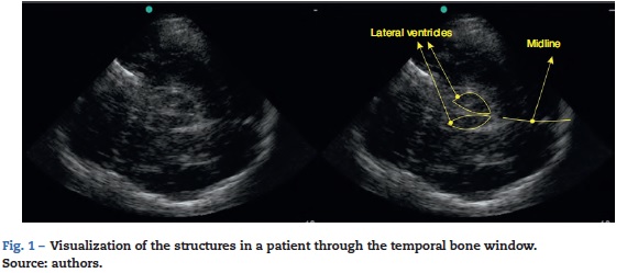
Shunts may be localized using this technique.14,15 It is important to remember that the ultrasound window is not good in up to 15% of cases, preventing the visualization of the brain structures.16
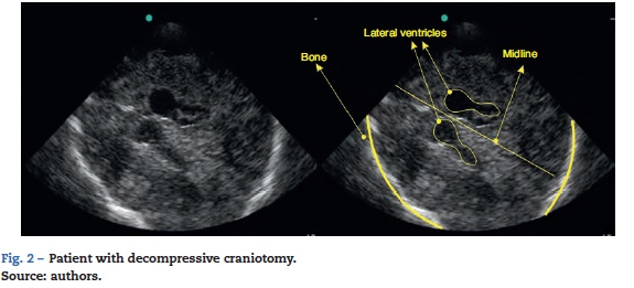
In patients taken to decompressive craniotomy (Fig. 2), the brain parenchyma may be visualized more readily given the absence of artefacts and bone acoustic shadowing. In these patients, the transducer is placed on sites with no bone tissue present, and no specific anatomic landmark is required. There are reports of the ability to follow-up on the size of intracranial haematomas, position shunt catheters,17 monitor midline shifts, or follow-up after treatment.
Optic nerve measurement and correlation with intracranial hypertension
Intracranial hypertension is a life-threatening condition. It is measured with the help of intracranial devices, but this is associated with complications such as infection, bleeding or dysfunction.19
The search for diagnostic tools associated with less morbidity has focused on non-invasive methods such as nuclear magnetic resonance, computed axial tomography and transcranial Doppler ultrasound. However, there is limited correlation between these methods and specific intracranial pressure values.20,21
The use of ocular ultrasound was first reported in 1965.22 It has recently been proposed that measurement of the optic nerve sheath diameter through the ocular window may be a non-invasive method for the detection of intracranial hypertension.19,23 Moreover, good intra- and inter-observer reproducibility has been demonstrated.24
This measurement is based on the fact that the most distal portion of the nerve has a dural covering known as the optic nerve sheath,25 which becomes dilated as intracranial pressure rises and CSF is distributed through along the dura. These changes are greater in the anterior portion of the nerve sheath, just behind the eyeball, an area that is easily accessible by ultrasound.26-28
Given the late onset of clinical signs in cases of intracranial hypertension, early ultrasound detection allows for prompt therapeutic action, thus contributing to improved outcomes.29,30 Ultrasound is less time consuming when compared with other neuroimaging studies, and eliminates the need to transfer critically ill patients. Moreover, it offers the possibility of assessing response to treatment by means of serial measurements.31-33
A learning curve of 10 measurements with 3 abnormal scans is proposed for physicians with experience in ultrasound, and 25 scans may be adequate for a non-experienced ultrasonographer.32
How to measure the optic nerve sheath
A high-frequency (7-10 MHz) linear transducer is required.22 The ultrasound machine is set to visualize structures up to 5-6 cm deep. The transducer is placed over the closed eyelid after generous gel application.
The optic nerve is identified as the hypoechoic structure traversing along a regular course behind the eyeball. For measurement, a vertical line is drawn from the junction between the optic nerve and the eyeball. This line serves just as a reference and must be 3 mm long. Once the 3 mm length is established, a horizontal line is drawn across the optic nerve. This second line provides the measurement of the optic nerve in mm (Figs. 3 and 4).32,33
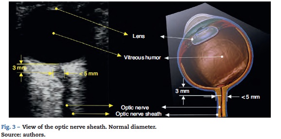
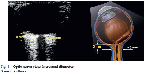
For most of the authors included in the review, 5 mm is the cut-off point for determining that the scan is positive for intracranial hypertension; other authors propose different values (Table 1).
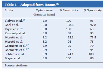
Worth mentioning is a systematic review published by Dubourg et al.33 in 2011 in which the authors assess the diagnostic accuracy of optic nerve measurement compared with the invasive measurement of the intra-parenchymal pressure as the gold standard; 6 prospective cohort studies totalling 231 patients were included, but no significant heterogeneity was found for the sensitivity or the specificity of the ultrasound measurement.33 The sensitivity and specificity of the optic nerve sheath measurement are 0.90 (95% CI, 0.80-0.95) and 0.85 (95% CI, 0.73-0.93), respectively. This review also showed a reliability of 0.2-0.3 mm among reviewers. The problem was the lack of an accurate cut-off point for defining optic nerve sheath dilatation in all the studies. The measurement of the optic nerve sheath diameter has been found to have good diagnostic accuracy for detecting intracranial hypertension and influences the decision of patient referral to specialized centres.33
There are authors who are against the use of optic nerve measurement for determining intracranial hypertension.34 However, the absence of correlation so far does not seem to be associated with the technique or the pathophysiological process but rather with the lack of a standard cut-off point suggesting the boundary between normalcy and hypertension.35 There is also a need to standardize the scanning technique because, as a sphere, the eye may be scanned longitudinally or cross-sectionally and there is no way of knowing how this may affect the results.24,35-37
To solve this issue, the PROSPERO initiative was created in 2013 with the aim of assessing the diagnostic accuracy of ultrasound measurement of the optic nerve sheath for the detection of intracranial hypertension and the establishment of an accurate cut-off point for the creation of an individualized patient database.38
Transcranial colour Doppler
The sonographic approach to the intracranial arteries poses greater difficulties when compared with optic nerve or midline assessment. The learning curve is steep and a good sonograhic window will not be achieved in 15% of patients, making access to that form of imaging difficult.16
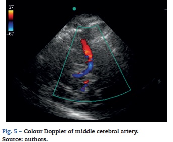
This may be overcome in part with the use of transcranial colour Doppler, which requires a machine equipped with a 1-5 MHz microconvex transducer and colour Doppler functionality. The sonographic window is obtained through the squama of the temporal bone (Fig. 5). A second window may be performed through the eye in order to find the ophthalmic artery, with a third window through the post-auricular area in order to access the posterior cerebral artery. The use of the colour function allows visualization of the intracranial arteries, in particular the middle cerebral artery38 (Fig. 5).
The two most widely accepted applications for transcra-nial Doppler ultrasound are vasospasm control in patients with subarachnoid haemorrhage (Class Ila evidence),16,38-47 and brain death confirmation (Class Ila evidence).39-48 Other applications, although with limited evidence, include the use of flow velocities in the main cerebral arteries as a means to monitor brain haemodynamics.42-45 The use of the relationship between the middle cerebral artery flow and velocities as measured by Doppler and the mean arterial pressure has been proposed for assessing the integrity of cerebral autoregulation.46
Of interest in neurocritical care and neuroanaesthesia has been the possibility of having a way to monitor critically ill patients in the intensive care room or in the operating room. Transcranial colour Doppler has the invaluable potential of providing non-invasive, real time and dynamic information about modifiable brain haemodynamic variables that may have an impact on patient outcomes.49,50
Conclusions
Central nervous system ultrasound performed by non-radiologist specialists is an increasingly available low-cost, versatile and accurate approach that helps improve timeliness and objective decision-making. These applications are expected to expand greatly in the near future and to offer improved correlation with specific pathophysiological processes that influence decision-making in neurocritical patients.
Conflicts of interest
The authors have no conflicts of interest to declare.
Funding
The authors did not receive sponsorship to undertake this article.
References
1. Gallagher RA, Levy JA. Advances in point-of-care ultrasound in pediatric emergency medicine. Curr Opin Pediatr. 2014;26:265-71. [ Links ]
2. Andruszkiewicz P, Sobczyk D. Ultrasound in critical care. Anaesthesiol Intensive Ther. 2013;45:177-81. [ Links ]
3. Hocking G, Mitchell CH. Optimizing the safety and practice of ultrasound-guided regional anesthesia: the role of echogenic technology. Curr Opin Anaesthesiol. 2012;25:603-9. [ Links ]
4. Heinrichs J, Fritze Z, Vandermeer B, Klassen T, Curtis S. Ultrasonographically guided peripheral intravenous cannulation of children and adults: a systematic review and meta-analysis. Ann Emerg Med. 2013;61:444-54. [ Links ]
5. Stengel D, Bauwens K, Rademacher G, Ekkernkamp A, Guthoff C. Emergency ultrasound-based algorithms for diagnosing blunt abdominal trauma. Cochrane Database Syst Rev. 2013;31:7. [ Links ]
6. Oren-Grinberg A, Talmor D, Brown SM. Focused critical care echocardiography. Crit Care Med. 2013;41:2618-26. [ Links ]
7. Taylor RA, Oliva I, Van Tonder R, Elefteriades J, Dziura J, Moore CL. Point-of-care focused cardiac ultrasound for the assessment of thoracic aortic dimensions, dilation, and aneurysmal disease. Acad Emerg Med. 2012;19:244-7. [ Links ]
8. Zengin S, Al B, Genc S, Yildirim C, Ercan S, Dogan M, et al. Role of inferior vena cava and right ventricular diameter in assessment of volume status: a comparative study: ultrasound and hypovolemia. Am J Emerg Med. 2013;31:763-7. [ Links ]
9. Liechtenstein D. Fluid administration limited by lung sonography: the place of lung ultrasound in assessment of acute circulatory failure (the FALLS-protocol). Expert Rev Respir Med. 2012;6:155-62. [ Links ]
10. Wu T. The CORE scan concentrated overview of resuscitative efforts. Crit Care Clin. 2014:151-75. [ Links ]
11. White H, Venkatesh B. Applications of transcranial Doppler in the ICU: a review. Intensive Care Med. 2006;32:981-94. [ Links ]
12. Schott ML, Pierog JE, Williams SR. Pitfalls in the use of ocular ultrasound for evaluation of acute vision loss. J Emerg Med. 2013;44:1136-9. [ Links ]
13. Rincon F. Bedside transcranial sonography: a promising tool for the neurointensivist. Crit Care Med. 2012;40:1969-70. [ Links ]
14. Caricato A, Mignani V, Bocci MG, Pennisi MA, Sandroni C, Tersali A. Usefulness of transcranial echography in patients with decompressive craniectomy: a comparison with computed tomography scan. Crit Care Med. 2012;40: 1745-52. [ Links ]
15. Becker G, Berg D. Neuroimaging in basal ganglia disorders: perspectives for transcranial ultrasound. Mov Disord. 2001;16:23-32. [ Links ]
16. Steiner T, Juvela S, Unterberg A, Jung C, Forsting M, Rinkel G, European Stroke Organization. European Stroke Organization guidelines for the management of intracranial aneurysms and subarachnoid haemorrhage. Cerebrovasc Dis. 2013;35:93-112. [ Links ]
17. Phillips SB, Gates M, Krishnamurthy S. Strategic placement of bedside ventriculostomies using ultrasound image guidance: report of three cases. Neurocrit Care. 2012;17:255-9. [ Links ]
18. Harrer JU, Eyding J, Ritter M, Schminke U, Schulte-Altedorneburg G, Kohrmann M. The potential of neurosonography in neurological emergency and intensive care medicine: monitoring of increased intracranial pressure brain death diagnostics, and cerebral autoregulation - part 2. Ultraschall Med. 2012;33:320-31. [ Links ]
19. Tayal VS, Neulander M, Norton HJ, Foster T, Saunders T, Blaivas M. Emergency department sonographic measurement of optic nerve sheath diameter to detect findings of increased intracranial pressure in adult head injury patients. Ann Emerg Med. 2007;49:508-14. [ Links ]
20. Winkler F, Kastenbauer S, Yousry TA, Maerz U, Pfister HW. Discrepancies between brain CT imaging and severely raised intracranial pressure proven by ventriculostomy in adults with pneumococcal meningitis. J Neurol. 2002;249:1292-7. [ Links ]
21. Schmidt B, Czosnyka M, Raabe A, Yahya H, Schwarze JJ, Sackerer D. Adaptive noninvasive assessment of intracranial pressure and cerebral autoregulation. Stroke. 2003;34: 84-9. [ Links ]
22. Sasson GP, Ubiergo SU. Ultrasonido en Emergencia y Cuidado Critico. 1st ed. Editorial Distribuna; 2013. [ Links ]
23. Qayyum H, Amlakhan S. Can ocular ultrasound predict intracranial hypertension? A pilot diagnostic accuracy evaluation in a UK Emergency Department. Eur J Emerg Med. 2013;20:91-7. [ Links ]
24. Bauerle J, Lochner P, Kaps M, Nedelmann M. Intra- and interobsever reliability of sonographic assessment of the optic nerve sheath diameter in healthy adults. J Neuroimaging. 2012;22:42-5. [ Links ]
25. Fledelius HC. Ultrasound in ophthalmology. Ultrasound Med Biol. 1997;23:365-75. [ Links ]
26. Helmke H, Hansen HC. Fundamentals of transorbital sonographic evaluation of optic nerve sheath expansion under intracranial hypertension. Patient study. Pediatr Radiol. 1996;26:706-10. [ Links ]
27. Geeraerts T, Newcombe VF, Coles JP, Abate MG, Perkes IP, Hutchinson PJ, et al. Use of T2-weighted magnetic resonance imaging of the optic nerve sheath to detect raised intracranial pressure. Crit Care. 2008;12:1-7. [ Links ]
28. Geeraerts T, Mercenron S, Benhamou D, Vigue B, Duranteau J. Non-invasive assessment of intracranial pressure using ocular sonography in neurocritical care patients. Intensive Care Med. 2008;34:2062-7. [ Links ]
29. Girisgin AS, Kalkan E, Kocak S, Cander B, Gul M, Seniz M. The role of optic nerve ultrasonography in the diagnosis of elevated intracranial pressure. Emerg Med J. 2007;24:251-1. [ Links ]
30. Newman WD, Hollman AS, Duttongn, Carachi R. Measurement of optic nerve sheath diameter by ultrasound: a means of detecting acute raised intracranial pressure in hydrocephalus. Br J Ophthalmol. 2002;86:1109-13. [ Links ]
31. Strumwasser A, Kwan RO, Yeung L, Miraflor E, Ereso A, Castro-Moure F, et al. Sonographicoptic nerve sheath diameter as an estimate of intracranial pressure in adult trauma. J Surg Res. 2011;170:265. [ Links ]
32. Dubourg J, Javouhey E, Geeraerts T, Messerer M, Kassai B. Ultrasonography of optic nerve sheath diameter for detection of raised intracranial pressure: a systematic review and meta-analysis. Intensive Care Med. 2011;37:1059-68. [ Links ]
33. Dubost C, Geeraerts T. Possible pitfalls when measuring the optic nerve sheath with sonography. J Surg Res. 2012;2: e43-5. [ Links ]
34. Hansen HC, Lagreze W, Krueger O, Helmke K. Dependence of the optic nerve sheath diameter on acutely applied subarachnoidal pressure - an experimental ultrasound study. Acta Ophthalmol. 2011;89:528. [ Links ]
35. Geeraerts T, Berges O, Merceron S, Launey Y, Benhamou D, Vigue B, et al. Reply to Copetti and Cattarossi. Intensive Care Med. 2009;35:1490. [ Links ]
36. Blehar DJ, Gaspari RJ, Montoya A, Calderon R. Correlation of visual axis and coronal axis measurements of the optic nerve sheath diameter. J Ultrasound Med. 2008:407. [ Links ]
37. Dubourg J, Messerer M, Karakitsos D, Rajajee V, Antonsen E, Javouhey E, et al. Individual patient data systematic review and meta-analysis of optic nerve sheath diameter ultrasonography for detecting raised intracranial pressure: protocol of the ONSD research group. Syst Rev. 2013;2:1-5. [ Links ]
38. Toi H, Matsumoto N, Yokosuka K, Matsubara S, Hirano K, Uno M. Prediction of cerebral vasospasm using early stage transcranial Doppler. Neurol Med Chir (Tokyo). 2013;53:396-402. [ Links ]
39. Lange MC, Zetola VH, Miranda-Alves M, Moro CH, Silvado CE, Rodrigues DL, et al. Brazilian guidelines for the application of transcranial ultrasound as a diagnostic test for the confirmation of brain death. Task Force Group of the Neurosonology Department, Brazilian Academy of Neurology. Arq Neuropsiquiatr. 2012;70:373-80. [ Links ]
40. Lovrencic-Huzjan A, Vukovic V, Gopcevic A, Vucic M, Kriksic V, Demarin V. Transcranial Doppler in brain death confirmation in clinical practice. Ultraschall Med. 2011;32:62-6. [ Links ]
41. Marinoni M, Alari F, Mastronardi V, Peris A, Innocenti P. The relevance of early TCD monitoring in the intensive care units for the confirming of brain death diagnosis. Neurol Sci. 2011;32:73-7. [ Links ]
42. Turek R, Linzer P, Filip M, Samal F, Jurek P. Application of transcranial color-coded sonography in severe brain injury. Acta Neurochir Suppl. 2013;118:265-7. [ Links ]
43. Shiogai T, Koyama M, Yamamoto M, Hashimoto H, Yoshikawa K, Nakagawa M. Monitoring of brain tissue perfusion utilizing a transducer holder for transcranial color duplex sonography. Acta Neurochir Suppl. 2013;118:229-33. [ Links ]
44. Bilotta F, Dei Giudici L, Lam A, Rosa G. Ultrasound-based imaging in neurocritical care patients: a review of clinical applications. Neurol Res. 2013;35:149-58. [ Links ]
45. Sharma V. Role of transcranial Doppler in traumatic brain injury. Turk Neurosurg. 2012;22:525-6. [ Links ]
46. Lewis PM, Smielewski P, Rosenfeld JV, Pickard JD, Czosnyka M. Monitoring of the association between cerebral blood flow velocity and intracranial pressure. Acta Neurochir Suppl. 2012;114:147-51. [ Links ]
47. Connolly ES Jr, Rabinstein AA, Carhuapoma JR, Derdeyn CP, Dion J, Higashida RT, et al. Guidelines for the management of aneurysmal subarachnoid hemorrhage: a guideline for healthcare professionals from the American Heart Association/American Stroke Association. Stroke. 2012;43:1711-37. [ Links ]
48. Lange MC, Zétola VH, Miranda-Alves M, Moro CH, Silvado CE, Rodrigues DL, et al. Brazilian guidelines for the application of transcranial ultrasound as a diagnostic test for the confirmation of brain death. Arq Neuropsiquiatr. 2012;70:373-80. [ Links ]
49. Karakitsos D, Poularas J, Karabinis A, Dimitriou V, Cardozo A, Labropoulos N. Considerations for the utilization of transcranial Doppler sonography in the study of progression towards cerebral circulatory arrest. Intensive Care Med. 2011;37:368-70. [ Links ]
50. Topcuoglu MA. Transcranial Doppler ultrasound in neurovascular diseases: diagnostic and therapeutic aspects. J Neurochem. 2012;123 (Suppl 2):39-51. [ Links ]
