


Services on Demand
Journal
Article
 text in
  English (pdf)
 Article in xml format
 Article references
 Send this article by e-mail
Indicators
-
 Cited by SciELO
 -
 Access statistics
Related links
-
 Cited by Google
 -
 Similars in
SciELO
Similars in
SciELO -
 Similars in Google
Share

 Permalink
Colombian Journal of Anestesiology
Print version ISSN 0120-3347
Rev. colomb. anestesiol. vol.43 no.4 Bogotá Oct./Dec. 2015
Review
Ergonomics in ultrasound-guided nerve blocks*
Ergonomía en los bloqueos nerviosos guiados por ultrasonografía
Oscar David Aguirre-Ospinaa,**, Julián Felipe González-Maldonadob, Ángela María Ríos-Medinac
a Specialist in Anaesthesia and Resuscitation, Professor at Universidad de Caldas, Anaesthetist at SES Hospital, Manizales, Colombia
b Anaesthesia and Resuscitation Resident, Universidad Nacional de Colombia, Bogotá, Colombia
c Specialist in Anaesthesia and Resuscitation, Professor at Universidad de Caldas, Anaesthetist, Clínica Comfamiliar y Liga Contra el Cancer, Pereira, Colombia
* Please cite this article as: Aguirre-Ospina OD, González-Maldonado JF, Ríos-Medina ÁM. Ergonomía en los bloqueos nerviosos guiados por ultrasonografía. Revisión no sistemática de la literatura. Rev Colomb Anestesiol. 2015;43:331-339.
** Corresponding author at: Calle 10 No. 12B-30 Apartamento 704 Torre 2, Pereira, Colombia. E-mail address: oscardavid.celta@gmail.com (O.D. Aguirre-Ospina).
Article info
Article history: Received 24 February 2015 Accepted 4 June 2015 Available online 10 August 2015
Abstract
Introduction: Performing ultrasound-guided nerve blocks has proven to be safe, but ergonomic considerations are essential.
Methods: A non-systematic literature search was conducted in the Medline/Pubmed and Embase databases.
Objective: To describe the influence of operator position, monitor location, and hand position in performing ultrasound-guided nerve blocks.
Results: Poor ergonomics is a key factor of error when performing nerve blocks under ultrasound guidance. "In-plane" needle insertion increases success. An inappropriate position may be a cause of muscle-skeletal disorders. Based on these results, we propose specific element location and operator position for the performance of certain nerve blocks. Conclusion:Performing ultrasound-guided nerve blocks requires an ergonomic work area allowing for greater comfort and effectiveness, thus reducing the occurrence of muscle-skeletal disorders.
Keywords: Ultrasonography, Nerve block, Human engineering, Analgesia, Anesthesia.
Resumen
Introducción: Los bloqueos guiados por ultrasonografía han demostrado ser eficaces, pero requieren considerar aspectos como la ergonomía.
Objetivo: Describir influencia de la postura del operador, ubicación del monitor y posición de las manos, al realizar bloqueos con ultrasonografía.
Métodos: Se realizó una búsqueda no sistemática de literatura en Medline/Pubmed y Embase.
Resultados: Una mala ergonomía es un factor de error al realizar bloqueos con ultrasonido. La inserción de la aguja "en plano" aumenta el éxito. Una posición inapropiada puede producir trastornosmusculo-esqueléticos en el operador. Basados en esto, se propone una ubicación de elementos y posición del operador para realizar algunos bloqueos.
Conclusión: Realizar bloqueos con ultrasonido requiere un área de trabajo ergonómica para lograr mayor eficacia, y disminuir aparición de trastornos músculo-esqueléticos.
Palabras clave: Ultrasonografía, Bloqueo nervioso, Ingeniería humanaAnalgesia, Anesthesia.
Introduction
The use of ultrasound (US) in perioperative regional anaesthesia and analgesia has evolved rapidly,1,2 and its use has grown significantly in anaesthetic practice.3,4 Its main advantage is the ability to identify anatomical structures in real time, allowing the precise administration and distribution of the local anaesthetic (LA) around the nerve structure.5,6
Increasingly more data are being published at the present time suggesting higher efficacy and safety with the use of US-guided nerve blocks,7,8 specifically for interscalene,9,10 supraclavicular,11 infraclavicular,12,13 axillary,14-16 femoral,17 and sciatic popliteal17-20 blocks. A meta-analysis found that when US is compared with peripheral nerve (PN) stimulation, the risk of failure is lower (RR 0.41, 95% CI 0.26-0.66, p = 0.001), the procedure is faster (1 min less in average with ultrasound), latency is shorter (onset time is reduced by a mean of 29%), duration is longer (by an average of 25%), and there is a lower risk of vascular puncture (RR 0.16,95% CI 0.05-0.47, p = 0.001).21
After analysing a database of 20,021 patients undergoing 25,336 PN blocks, Barrington and Kluger22 reported that the use of US, compared to PN stimulations, reduced the frequency of LA-related toxicity by up to 65% (0.59 events/1000 blocks in those cases where US was used vs. 2.1 events/1000 blocks using PN stimulation).22 However, as was mentioned by Hadzic et al.,23 the use of US in expert hands may reduce, although not eliminate, the most common complications of regional anaesthesia (RA) such as blood vessel punctures and inadvertent intraneural or intravascular injections.23
The growing use of US in RA3,4 has created the need to consider aspects that are relevant to this practice, including ergonomics and the adequate location of all the necessary elements in space.24 Multiple authors suggest that these variables influence the performance of US-guided blocks25,26 in terms of outcomes for the patient and of operator wellbeing.27-30 This prompted us to conduct a non-systematic review of the literature in the Medline/Pubmed and Embase databases to look for current evidence regarding ergonomics in RA.
Is there a relationship between ergonomics and nerve blocks?
Ergonomics is the science of the physical interaction between individuals and their working environment, including equipment design and operator training in terms of motor, visual, spatial, and hearing skills, and abilities.31 When ergonomic strategies are applied, task performance and efficacy can be optimized and human wellbeing may be maximized.27
Given the advances in biomedical knowledge and technology, healthcare professionals currently find themselves working in increasingly complex and demanding environments. An example of this is the work of the anaesthetist in the modern operating theatre where he/she is exposed to multiple visual, tactile and auditory stimuli, creating a greater clinical and ergonomic challenge, especially when it comes to performing procedures.27 There is growing recognition of the importance of ergonomics in the practice of anaesthesia.32,33 To this date, ergonomics is not taught in most academic training programmes despite the recognition of its potential benefits.26,34,35
The use of US in RA requires education and training in order for the anaesthetist to acquire concepts and develop psychomotor skills, including the following26,36:
-
Achieving adequate imaging and visualization of the nerve structure.37
-
Alignment of the needle with the US beam.38
-
Identifying the advancement of the needle in real time.37
-
Placing the tip of the needle on the target point.38
-
Identifying the appropriate distribution of the LA around the nerve.37
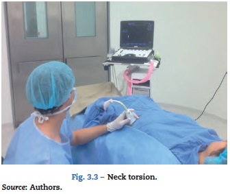
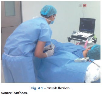
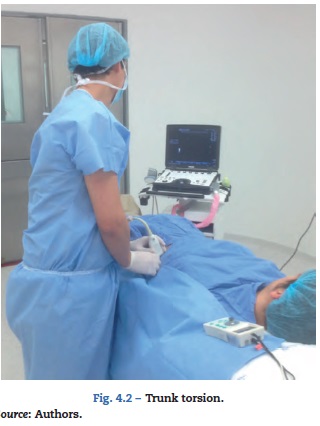
If any of these steps is performed suboptimally, there is a lower probability of achieving a successful nerve block. Sites et al.26,39 found that poor ergonomics (defined as turning the trunk, holding the needle with the non-dominant hand and turning the head 45 degrees or more) is a source of error among anaesthesia residents and novice operators when performing US-guided blocks, and is associated with fatigue and lower performance.26,39
In view of the above, it is suggested that adopting the inadequate ergonomics may have a dual relevant impact: first, block failure due to lower accuracy of hand motions, either of the hand holding the needle or the hand holding the probe26 and difficulty aligning with the US bema,40 leading to impaired viewing of needle advancement and LA distribution; and, second, the potential development of muscle-skeletal disorders in the operator.41,42
Does ergonomics have an impact on the success of a nerve block?
As has been previously determined, US-guided RA is a safe and effective technique for performing nerve blocks,8,21,22 provided perfect visualization of the nerve structure is achieved.43 The needle may be advanced towards the target perpendicular or parallel to the US beam, in what is known "out-of-plane" insertion (OPI) or "in-plane insertion (IPI), respectively. With the in-plane technique it is possible to watch the needle as it moves towards the target, but it requires skill and may create a false sense of security.25,44 OPI is a more complex technique aimed at visualizing the tip of the needle or watching for indirect signs such as the movement of the adjacent tissue as the needle advances, or hydrolocalization,45 but it has the advantage of providing a length of insertion which is three times shorter and more comfortable for the patient.46
Chapman et al.25 state that "The operator must focus on the image on the monitor and on the patient, hence the importance of having the anatomical area and the US machine in the same line of view. This requires placing the screen at eye level on the opposite side of the operator".25 Using this hypothesis, Langford et al., in a trial with 31 anaesthetists, found that placement of the US machine in front of the operator improved accuracy with in-plane needle advancement when compared with the placement of the machine perpendicular to the operator, but found no difference in terms of the time for performing the task.28 The importance of these results relates to the fact that "accuracy in needle placement is more important than the speed in reaching the target because it improves the odds of a successful block and reduces complications associated with inadvertent damage to the non-target structures".28
Speer et al.29 describe two ways to position the hands in relation to the visual axis of the operator while performing "in-plane" needle insertion29:
-
"ALONG the visual axis" (ALVA): the long axis of the transducer with the needle parallel to the visual axis (Fig. 1A).
-
"ACROSS the visual axis" (ACVA): locating the long axis of the transducer and placing the needle perpendicular to the visual axis (Fig. 1B).
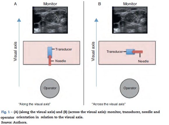
A randomized clinical trial using simulation with phantom-type models analyzed the performance of 24 novice operators as they advanced the needle towards a target under ultrasound guidance. Each participant performed 5 ALVA attempts and 5 ACVA attempts, and it was found that when the 2 axes are parallel there is a significant reduction in the time to perform the task and improved image quality.29
Wilson et al.30 compared the use of the ALVA and ACVA techniques in medical students and found that the use of the IPI approach with the former technique was associated with better visualization, shorter time to complete the task, and higher success; moreover, the participants reported a higher preference for this technique.30 The authors suggest that in novice operators, needle advancement with the ALVA technique minimizes the need for needle relocations, reduces the time to perform the procedure, improves ergonomic performance and enhances needle tip viewing, thus improving safety and the rate of successful outcomes.30
May inappropriate ergonomics when performing US-guided nerve blocks give rise to muscle-skeletal disorders in the operator?
The importance of an ergonomic ultrasound workstation to reduce the incidence of muscle-skeletal disorders has been widely described.42 Although this aspect has not been studied specifically for US-guided nerve blocks, similar considerations could be extrapolated and applied.
The physical factors most commonly associated with the development of muscle-skeletal disorders include vibrations, excess force and tension, strong or uncomfortable motions, poor posture, repetitive motions and prolonged pressure time.47 Except for vibrations, all of these factors may be present when performing ultrasound scans, in particular in obese patients who require the use of higherforce on the probe in order to reduce the thickness of the fat layers and improve image quality.42,48
The parts of the body most commonly affected as a result of poor posturing are the shoulders (76%), neck (74%), wrists (59%), back (58%) and hands (55%).49 Although many of the symptoms are mild, if left unaddressed they may lead to serious and debilitating chronic disorders.42 The most frequent diagnoses are tendinitis and tenosynovitis affecting shoulders, hands and wrists.50
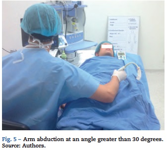
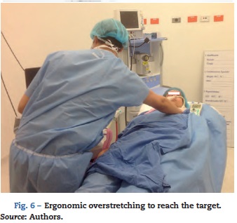
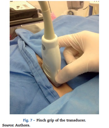
The positions most frequently associated with inappropriate ergonomics when performing ultrasound scans are shown below42:
-
Arm abduction (angle >30 degrees) (Fig. 5).
-
Ergonomic overstretching to reach the target (Fig. 6).
-
Pinch grasp of the probe (there is up to a five-fold reduction in the force applied when the volar grip is used42) (Fig. 7).
Operator posture and interaction with all the pieces of equipment have to be considered for an appropriate scan.51 An ergonomic work area must include the following:
-
A table with lateral retractile supports in order to avoid creating a larger distance to the patient, with powered height adjustment because manual controls interfere with the chair and operator location.42
-
A chair with wheels, adjustable from the seating position.42,51
-
A spacious examination room with indirect lighting that does not interfere with the display.42,51
Appropriate ergonomics for nerve blocks
Based on the information above, approaches to ensure appropriate ergonomics are proposed24:
-
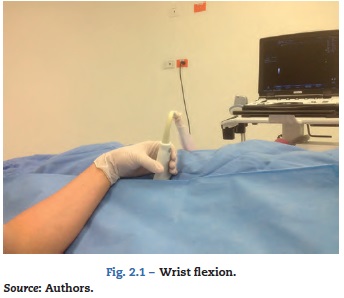
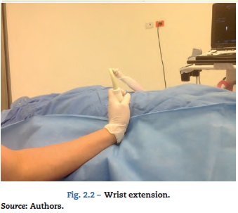
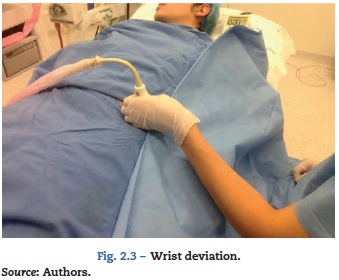
Avoiding inadequate positioning of the hands, wrists, neck, trunk and shoulders (Figs. 2-7).
-
Placing the US machine on the opposite side of the side of the body where the nerve block will be performed.
-
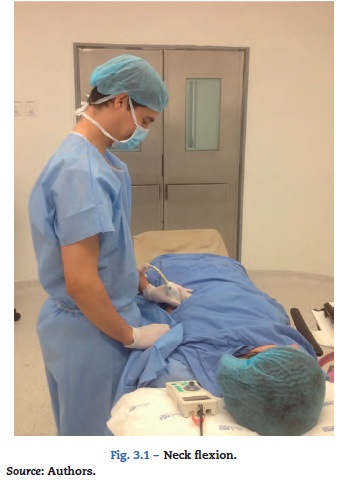
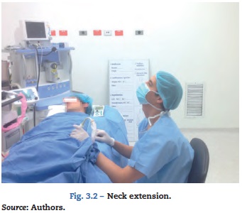
Avoid turning the head 45 degrees or more.
-
Aligning the monitor with the operator's visual axis.
-
ALVA alignment of the probe and needle.
-
Working from a sitting position with the arm supported on the bed.
-
Adjusting the height of the bed.
-
No hand crossing (transducer and needle).
-
Holding the needle with the dominant hand.
-
Avoid the pinch grip when holding the transducer (Fig. 7).
-
Holding the probe from the bottom and using the patient's skin as support for the fingers in order to gain greater stability.
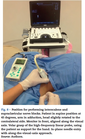
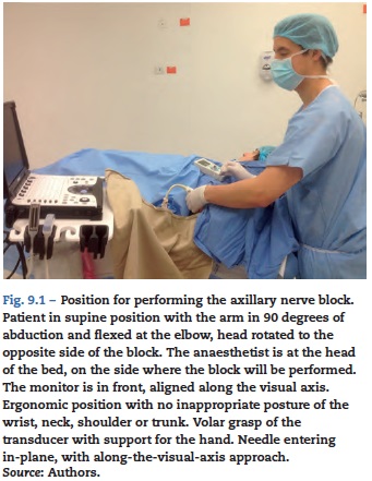
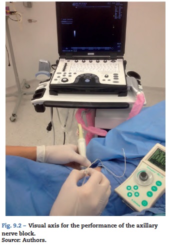
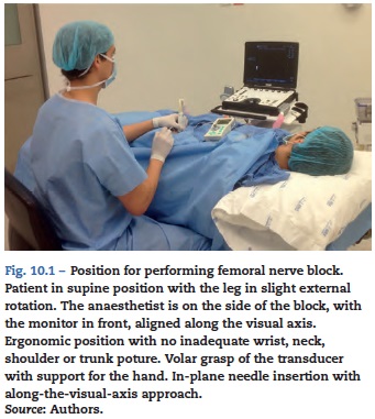
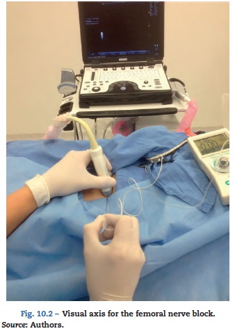
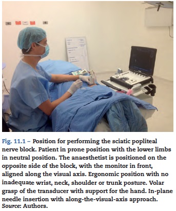
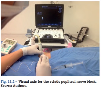
A proposal for ergonomic performance of interscalene and supraclavicular nerve blocks (Fig. 8), axillary blocks (Figs. 9.1 and 9.2), femoral blocks (Figs. 10.1 and 10.2) and sciatic popliteal blocks (Figs. 11.1 and 11.2) is presented below.
The growing use and development of US in RA are driven by the ability to achieve real-time visualization, thus improving efficacy and safety. Because it is "operator-dependent", its use does not guarantee success or absence of complications in cases of insufficient skill or knowledge of the anatomy. The main psychomotor skills that need to be developed include the following: achieving an image of the area of interest, aligning the needle with the US beam, advancing the tip of the needle accurately to the ideal site for injecting the LA, and recognizing adequate distribution. As part of developing and maintaining these skills, an ergonomic work area helps adopt the appropriate postures to reduce the occurrence of muscle-skeletal disorders and to improve comfort and effectiveness when performing the procedure. The latter is based on data suggesting that the alignment of the transducer, needle and screen in the same parallel plane and visual axis, improves of visualization and needle advancement accuracy, which is key to a successful nerve block.
Funding
The authors did not receive sponsorship to undertake this article.
Conflict of interest
The authors have no conflict of interest to declare
References
1. Mariano ER, Marshall ZJ, Urman RD, Kaye AD. Ultrasound and its evolution in perioperative regional anesthesia and analgesia. Best Pract Res Clin Anaesthesiol. 2014;28:29-39. [ Links ]
2. Choquet O, Abbal B, Capdevila X. The new technological trends in ultrasound-guided regional anesthesia. Curr Opin Anaesthesiol. 2013;26:605-12. [ Links ]
3. Onishi E, Yamauchi M. The trends of ultrasound-guided neuraxial block. Masui. 2014;63:1011-7. [ Links ]
4. Marhofer P, Harrop-Griffiths W, Kettner SC, Kirchmair L. Fifteen years of ultrasound guidance in regional anaesthesia: part 1. Br J Anaesth. 2010;104:538-46. [ Links ]
5. Horlocker TT, Wedel DJ. Ultrasound-guided regional anaesthesia: the search for the holy grail. Anesth Analg. 2007;104:1009-11. [ Links ]
6. McCartney CJ, Dickinson V, Dubrowski A, Riazi S, McHardy P, Awad IT. Ultrasound provides a reliable test of local anesthetic spread. Reg Anesth Pain Med. 2010;35:361-3. [ Links ]
7. Salinas FV, Hanson NA. Evidence-based medicine for ultrasound-guided regional anesthesia. Anesthesiol Clin. 2014;32:771-87. [ Links ]
8. Gelfand HJ, Ouanes JP, Lesley MR, Ko PS, Murphy JD, Sumida SM, Isaac GR, Kumar K, Wu CL. Analgesic efficacy of ultrasound-guided regional anesthesia: a meta-analysis. J Clin Anesth. 2011;23:90-6. [ Links ]
9. Kapral S, Greher M, Huber G, Willschke H, Kettner S, Kdolsky R, et al. Ultrasonographic guidance improves the success rate of interscalene brachial plexus blockade. Reg Anesth Pain Med. 2008;33:253-8. [ Links ]
10. Liu SS, Zayas VM, Gordon MA, Beathe JC, Maalouf DB, Paroli L, et al. A prospective, randomized, controlled trial comparing ultrasound versus nerve stimulator guidance for interscalene block for ambulatory shoulder surgery for postoperative neurological symptoms. Anesth Analg. 2009;109:265-71. [ Links ]
11. Williams SR, Chouinard P, Arcand G, Harris P, Ruel M, Boudreault D, et al. Ultrasound guidance speeds execution and improves the quality of supraclavicular block. Anesth Analg. 2003;97:1518-23. [ Links ]
12. Sauter AR, Dodgson MS, Stubhaug A, Halstensen AM, Klaastad 0. Electrical nerve stimulation or ultrasound guidance for lateral sagittal infraclavicular blocks: a randomized, controlled, observer-blinded, comparative study. Anesth Analg. 2008;106:1910-5. [ Links ]
13. Ponde VC, Diwan S. Does ultrasound guidance improve the success rate of infraclavicular brachial plexus block when compared with nerve stimulation in children with radial club hands? Anesth Analg. 2009;108:1967-70. [ Links ]
14. Chan VW, Perlas A, McCartney CJ, Brull R, Xu D, Abbas S. Ultrasound guidance improves success rate of axillary brachial plexus block. Can J Anaesth. 2007;54:176-82. [ Links ]
15. Casati A, Danelli G, Baciarello M, Corradi M, Leone S, Di Cianni S, et al. A prospective, randomized comparison between ultrasound and nerve stimulation guidance for multiple injection axillary brachial plexus block. Anesthesiology. 2007;106:992-6. [ Links ]
16. Yu W, Xu X, Wu DS, Guo XY, Huang PT. Efficacy of axillary approach brachial plexus blocking by ultrasound-guided four points via one-puncture technique. Zhonghua Yi Xue Za Zhi (Natl Med J China). 2007;87:740-5. [ Links ]
17. Oberndorfer U, Marhofer P, Bosenberg A, Willschke H, Felfernig M, Weintraud M, et al. Ultrasonographic guidance for sciatic and femoral nerve blocks in children. Br J Anaesth. 2007;98:797-801. [ Links ]
18. Perlas A, Brull R, Chan VW, McCartney CJ, Nuica A, Abbas S. Ultrasound guidance improves the success of sciatic nerve block at the popliteal fossa. Reg Anesth Pain Med. 2008;33:259-65. [ Links ]
19. Danelli G, Fanelli A, Ghisi D, Moschini E, Rossi M, Ortu A, et al. Ultrasound vs nerve stimulation multiple injection technique for posterior popliteal sciatic nerve block. Anaesthesia. 2009;64:638-42. [ Links ]
20. Van Geffen GJ, van den Broek E, Braak GJ, Giele JL, Gielen MJ, Scheffer GJ. A prospective randomised controlled trial of ultrasound guided versus nerve stimulation guided distal sciatic nerve block at the popliteal fossa. Anaesth Intensive Care. 2009;37:32-7. [ Links ]
21. Abrahams MS, Aziz MF, Fu RF, Horn JL. Ultrasound guidance compared with electrical neurostimulation for peripheral nerve block: a systematic review and meta-analysis of randomized controlled trials. Br J Anaesth. 2009;102:408-17. [ Links ]
22. Barrington MJ, Kluger R. Ultrasound guidance reduces the risk of local anesthetic systemic toxicity following peripheral nerve blockade. Reg Anesth Pain Med. 2013;38:289-97. [ Links ]
23. Hadzic A, Sala-Blanch X, Xu D. Ultrasound guidance may reduce but not eliminate complications of peripheral nerve blocks. Anesthesiology. 2008;108:557-8. [ Links ]
24. Paloma Morillas-Sendin MD, Alejandro Ortega-Romero MD, Concepción del-Olmo MD. Basic considerations before injections and scanning techniques. Tech Reg Anesth Pain Manag. 2013;17:53-63. [ Links ]
25. Chapman GA, Johnson D, Bodenham AR. Visualization of needle position using ultrasonography. Anesthesia. 2006;61:148-58. [ Links ]
26. Sites BD, Spence BC, Gallagher JD, Wiley CW, Bertrand ML, Blike GT. Characterizing novice behavior associated with learning ultrasound-guided peripheral regional anesthesia. Reg Anesth Pain Med. 2007;32:107-15. [ Links ]
27. Ajmal M, Power S, Smith T, Shorten GD. Ergonomic task analysis of ultrasound-guided femoral nerve block: a pilot study. J Clin Anesth. 2011;23:35-41. [ Links ]
28. Langford RA, Hockey B, Leslie K. Monitor position and the accuracy and speed of ultrasound-guided nerve blocks. Anaesthesia. 2009;64:845-9. [ Links ]
29. Speer M, McLennan N, Nixon C. Novice learner in-plane ultrasound imaging: which visualization technique? Reg Anesth Pain Med. 2013;38:350-2. [ Links ]
30. Wilson JM, Germain G, Vaghadia H, Tang R, Sawka A. In-plane ultrasound-guided needle insertion ALONG or ACROSS the visual axis hand positions. Br J Anaesth. 2014;113:717-8. [ Links ]
31. Stone R, McCloy R. Ergonomics in medicine and surgery. BMJ. 2004;328:1115-8. [ Links ]
32. Berguer R. The application of ergonomics in the work environment of general surgeons. Rev Environ Health. 1997;12:99-106. [ Links ]
33. Friesdorf W, Konichezky S, Gross-Alltag F, Schwilk B. Ergonomics applied to anaesthesia record keeping. J Clin Monit Comput. 1993;10:251-9. [ Links ]
34. Smith AF, Pope C, Goodwin D, Mort M. What defines expertise in regional anaesthesia? An observational analysis of practice. Br J Anaesth. 2006;97:401-7. [ Links ]
35. Fletcher GC, McGeorge P, Flin RH, Glavin RJ, Maran NJ. The role of non-technical skills in anaesthesia: a review of current literature. Br J Anaesth. 2002;88:418-29. [ Links ]
36. Sites BD, Chan VW, Neal JM, Weller R, Grau T, Koscielniak-Nielsen ZJ, et al. American Society of Regional Anesthesia and Pain Medicine, European Society of Regional Anaesthesia and Pain Therapy Joint Committee: the American Society of Regional Anesthesia and Pain Medicine and the European Society of Regional Anaesthesia and Pain Therapy Joint Committee recommendations for education and training in ultrasound-guided regional anesthesia. Reg Anesth Pain Med. 2010;35:S74-80. [ Links ]
37. Marhofer P, Chan VWS. Ultrasound-guided regional anaesthesia: current concepts and future trends. Anaesth Analg. 2007;104:1265-9. [ Links ]
38. de Oliveira Filho GR, Helayel PE, da Conceicao DB, Garzel IS, Pavei P, Ceccon MS. Learning curves and mathematical models for interventional ultrasound basic skills. Anesth Analg. 2008;106:568-73. [ Links ]
39. Sites BD, Gallagher JD, Cravero J, Lundberg J, Blike G. The learning curve associated with a simulated ultrasound-guided interventional task by inexperienced anesthesia residents. Reg Anesth Pain Med. 2004;29: 544-8. [ Links ]
40. Chin KJ, Tse C, Chan V, Tan JS, Lupu CM, Hayter M. Hand motion analysis using the imperial college surgical assessment device: validation of a novel and objective performance measure in ultrasound-guided peripheral nerve blockade. Reg Anesth Pain Med. 2011;36:213-9. [ Links ]
41. Esser AC, Koshy JG, Randle HW. Ergonomics in office-based surgery: a survey-guided observational study. Dermatol Surg. 2007;33:1304-14. [ Links ]
42. Baker JP, Coffin CT. The importance of an ergonomic workstation to practicing sonographers. J Ultrasound Med. 2013;32:1363-75. [ Links ]
43. Koscielniak-Nielsen ZJ. Ultrasound-guided peripheral nerve blocks: what are the benefits? Acta Anaesthesiol Scand. 2008;52:727-37. [ Links ]
44. Chin KJ, Perlas A, Chan VW, Brull R. Needle visualization in ultrasound-guided regional anesthesia: challenges and solutions. Reg Anesth Pain Med. 2008;33:532-44. [ Links ]
45. Bloc S, Ecoffey C, Dhonneur G. Controlling needle tip progression during ultrasound guided regional anesthesia using the hydrolocalization technique. Reg Anesth Pain Med. 2008;33:382-3. [ Links ]
46. Marhofer P, Greher M, Kapral S. Ultrasound guidance in regional anaesthesia. Br J Anaesth. 2005;94:7-17. [ Links ]
47. Occupational Safety and Health Administration. Clinical services: sonography. Occupational Safety and Health Administration; 2008 [Cited 2014 Dec 15]. Available from: http://www.osha.gov/SLTC/etools/hospital/sonography/sonography.html. [ Links ]
48. Brodsky JB, Mariano ER. Regional anaesthesia in the obese patient: lost landmarks and evolving ultrasound guidance. Best Pract Res Clin Anaesthesiol. 2011;25:61-72. [ Links ]
49. Evans K, Roll S, Baker J. Work-related musculoskeletal disorders (WRMSD) among registered diagnostic medical sonographers and vascular technologists: a representative sample. J Diagn Med Sonography. 2009;25:287-99. [ Links ]
50. Evans K, Roll SC, Hutmire C, Baker JP. Factors that contribute to wrist-hand-finger discomfort in diagnostic medical sonographers and vascular technologists. J Diagn Med Sonography. 2010;26:121-9. [ Links ]
51. Society of Diagnostic Medical Sonography. Industry standards for the prevention of work-related musculoskeletal disorders in sonography. Society of Diagnostic Medical Sonography; 2003 [Cited 2014 Dec 15]. Available from: http://www.sdms.org/pdf/wrmsd2003.pdf. [ Links ]
