SciELO - Scientific Electronic Library Online

 
vol.46 suppl.1Airway burn or inhalation injury: should all patients be intubated?Ischemia and perioperative infarction author indexsubject indexarticles search
Home Pagealphabetic serial listing  

Services on Demand

Journal

Article

Indicators

Related links

  • On index processCited by Google
  • Have no similar articlesSimilars in SciELO
  • On index processSimilars in Google

Share


Colombian Journal of Anestesiology

Print version ISSN 0120-3347

Rev. colomb. anestesiol. vol.46  supl.1 Bogotá Dec. 2018

https://doi.org/10.1097/cj9.0000000000000043 

Review article

Ultrasound for central vascular access. A safety concept that is renewed day by day: review

Andrés Fabricio Caballeroa 

Karen Villarrealb  * 

a Anaesthesia Graduate Program, Universidad del Valle, Cali, Colombia.

b Anaesthesiology Department, Universidad del Valle, Cali, Colombia.


Abstract

Background:

Ultrasound (US)-guided central venous catheterization is intended to reduce complications, enhance success rates on the first attempt, and increase accuracy, thus becoming a standard in clinical practice.

Objectives:

To review the relevant literature on the importance of US as a guide to central venous access and to describe the benefits of this tool and the impact of its use on the safety of the procedure.

Methods:

A narrative review of various medical literature databases and recognized guidelines (National Guideline Clearinghouse, New Zealand Guidelines, National Institute for Clinical Excellence, Cochrane, and JAMA).

Results:

High-quality evidence recommends US-guided inter nal jugular vein access as the preferred approach in routine practice. However, different other anatomical sites may be necessary for vascular access depending on the clinical setting. Related complications associated with landmark-guided techni ques range between 0.3% and 18.8% and depend on multiple conditions such as patient characteristics and access site. US has been associated with a reduction in the relative risk of complica tions, failed attempts, and failed first attempt of 57%, 86%, and 41%, respectively.

Conclusion:

US should be used routinely in central vascular access. Current evidence supports this recommendation for the internal jugular vein approach, but no so for the other approaches, in cases of difficult or failed access using conventional approaches.

Keywords: Catheterization Central Venous; Subclavian Vein; Catheters; Ultrasonography; Education; Medical

Resumen

Introducción:

El catéter guiado por ultrasonido busca impactar en la incidencia de complicaciones, aumentar las tasas de éxito en el primer intento, e incrementar la precisión; convirtiéndose en estándar en la práctica clínica.

Objetivo:

Elaborar una revisión de la literatura más relevante sobre la importancia del ultrasonido (US) como guía para la canulación de accesos vasculares centrales y hacer una descripción sobre los beneficios de esta herramienta y cómo impacta en la seguridad del procedimiento

Métodos:

Revisión narrativa; se consultaron diferentes bases de datos, como National Guideline Clearinghouse, New Zeland Guidelines, Medline, NICE, Cochrane, JAMA.

Resultados:

El abordaje para un catéter venoso central que tiene suficiente evidencia para recomendar su uso rutinario guiado por US, es en vena yugular interna, pero no quiere decir que ésta deba prevalecer sobre situaciones en las cuales esté indicado un catéter venoso central por otra vía. Las complicaciones de las técnicas guiadas por referencias anatómicas oscilan entre 0,3% a 18,8%, por múltiples variables como la población de pacientes, sitio de inserción; se han impactado con el uso del US en reducción del riesgo relativo de complicaciones, intentos fallidos y fracaso primer intento en 57%, 86% y 41%, respectiva mente.

Conclusión:

El ultrasonido debe ser rutinario para obtener un acceso vascular central, la evidencia actual soporta esta reco mendación en vena yugular interna, no tanto así para los demás abordajes; en aquellos casos de canalización difícil o fallida por métodos convencionales, en cualquiera de las vías descritas hay que utilizar el ecógrafo como herramienta para garantizar el éxito.

Palabras clave: Cateterismo venoso Central; Vena Subclavia; Catéteres; Ultrasonografía; Educación Médica

Introduction

Catheter placement in the internal jugular and subclavian veins under ultrasound (US) guidance was first described in 1975, and the initial attempts at the use of Doppler for percutaneous angiographic guides were made in 1973.1

Today, the primary aim of US-guided catheter placement is to impact the incidence of complications, increase success rates on first attempt, and increase accuracy, thus becoming a standard in clinical practice.

The traditional landmark technique for central venous catheter placement (local anatomy and palpation of arteries close to veins) does not allow to identify anatomic variants at insertion sites. Moreover, venous thrombosis may occur, particularly in cancer and critical patients, making it impossible to place a central venous catheter. 2,3 Technical breakthroughs and improvements in image quality-US-guided access-allow to identify vessel location, optimal puncture site, and anatomic variants. To a large extent, this helps to avoid venous thrombosis, among other complications. 1 However, the level of benefit varies depending on the skill, training, and learning curve of the operator, as well as the anatomical site. 3 consequently, the type of patient, the interventional team, and the US machine can be considered the cornerstones of US-guided vascular interventions. 1

In general, central venous catheterization may be required for hemodynamic monitoring, delivery of vaso-active drugs and hyperosmolar fluids, and volumetric resuscitation; and in oncologic and hematological patients for blood sampling and preparation of peripheral stem cells, and for the administration of blood products, chemotherapy, or other medications. 4 Therefore, the advantages of US in critically ill patients, 5 ventilated patients, 6 or in situations requiring parenteral nutrition or hemodialysis, 7 are clear today and result in positive outcomes. 8

The aim of this review is to provide a detailed description of the most relevant literature on the impor tance of US guidance for central venous catheterization, as well as on its impact on the safety of the procedure.

Materials and methods

Narrative review of the medical literature related to US-guided central venous catheterization. Various databases were queried, including the National Guideline Clearing house, New Zealand Guidelines, Australian Guidelines, Oxford Journals, Medline, National Institute for Clinical Excellence (NICE), Cochrane, BMJ, JAMA, Science Direct, and OVID. The MeSH terms and key words used for the search map were evidence-based clinical practice guide lines, central venous catheterization indications, US guidance, vascular access, central venous catheter, con ventional landmark guidance.

After applying the selected filters, 91 articles were reviewed, and the final sources were 16 publications with the best level of evidence, because of their designs as clinical practice guidelines, systematic reviews, and meta-analyses.

Results and discussion

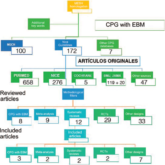
Figure 1 shows the results according to the search criteria established for the review.

Source: Authors.

Figure 1 Search and results flowchart. 

The only central venous catheter approach with sufficient evidence for recommending its use under US guidance routinely is the internal jugular vein, but it does not mean that this approach should prevail in situations in which the subclavian vein approach for central venous catheterization is indicated. 3,9

This is important as the subclavian approach is better in terms of the percentages of infection, bacteremia, and anatomic variants. 10 However, according to the variables already mentioned, the internal jugular vein and the right femoral approaches provide a more direct route to central veins, facilitating catheter placement without the need for control chest radiographs. 11

Once the need for a central venous access is identified, the next step is to define the approach. The approach that should be considered in the first instance because of its association with a lower percentage of infection is the subclavian vein. If access cannot be achieved through that approach, the next step is to move toward the internal jugular vein, associated with a higher possibility of infection. 12

The internal jugular vein approach has advantages that prompt its selection, including the lower incidence of complications in adjacent structures13; a lower risk of mechanical complications compared with the subclavian approach2; and finally, ease for catheter placement without the need for control chest radiograph. 14

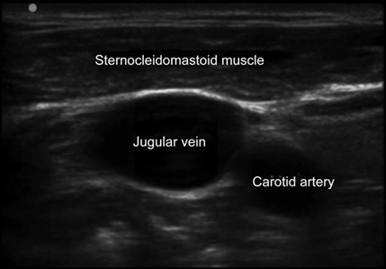
Technological sophistication of US machines varies significant, although the most basic models are good for vascular accesses. US modes for identifying and localizing vessels for catheterization are B mode (bidimensional) and Doppler for flow analysis (arterial or venous). Selecting the appropriate linear high-frequency (5-12MHz) probe is critical for obtaining high-quality images; high-frequency provides better resolution of the tissues lying close to the skin surface, which is ideal for vessel visualization10 (Fig. 2).

Source: Authors.

Figure 2 Ultrasound window showing the relationship between the jugular vein and its neighboring structures. 

The structures covered by the US beam underneath the left side of the probe marker (indicated by a light or notch on the side of the probe) always come up on the screen from left to right. Therefore, probes have markings that help with correct orientation. Moving the probe to the left or to the right while watching the image should help confirm the adequate orientation. 15

Transverse and/or longitudinal planes can be used for localization of the selected vein and catheter placement. 16 The transverse position (short axis) is useful for accessing vessels near the skin surface such as the internal jugular vein. 17,18 For deeper vessels, as is the case with the common femoral vein, a combined approach may be used, starting with the transverse position to identify the vessel, followed by rotation to the longitudinal view, initial needle advancement, and cannulation completion. 19

Additional steps are required when using US for central venous access such as the use of the stockinette and the sterile conductor gel. 20

Generally, the patient is placed at 10 and 20 degrees of Trendelenburg to reduce the risk of embolism and promote vein dilation for jugular access; the head is rotated to the opposite side of the puncture, bilateral anatomic recognition of the vascular structures is per formed in a medial-to-lateral direction, making sure there is no thrombosis, and the best site for puncture in the neck or subclavian vessels is identified. Once the puncture site is selected, the area is prepared with asepsis and antisepsis with chlorhexidine for 60 second, sterile drapes are placed, catheters are purged with saline solution, and the probe is covered with the sterile stockinette. 19,20

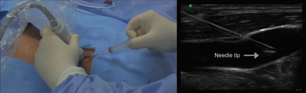
An illustrative academic video of the techniques was prepared to provide a graphic description of US-guided in plane and oblique longitudinal internal jugular access and US-guided in-plane longitudinal subclavian vascular access (Fig. 3) (Watch the video here: http://links.lww.com/RCA/A57).

Source: Authors.

Figure 3 In-plane long-axis internal jugular access. 

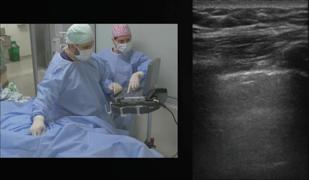
In addition, having an US machine allows us to run several checks to ensure the safety of the procedure, such as an US scan of the pleura and the lung to determine the presence of pleural sliding and rule out pneumothorax, providing higher positive predictive and negative predictive values than chest X-ray14,21,22; look for free fluid to rule out haemothorax23; and finally, determine the correct placement of the catheter in the right atrium, with an apical subxiphoid window and bolus in the distal catheter line with 10cm of saline solution24 (Fig. 4).

Source: Authors.

Figure 4 Ultrasound scan of the pleura and lung. 

When the traditional landmark approach is used, complications range between 0.3% and 18.8%, due to multiple variables such as patient population, insertion site, time used, and number of attempts. 25-27

Both Randolph et al4 in a 1996 meta-analysis as well as Rabindranath et al7 in a systematic review and meta-analysis conducted in 2011 show that the use of US for central venous catheterization through the internal jugular vein and subclavian vein in adults is associated with a significantly lower failure rate when compared with the traditional "blind" approach, and also with a reduction in the rate of complications, a shorter approach time and a lower number of attempts at successful access.

Figures for US and the approach to the internal jugular vein are strong. A meta-analysis by Hind et al28 conducted by the British NICE29 found a reduction of the relative risk of complications, failed attempts, and failed first attempt of 57%, 86%, and 41%, respectively. Therefore, the risks of inadvertent arterial puncture and local hematoma, among others, are lower with the use of US for the placement of a central venous catheter in the internal jugular vein.

However, it is important to recognize the adverse events that my occur with US-guided catheterization, including incorrect placement, arterial puncture, local hematoma, air embolism, or nerve injuries. 14,30

Pneumothorax and/or hemothorax are rare events; the majority of cases are recognized by US at the time of the intervention. 31,32 Thrombosis, arteriovenous fistula, and pseudoaneurysms are potential long-term complications and may be readily identified by US. 14,21

In the study by Kaye et al33 (n = 325 patients taken to cardiovascular surgery), the rate of complications follow ing the placement of a central venous catheter (including carotid puncture and pneumothorax) was significantly greater in the group in which no US was used as compared with the group in which US was used.

When US was used as a guide for the central venous catheter, Cavanna et al34 reported symptomatic deep vein thrombosis in the upper limbs in 2.4% of cases and catheter-related infections in 9.96% of the catheters inserted. Catheter removal due to complications was required only in 2.9% of the cases, and there were no reports of nerve injury, major bleeding, or pneumothorax.

Even with the help of US, puncture of the posterior vascular wall may still occur as a complication of venous catheterization. 35,36 The factors that influence the risk of posterior wall penetration include the approach technique, in particular short axis versus long axis, speed of needle insertion, distance between needle entry and the transducer, and insertion angle. 37

In their systematic review, Brass et al mention, regarding internal jugular, subclavian, and femoral vein accesses, complications of nearly 13.5% with the conventional technique, compared with 4% for US-guided catheterization of the internal jugular vein3,9; additionally, the success rate is greater with US, with shorter access time and a lower number of attempts. 1,38

Regarding the subclavian and femoral veins, although some data favor US for these approaches, meta-analyses have not found statistically significant differences when compared with the landmark-guided technique. 18,39-41

In experienced hands, the rate of failure for the landmark technique in the subclavian vein ranges between 9% and 13%.17 Standard approaches have been assessed (supraclavicular, infraclavicular, and, more recently, the axillary approach), but the results are inconclusive. 42-44

Based on these findings, whenever there are difficulties establishing a central vascular access in an adult patient, the suggestion is to use US to help guide the access, provided the operator is experienced. 45-50

Dietrich et al1 mention the following recommendations to help avoid risks and complications:

Optimize the target image in B mode.

Adjust patient position (Trendelenburg) and the position of the examiner and the US machine in relation to the puncture site (comfort for the interventional operator).

Position the head appropriately for localizing the target vein laterally, instead of anterior to the artery.

Get training in viewing skills using adequate practice models such as phantoms, and also in normal patient conditions, and emergency situations.

In hypovolemia, administer intravenous fluid before the puncture.

Obtain training that is certified or has a steep learning curve in simulators or in real life with an instructor to attain international success standards and reduce complications.

Conclusion

US must be used routinely for establishing a central venous access. Current evidence supports this recommendation for the internal jugular vein, but not so for the other approaches; however, in cases of difficult or failed catheterization using conventional methods, US must be used for any of the approaches described as a tool to ensure success. 1,2

Practitioners must be adequately trained in the use of the US machine and must have experience using a high resolution device. Time and practice are needed to become fully competent in these techniques and processes must be reviewed and audited to improve confidence and skill in vascular access, and compliance with standards must be appraised.

In the near future, as a result of the growing use of US for central venous catheterization, new research, brands, and more echogenic catheter materials will become available in the market. The need for these new materials is clearly implicit, considering that they will have a direct impact on the location of the catheter tip, helping reduce the percentage of complications and eliminating the need for verification by means of postpuncture chest radio graphs or fluoroscopy, among other advantages.

Ethical disclosures

Protection of human and animal subjects. The authors declare that no experiments were performed on humans or animals for this study.

Right to privacy and informed consent. The authors declare that no patient data appear in this article.

References

1. Dietrich CF, Horn R, Morf S, Chiorean L, Dong Y, Cui X-W, et al. Ultrasound-guided central vascular interventions, comments on the European Federation of Societies for Ultrasound in Medicine and Biology guidelines on interventional ultrasound. J Thorac Dis 2016;8:E851"E868. [ Links ]

2. Bodenham Chair A, Babu S, Bennett J, Binks R, Fee P, Fox B, et al. Association of Anaesthetists of Great Britain and Ireland: Safe vascular access 2016. Anaesthesia 2016;71:573-585. [ Links ]

3. Brass P, Hellmich M, Kolodziej L, Schick G, Smith AF. Ultrasound guidance versus anatomical landmarks for subclavian or femoral vein catheterization. Cochrane Database Syst Rev 2015;1: CD011447. [ Links ]

4. Randolph AG, Cook DJ, Gonzales CA, Pribble CG. Ultrasound guidance for placement of central venous catheters: a meta-analysis of the literature. Crit Care Med 1996;24:2053-2058. [ Links ]

5. Fragou M, Gravvanis A, Dimitriou V, Papalois A, Kouraklis G, Karabinis A, et al. Real-time ultrasound-guided subclavian vein cannulation versus the landmark method in critical care patients: a prospective randomized study. Crit Care Med 2011; 39:1607-1612. [ Links ]

6. Hayashi H, Amano M. Does ultrasound imaging before puncture facilitate internal jugular vein cannulation? Prospective randomized comparison with landmark-guided puncture in ventilated patients. J Cardiothorac Vasc Anesth 2002;16:572-575. [ Links ]

7. Rabindranath KS, Kumar E, Shail R, Vaux E. Use of real-time ultrasound guidance for the placement of hemodialysis catheters: a systematic review and meta-analysis of randomized controlled trials. Am J Kidney Dis 2011;58:964-970. [ Links ]

8. Rando K, Castelli J, Pratt JP, Scavino M, Rey G, Rocca ME, et al. Ultrasound-guided internal jugular vein catheterization: a randomized controlled trial. Heart Lung Vessel 2014;6:13-23. [ Links ]

9. Brass P, Hellmich M, Kolodziej L, Schick G, Smith AF. Ultrasound guidance versus anatomical landmarks for internal jugular vein catheterization. Cochrane Database of Systematic Reviews 2015;1: CD006962. [ Links ]

10. Jenssen C, Brkljacic B, Hocke M, Ignee A, Piscaglia F, Radzina M, et al. EFSUMB Guidelines on Interventional Ultrasound (INVUS), Part VI - Ultrasound-Guided Vascular Interventions. Ultraschall Med 2016;37:473-476. [ Links ]

11. Matsushima K, Frankel HL. Bedside ultrasound can safely eliminate the need for chest radiographs after central venous catheter placement: CVC sono in the surgical ICU (SICU). J Surg Res 2010;163:155-161. [ Links ]

12. Abboud PA, Kendall JL. Ultrasound guidance for vascular access. Emerg Med Clin North Am 2004;22:749-773. [ Links ]

13. Benter T, Teichgraber UK, Kluhs L, Papadopoulos S, Kohne CH, Felix R, et al. Anatomical variations in the internal jugular veins of cancer patients affecting central venous access. Anatomical variation of the internal jugular vein. Ultraschall Med 2001; 22:23-26. [ Links ]

14. Vezzani A, Manca T, Vercelli A, Braghieri A, Magnacavallo A. Ultrasonography as a guide during vascular access procedures and in the diagnosis of complications. J Ultrasound 2013;16:161-170. [ Links ]

15. Pittiruti M, Hamilton H, Biffi R, MacFie J, Pertkiewicz M. EspenESPEN Guidelines on Parenteral Nutrition: central venous catheters (access, care, diagnosis and therapy of complications). Clin Nutr 2009;28:365-377. [ Links ]

16. Ball RD, Scouras NE, Orebaugh S, Wilde J, Sakai T. Randomized, prospective, observational simulation study comparing residents’ needle-guided vs free-hand ultrasound techniques for central venous catheter access. Br J Anaesth 2012;108:72-79. [ Links ]

17. Shrestha GS, Gurung A, Koirala S. Comparison between long- and short-axis techniques for ultrasound-guided cannulation of internal jugular vein. Ann Card Anaesth 2016;19:288-292. [ Links ]

18. Batllori M, Urra M, Uriarte E, Romero C, Pueyo J, Lopez-Olaondo L, et al. Randomized comparison of three transducer orientation approaches for ultrasound guided internal jugular venous cannulation. Br J Anaesth 2016;116:370-376. [ Links ]

19. Tokumine J, Lefor AT, Yonei A, Kagaya A, Iwasaki K, Fukuda Y. Three-step method for ultrasound-guided central vein catheterization. Br J Anaesth 2013;110:368-373. [ Links ]

20. Merz E. [Transducer hygiene - an underrated topic?]. Ultraschall Med 2005;26:7-8. [ Links ]

21. Maury E, Guglielminotti J, Alzieu M, Guidet B, Offenstadt G. Ultrasonic examination: an alternative to chest radiography after central venous catheter insertion? Am J Respir Crit Care Med 2001;164:403-405. [ Links ]

22. Lichtenstein DA. Pneumothorax and the A'-Proile. Lung Ultrasound in the Critically Ill: The BLUE Protocol. 2016; Cham: Springer International Publishing, pp. 97-108. [ Links ]

23. Lanza C, Russo M, Fabrizzi G. Central venous cannulation: are routine chest radiographs necessary after B-mode and colour Doppler sonography check? Pediatr Radiol 2006;36:1252-1256. [ Links ]

24. Weekes AJ, Johnson DA, Keller SM, Efune B, Carey C, Rozario NL, et al. Central vascular catheter placement evaluation using saline flush and bedside echocardiography. Acad Emerg Med 2014;21: 65-72. [ Links ]

25. Sznajder JI, Zveibil FR, Bitterman H, Weiner P, Bursztein S. Central vein catheterization. Failure and complication rates by three percutaneous approaches. Arch Intern Med 1986;146:259-261. [ Links ]

26. Mansield PF, Hohn DC, Fornage BD, Gregurich MA, Ota DM. Complications and failures of subclavian-vein catheterization. N Engl J Med 1994;331:1735-1738. [ Links ]

27. Merrer J, De Jonghe B, Golliot F, Lefrant JY, Raffy B, Barre E, et al. Complications of femoral and subclavian venous catheterization in critically ill patients: a randomized controlled trial. JAMA 2001;286:700-707. [ Links ]

28. Hind D, Calvert N, McWilliams R, Davidson A, Paisley S, Beverley C, et al. Ultrasonic locating devices for central venous cannulation: meta-analysis. BMJ 2003;327:361. [ Links ]

29. National Institute for Clinical Excellence (NICE). Guidance on the Use of Ultrasound Locating Devices for Placing Central Venous Catheters. London UK: NICE; 2002. Technology appraisal guidance no. 49. [ Links ]

30. Theodoro D, Krauss M, Kollef M, Evanoff B. Risk factors for acute adverse events during ultrasound-guided central venous cannulation in the emergency department. Acad Emerg Med 2010;17:1055-1061. [ Links ]

31. Wu SY, Ling Q, Cao LH, Wang J, Xu MX, Zeng WA. Real-time two-dimensional ultrasound guidance forcentral venous cannulation: a meta-analysis. Anesthesiology 2013;118:361-375. [ Links ]

32. Mehta N, Valesky WW, Guy A, Sinert R. Systematic review: is real-time ultrasonic-guided central line placement by ED physicians more successful than the traditional landmark approach? Emerg Med J 2013;30:355-359. [ Links ]

33. Kaye AD, Fox CJ, Hymel BJ, Gayle JA, Hawney HA, Bawcom BA, et al. The importance of training for ultrasound guidance in central vein catheterization. Middle East J Anaesthesiol 2011;21:61-66. [ Links ]

34. Cavanna L, Civardi G, Vallisa D, Di Nunzio C, Cappucciati L, Berte R, et al. Ultrasound-guided central venous catheterization in cancer patients improves the success rate of cannulation and reduces mechanical complications: a prospective observational study of 1,978 consecutive catheterizations. World J Surg Oncol 2010;8:91. [ Links ]

35. Blaivas M, Brannam L, Fernandez E. Short-axis versus long-axis approaches for teaching ultrasound-guided vascular access on a new inanimate model. Acad Emerg Med 2003;10:1307-1311. [ Links ]

36. Moon CH, Blehar D, Shear MA, Uyehara P, Gaspari RJ, Arnold J, et al. Incidence of posterior vessel wall puncture during ultrasound-guided vessel cannulation in a simulated model. Acad Emerg Med 2010;17:1138-1141. [ Links ]

37. Stone MB, Moon C, Sutijono D, Blaivas M. Needle tip visualization during ultrasound-guided vascular access: short-axis vs long-axis approach. Am J Emerg Med 2010;28:343-347. [ Links ]

38. Lalu MM, Fayad A, Ahmed O, Bryson GL, Fergusson DA, Barron CC, et al. Ultrasound-Guided Subclavian Vein Catheterization: A Systematic Review and Meta-Analysis. Crit Care Med 2015; 43:1498-1507. [ Links ]

39. Mozersky DJ, Olson RM, Coons HG, Hagood COJr. Doppler-controlled needle director: a useful adjunct to angiography. Radiology 1973;109:221-222. [ Links ]

40. Hoffman T, Du Plessis M, Prekupec MP, Gielecki J, Zurada A, Tubbs RS, et al. Ultrasound-guided central venous catheterization: A review of the relevant anatomy, technique, complications, and anatomical variations. Clin Anat 2017;30:237-250. [ Links ]

41. Milone M, Di Minno G, Di Minno MN, Salvatore G, Iacovazzo C, Policastro C, et al. The real effectiveness of ultrasound guidance in subclavian venous access. Ann Ital Chir 2010;81:331-334. [ Links ]

42. Stone MB, Wang R, Price DD. Ultrasound-guided supraclavicular brachial plexus nerve block vs procedural sedation for the treatment of upper extremity emergencies. Am J Emerg Med 2008;26:706-710. [ Links ]

43. Taylor BL, Yellowlees I. Central venous cannulation using the infraclavicular axillary vein. Anesthesiology 1990;72:55-58. [ Links ]

44. Nickalls RW. A new percutaneous infraclavicular approach to the axillary vein. Anaesthesia 1987;42:151-154. [ Links ]

45. Turba UC, Uflacker R, Hannegan C, Selby JB. Anatomic relationship of the internal jugular vein and the common carotid artery applied to percutaneous transjugular procedures. Cardiovasc Intervent Radiol 2005;28:303-306. [ Links ]

46. Troianos CA, Hartman GS, Glas KE, Skubas NJ, Eberhardt RT, Walker JD, et al. Special articles: guidelines for performing ultrasound guided vascular cannulation: recommendations of the American Society of Echocardiography and the Society Of Cardiovascular Anesthesiologists. Anesth Analg 2012;114:46-72. [ Links ]

47. Alderson PJ, Burrows FA, Stemp LI, Holtby HM. Use of ultrasound to evaluate internal jugular vein anatomy and to facilitate central venous cannulation in paediatric patients. Br J Anaesth 1993; 70:145-148. [ Links ]

48. Denys BG, Uretsky BF. Anatomical variations of internal jugular vein location: impact on central venous access. Crit Care Med 1991;19:1516-1519. [ Links ]

49. Troianos CA, Kuwik RJ, Pasqual JR, Lim AJ, Odasso DP. Internal jugular vein and carotid artery anatomic relation as determined by ultrasonography. Anesthesiology 1996;85:43-48. [ Links ]

50. Aponte H, Acosta S, Rigamonti D, Sylvia B, Austin P, Samolitis T. The use of ultrasound for placement of intravenous catheters. AANA J 2007;75:212-216. [ Links ]

How to cite this article: Caballero AF, Villarreal K. Ultrasound for central vascular access. A safety concept that is renewed day by day: review. Rev Colomb Anestesiol. 2018;46:32-38.

Funding No sponsorship was received for this article.

Conflict of interest The authors declare having no conflict of interest in relation to this publication.

* Correspondence: Departamento de Anestesiología, Universidad del Valle, Hospital Universitario del Valle, Calle 5 No 36-08 piso 4, Cali, Colombia. E-mail: karenvillarrealmd@gmail.com

Creative Commons License This is an open-access article distributed under the terms of the Creative Commons Attribution License