


 text in
  English (pdf)
 Article in xml format
 Article references
 Send this article by e-mail
 Cited by SciELO
  Cited by Google
  Similars in
SciELO
Similars in
SciELO  Similars in Google
 Permalink
Wellens syndrome is an uncommon perioperative occurrence. It is associated with critical proximal left anterior descending (LAD) artery stenosis and impending massive anterior wall myocardial infarction.1,2 Two electrocardiographic patterns have been described: Types A and B.
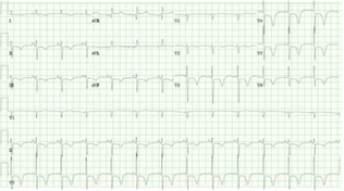
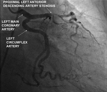
Type A Wellens syndrome is characterized by progressive deep T-wave inversions in precordial leads V3 to V5 (Fig. 1). In addition to absence of ST elevation and Q waves, normal precordial R-wave progression is present. These changes, accompanied by history of anginal chest pain and normal or minimal troponin elevation, define Wellens syndrome. This electrocardiographic pattern occurs during pain-free periods in patients experiencing intermittent angina and represents reperfusion between ischemic episodes. In Type B Wellens syndrome, biphasic T waves are seen in the precordial leads instead of deeply inverted T waves. Absence of ST-segment changes and troponin elevation is falsely reassuring.1,2 Coronary angiography reveals severe proximal LAD stenosis (Fig. 2). These images are from a 70-year-old patient who presented for outpatient bronchoscopy under general anesthesia for a suspected pneumonia. The procedure was canceled and she was admitted for coronary angiography by cardiology. Territories perfused by the LAD artery may exhibit hypokinesia or akinesia on echocardiography (See Video 1a, http://links.lww.com/RCA/A871 and Video 1b, http://links.lww.com/RCA/A872 [Source: Sandeep Khanna.]).
Source: Authors.
Figure 1 A total of 12 lead electrocardiogram exhibiting deep T-wave inversions in precordial leads.
Source: Authors.
Figure 2 Coronary angiogram exhibiting proximal left anterior descending artery stenosis.
Anesthetic evaluation of such patients includes rapid assessment of airway, breathing, and circulation. In addition to performing an abbreviated history and physical examination, establishing intravenous access and monitoring is prioritized. Presence of resuscitation equipment is ensured. Serial electrocardiograms and troponin assessments are encouraged. Early cardiology consultation is prudent as urgent percutaneous intervention or Coronary Artery Bypass Grafting are treatment modalities of choice. Stress testing is contraindicated because it can precipitate massive myocardial infarction. If chest pain develops, oral aspirin, sublingual nitrates, beta blockers, statins, and opioid analgesics are commenced in the absence of contraindications.2,3
Ethical responsibilities
Protection of persons and animals. No experiments on people or animals were done.
Confidentiality of the information. All protocols at our institute were followed and patient or hospital identifiers have been removed from all images.
Right to privacy and informed consent. As patient and hospital identifiers have been removed, no informed consent was solicited for this production.

