


 text in
  English (pdf)
 Article in xml format
 Article references
 Send this article by e-mail
 Cited by SciELO
  Cited by Google
  Similars in
SciELO
Similars in
SciELO  Similars in Google
 Permalink
Introduction
High intraocular pressure (IOP) is the main risk factor for glaucoma and the only modifiable condition of this pathology; therefore, measuring IOP is essential to diagnose, follow-up and treat this illness.1 In pediatrics, this measuring is especially complex, as stillness and collaboration are required from patients during tonometry, conditions difficult to meet when it comes to children. In adults, the IOP values range from 9 to 21 mm Hg, but, in children, normal values are not that clear. Several authors have measured and established IOP values in healthy children and results range from 13.3 ± 3.4 to 17.7 ± 2.7 mm Hg.2-4 Glaucoma is a broad and varied pathology; it causes blindness at long-term and, in children, it characterizes by having a rapid progression, severe clinical condition and difficult management.5,6 Incidence of pediatric glaucoma is 1/10,000 in newborn alive children.7 According to the World Health Organization, it is part of the first 5 causes of blindness at world level in the general population,8 and the fourth in Colombia in pediatric population.9
Blindness in children ranges from 0.3 to 1.5 per 1000, according to the social and economic conditions of the population.10,11 Early diagnosis and appropriate control of the IOP are the treatment cornerstone, in order to prevent severe ocular morbidity.12-14 New non-contact tonometry technologies seem the ideal for pediatric patients, with acceptable accuracy and concordance in comparison with the gold standard of Goldmann's applanation tonometry; however, access to these technologies is yet limited in many scenarios.15,16
Due to their unability to cooperate, newborn, nursing babies, and children cannot tolerate the ophthalmoscopic examination and IOP measuring; therefore, general anesthesia guarantees an accurate evaluation of the eye. Anesthetic intervention may alter the accuracy of the IOP measurement; hypertension, increase in the central venous pressure, hypoxia, and hypoventilation increase IOP, while most anesthetics (inhaled, inductors, benzodiazepines, and opioids) reduce it.17,18 Some authors agree that ketamine may increase IOP or produce a negligible effect in it.19,20 Providing a balanced sedation, analgesia, low respiratory, and cardiovascular depression, ketamine is an interesting alternative to provide anesthesia to pediatric patients requiring an examination.21,22
Several authors support the hypothesis that the IOP after ketamine administration is the most accurate representation of IOP in awake calmed patients.17,23 The purpose of this study is describing changes in intraocular pressure values in pediatric patients after administering ketamine as general anesthetic.
Methods
A systematic review of literature was conducted by consulting the PubMed, LILACS, Ovid, Cochrane, and ScienceDirect databases, through search strategies that may be consulted in Annex 1. The research protocol was not recorded in databases (Prospective Register of Systematic Reviews); nonetheless, this was not subject to modifications during the execution of the project.
The inclusion criteria comprised articles published from 1970 to February 2019, observational descriptive studies, cases and controls, randomized controlled cohorts and clinical trials in English, Spanish, French, and Portuguese languages. Studies non-complying with these characteristics were excluded. The PICO structure of studies should comprise patients aged under 18 years who had been performed an ocular tonometry, intervention was the administration of ketamine intravenously (IV), intramuscularly (IM), orally (O), or intrarectally for sedation or anesthesia during the examination, placebo or the administration of another anesthetic (sevoflurane, halo-thane, propofol, etomidate, thiopental) (C), Intraocular pressure measured (O). This was compared with placebo or the administration of another anesthetic (sevoflurane, halothane, propofol, etomidate, thiopental), and intraocular pressure measured after the administration of medicine was established as outcome. Figure 1 shows the inclusion and exclusion of articles with their respective causes.
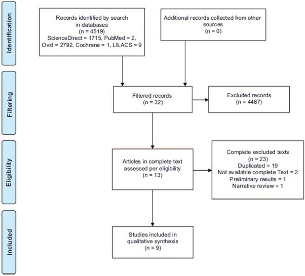
Source: Authors.
Figure 1 Preferred Reporting Items for Systematic Reviews and Meta-analysis Flowchart showing the identification, filtering and selection process of studies. No study included in meta-analysis is shown as these were not included in this systematic review.
Results from search in each database were compiled in a form previously created in Excel by the authors; such form contains authors, title, publication date, and abstract of each article. Independently, each author identified articles subject to evaluation according to their title and abstract. Disagreements were solved by consensus. Articles included are qualitatively summarized in Table 1. The absolute variation value of IOP after administering ketamine as described above was compiled as primary outcome; in case of absence of absolute values, the variation percentage of IOP would be used as alternative. In studies reporting safety data, safety variables on respiratory complications, psychomotor agitation, and sedation were used as secondary outcomes.
Table 1 Qualitative summary of included studies.
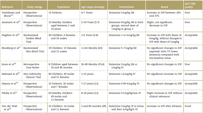
IM=intramuscular, IOP=intraocular pressure, IV=intravenous, QAT-NIH=Quality Assessment Tool from the National Institute of Health.
Source: Authors.
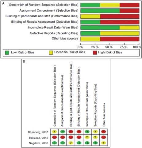
Observational studies were evaluated through the quality assessment tool from the National Institute of Health (QAT-NIH) for observational studies. In the case of randomized clinical trials, the QAT-NIH for randomized controlled studies was used. In addition, the Cochrane risk-of-bias tool was used (Fig. 2a and 2b). An additional search was made on the preliminary results of research and in-progress researches without final results, finding no further information for the systematic review. No study was subject to be included in a meta-analysis; therefore, a qualitative synthesis of literature was performed.
Results
A total of 4519 studies were found, of which 4487 did not correspond to planned characteristics for the systematic review as these were articles on trials conducted in adults, most of them did not include the administration of ketamine in their methodology, did not include measuring of intraocular pressure or were not presented in the languages described in the inclusion criteria (Fig. 1).
Nine studies were included, of which 3 were clinical trials, 2 of them conducted in patients without glaucoma diagnosis and 1 in patients with glaucoma or suspected glaucoma. General results and a summary on the studies are shown in Table 1.
The Yoshikawa and Murai24 study intended to assess the impact of ketamine on intraocular pressure in healthy children; patients aged 4 to 7 years were included and the intervention conducted consisted on administering ketamine doses at 5 mg/kg IM, without premedication. Authors explained to have taken a basal IOP before the injection, followed by measurements every 5 minutes up to 30 minutes after injection. Authors do not describe in their methodology the tests run to assess the statistical significance of results; nevertheless, they reported a statistically significant increase in IOP from minute 5 (18% increase in measurements, P < 0.001) up to minute 15 (37% increase in measurements, P < 0.005), and return to basal values in minute 30 with P < 0.4. This change was accompanied by increase in blood pressure and heart rate in the first 5 minutes after injection. A patient with strabismus was reported with an early drop in IOP after the administration of the drug. Authors did not describe respiratory, cardiovascular or behavioral complications during the study.
Ausinsch et al25 study sought to assess the impact of ketamine on IOP in children with healthy eyes; therefore, 10 children without ocular pathology evidence were recruited. All children were premedicated with atropine 0.02 mg/kg IM, and 5 of them received 1.1 mg/kg IM of meperidine and 4.4mg/kg IM of pentobarbital. Basal values were taken with ocular instillation of proparacaine hydrochloride ophthalmic solution in both eyes. The study was divided in 2 stages. Initially, all patients received 8 mg/kg of ketamine IM and measurements were taken at 5, 10, 15, and 20minutes. Then, a second dose of 1 mg/kg of ketamine was administered to all patients and 5 of them additionally received 0.8 mg/kg of d-tubocurarine IV, these were intubated 5 minutes later and received anesthetic treatment with nitrous oxide. IOP was measured at 5, 10, and 15 minutes after the first dose.
Authors found decrease between the average basal IOP and after induction with ketamine (22.2 ± 4.8 vs. 16.7 ± 3.3 mm Hg) which they described as statistically significant (P < 0.001); however, they do not mention the tests run in any other part of the article. Three out of 5 premedicated children cooperated with basal IOP measuring and they showed a value significantly lower than values of those patients who did not cooperate (17.5 ± 2.6 vs. 24± 3.5 mm Hg, P < 0.05). Subsequently, after administering a second dose lower than the induction dose, no significant change was reported in the IOP and the administration of neuromuscular relaxant did not produce clinically or statistically significant change. IOP values after induction with ketamine in stage I between premedicated and non-premedicated patients were similar, without statistically significant changes.
Nagdeve et al26 sought to answer the same question through a randomized double-blind clinical trial; therefore, 40 pediatric patients classified as category I by the American Society of Anesthesiologists (ASA) I, weighting more than 25 kg and with 30 to 90-minute scheduled surgeries under general anesthesia, were included. Patients with previously high IOP, endocranial hypertension, open ocular trauma, vascular aneurisms, psychiatric disorders, and seizure syndromes were dismissed. All patients received premedication 60 to 90 minutes before surgery with oral triclofos (100 mg/kg). Anesthetic induction was through inhalation with halothane up to 4%. Following the Güedel's planes, once patients reached surgical anesthesia, they continued administering halothane at 1%. 10 minutes after induction, patients were randomized to groups of low doses (3 mg/kg) or high doses (6 mg/kg) of ketamine. The drug was administered IM in the deltoid muscle and measuring of IOP was taken every 5 minutes. During 20 minutes no surgical stimulation was allowed in the patient. There was blinding of the observer taking measurements on the administered dose, who measured IOP in both eyes unless 1 of them were intended for surgery, in which case the eye to be operated was excluded.
Both groups were comparable in terms of demographic distribution (gender, weight, age). In each group, data were subject to repeated measures analysis of variance and compared with measurements defined as basal using the Student t test. The induction dose group reported increase in IOP 5 and 10-minute measurements (basal 10.8 ± 2.2 mm Hg, 5 minutes 12.6 ± 2.8 mm Hg and 10 minutes 11.9 ± 2.5 mm Hg, P < 0.001). The low-dose group reported no change in IOP. However, when comparing the values of IOP at minute 10 and 15 of both groups, no statistically significant difference was reported; results were as follows: basal 11.4± 2.0, 11.1 ± 2.2 mm Hg at 5 minutes, and 11.1 ± 2.2 mm Hg at 10 minutes. With respect to complications, the high-dose group reported a more frequent airway obstruction, with 18 patients, in comparison with 4 from the low-dose group (P < 0.001). Likewise, the postoperative sedation was higher in the higher dose group (P < 0.001). Two dissociative reactions at the moment of waking up were reported, both in the high-dose group.
Blumberg et al27 reported results from a randomized clinical trial published in 2007. This study included patients of any age with glaucoma or suspected glaucoma scheduled for tonometry with anesthesia due to low collaboration. Contingency tables were used to compare characteristics of children in the 2 arms of the study and changes were valued in several monitoring periods (2, 4, 6, and 8 minutes).
No standardization was conducted in the administration or not of premedication; some patients received midazolam O (0.5-1 mg/kg-max.: 20 mg), IV (0.05-0.1 mg/ kg-max.: 5 mg) or rectally (0.5-1 mg/kg-max.: 20 mg), and some also received atropine IM (0.02 mg/kg) or IV (0.01 mg/ kg). Authors applied an induction with sevoflurane at 8% and FiO2 at 100% and maintenance 1 to 2 minutes after with 2 to 4vol.%. Regarding the ketamine group, doses were reported between 5 and 7 mg/kg IM. One patient was excluded as he required intubation in the sevoflurane group, plus 3 patients whose IOP, after randomized, could not be measured. The IOP baseline measurement was taken immediately after induction (T1); therefore, values of awake patients were not collected. The T1 time ranged between 3 and 10 minutes after induction. Other values were recorded at minutes 2, 4, 6, and 8.
In order to approximate to the basal IOP, they made a regression of the best adjustment by extrapolating data backwards, and found the cross point of both variables between 2 and 4 minutes previous to the T1. Medians for IOP taken in T1 between the 2 groups reported no differences (P = 0.15). With sevoflurane, a greater and more consistent decrease was reported at minutes 2, 4, 6, and 8 after induction when compared to T1, of 11.5%, 19.2%, 18.5%, and 16.7%, respectively (P < 0.001). The ketamine group reported IOP decrease at times measured, but its variation with respect to T1 was not clinically significant (7.4%), even if the result was statistically relevant (P = 0.03), with a 95% assumed confidence interval (CI) with 5% of error. These data were not reported in the article.
Jones et al28 intended to describe changes in IOP after intramuscular or intravenous ketamine administration and final maintenance with sevoflurane, through retrospective review of cases. They reviewed clinical histories for a period of 3 years and assessed patients unable to cooperate subject to examinations under anesthesia (EUA) and unreliable measurements taken to patients awake. 5 mg/kg IM or 2 mg/kg IV of ketamine were administered. Measurements were averaged in groups of 3 (alternately between the 2 eyes). They found an average difference between ketamine measurements and sevoflurane measurements of 7.39 mm Hg (P < 0.001), 28.5% less with sevoflurane. The mean IOP value was 17.0 mm Hg in the sevoflurane group and 24.4 mm Hg in the ketamine group.
Drayna et al29 prospectively recruited patients to perform a descriptive analysis of the impact of different doses of ketamine on the IOP of pediatric patients scheduled for procedures under sedation with this drug. They calculated the basal IOP by measuring it in both eyes and averaging it before administering ketamine. They found no clinically or statistically significant increase between baseline and IOP measurements performed at minutes 1, 3, 5, 15, and 30 (P = 0.15). A Bland Altman's analysis showed an appropriate level of concordance between the measurements of each eye at different times. The association of ketamine doses with IOP was not statistically significant (P = 0.90); the average IV dose was 1.88 (mg/kg). The mean values were: baseline 14.66 mm Hg, 1minute 14.84mm Hg, 3minutes 15.18mm Hg, 5 minutes 15.46mm Hg, and 15 minutes 15.75mm Hg.
Halstead et al30 proposed a non-inferiority study including pediatric ASA I or II patients scheduled for procedures under sedation with ketamine for problems other than eye injury, in which all patients received ketamine at doses between 1 and 2 mg/kg IV, and were controlled with baseline values taken immediately after the medication was administered. According to their findings, there were no clinically significant changes in IOP (>15%, 2.6mm Hg); these results were applied to the 3 age groups in the study: 1 to 5, 6 to 10, and 11 to 15 years. In their results, the mean value of initial IOP was 17.5 and 18.9 mm Hg at 2.5 minutes, with an average difference between the 2 measurements of 1.4mm Hg.
Wadia et al,31 in their prospective observational study using a convenience sample that included children up to 18 years of age, performed baseline measurements of IOP before administering procedural sedation with ketamine in Emergency. The average increase in IOP was 3mm Hg (0-8 mm Hg). They reported the results descriptively, using 95% Clopper Pearson CIs, averages and medians, as applicable.
Van der Walt et al32 studied children with glaucoma or suspected glaucoma who required evaluation under anesthesia. Anesthesia was protocolized: induction with sevoflurane, peripheral vein channeling, IV administration of ketamine at 2 mg/kg and maintenance with ketamine infusion at 4mg/kg/h for15minutes. Comparing the mean IOP at time 0 (sevoflurane only) with time 15 minutes (ketamine only), an increase of 3.68 mm Hg in IOP (95% CI 1.35-6.02 mm Hg) (P = 0.002) was shown. Comparing the mean IOP at 15 minutes (ketamine alone) and 20 minutes (near awakening), there was an increase of 0.28 mm Hg (P=0.826).
The evaluation of the quality of the evidence made it possible to identify that the studies by Yoshikawa et al, Ausinsch et al, Nagdeve et al, Blumberg et al, and Jones et al have deficiencies in reporting the methodology used to recruit patients, determine losses and participation percentage, justify sample sizes and describe the statistical power sought. Some also presented difficulties in their methodology for measuring the effect of different drug doses (ketamine) on intraocular pressure (primary out-come). Only the study by Nagdeve et al speaks of double blinding, and in the other studies they do not report it or deny having done it in their manuscripts.
According to the evaluation carried out with the QAT-NIH instrument, it was concluded that the studies of Yoshikawa, Ausinsch, and Jones are of poor quality, those of Nagdeve, Blumberg, Halstead, Drayna, and Wadia are of acceptable quality, and that of Van der Walt is of good quality.
In terms of complications and safety, the first to report results were Nagdeve et al, who in their work found that higher doses (6 mg/kg IM) compared with doses of 3 mg/kg IM produced a greater number of airway complications (obstruction), with 18 patients vs. 4. Sedation at0 and 2hours after the procedure was higher in the high-dose group: for immediate postprocedure (0 hours) there were 13 patients compared with 8 in the low-dose group, and at 2 hours postprocedure there were 19 patients compared with 2 in the low-dose group. All these comparisons showed differences between the study groups (P< 0.005).
In the experience of Van der Walt et al, these authors refer in their methodology to having recorded the presentation of complications; however, there is no report in the results of their work on whether or not complications occurred during the study. No other studies included in the analysis of this manuscript accounted for or reported the occurrence of complications.
The bias risk of the Blumberg et al27 work assessed with the Cochrane risk-of-bias tool for individual studies identified as high-risk sources the lack of blinding of participants and staff and assessment of results. In the case of Halstead et al,30 the sources described above, plus the absence of a random sequence for the selection of patients and the concealment of the selection, made it a work with a high risk of bias, according to the same tool. For its part, the study by Nagdeve et al26 was the work with the lowest risk of bias among those evaluated; the only source identified as an uncertain risk of bias was the blinding of participants and staff, as its description was not completely clear in the manuscript. The results of these evaluations are shown in Fig. 2A and 2B.
Discussion
As previously mentioned, IOP measurement is of paramount importance for glaucoma diagnosis, follow-up, and treatment; however, in young children, who are unable to collaborate to make the measurement possible or at least 1 reliable measurement, it is necessary to perform it under anesthesia. The problem is that it has been widely described how sedative and anesthetic medications decrease intraocular pressure, and therefore it is important to find an option that produces no effect on it, for the measurement to be reliable and not affect the evaluation and adequate clinical analysis of patients and their therapeutic needs.
This systematic review aimed to assess the impact of ketamine administration on intraocular pressure in the pediatric population. The results, although of low quality of evidence, suggest that the administration of ketamine in pediatric patients to perform ocular tonometry has little or no effect on intraocular pressure. Of the 9 articles included in the review, 2 showed a statistically or clinically significant impact on the measurement of intraocular pressure following ketamine administration, and 7 others had results supporting their use in conducting these examinations. In studies that showed a significant impact of ketamine on IOP, doses of ketamine were higher (>4mg/kg). Doses of up to 0.96 mg/kg in bolus were also documented, which produced appropriate conditions for tonometry in terms of sedation.
The limitations of this review are due to the heterogeneity of the studies in terms of population size and doses used of ketamine, the high risk of bias of several of the components of the included studies, and the lack of good quality studies to determine recommendations with stronger evidence. The use of ketamine by different routes (IM and IV) causes doses and instauration times of clinical effect to vary for IOP measurement. The impossibility of performing a meta-analysis due to the difficulties described in the methodology is another limitation.
Ausinsch et al25 found a slight decrease in IOP after intramuscular administration of ketamine in the initial dose and in a second dose. Their population was small and they performed several cointerventions, a situation that negatively affects the strength of their methodology. They administered a small subgroup neuromuscular relaxation and further divided the main group into single and repeated doses of ketamine. It is difficult to assess the actual weight of the administration of a single drug on the outcome to be measured, and for this reason the final assessment of the quality of evidence in this study was not satisfactory.
For other studies evaluated in the review, such as Halstead et al30 and Jones et al,28 a major difficulty was the adequate identification of an IOP baseline to determine the actual impact of ketamine administration. In some cases, the patient was under the effects of inhaled anesthetics, and in others measuring intraocular pressure in uncooperative conscious children resulted in unreliable values sometimes even greater than the patient's normal values.
In the best quality studies (acceptable and good in QAT-NIH) no significant changes in IOP were reported at doses from 0.96 to 2 mg/kg IV, and from 3 to 5 mg/kg IM. However, no available evidence was found on the administration of ketamine by other routes to perform eye tonometry in children, and for this reason it is not possible to identify an appropriate dose for alternative routes with the evaluation of the present review; a systematic review of the literature identified the use of ketamine and its effectiveness intranasally for sedation, being between 3 and 9 mg/kg intranasally with good results in terms of sedation and analgesia.33
The results of this work do not allow us to recommend the use of ketamine as a general anesthetic for the measurement of intraocular pressure in children; although most of the results of the studies evaluated show little or no impact on this, there are no studies which methodological strength allows the recommendation to be made. Clinical trials with larger populations and greater methodological strength are still required.
Conclusion
Ketamine is a reliable anesthetic drug that may have less effect on intraocular pressure than other drugs used for the same purpose; however, the studies found do not have the necessary characteristics to consider assertively recommending this drug for EUA in children with glaucoma. It is therefore necessary to expand the evidence by conducting clinical trials with greater statistical force that would allow its use to be recommended in a protocol manner.
In case it is used among the different existing alternatives, clinicians should know that the available evidence, although of low quality, is inclined to show it as a safe and reliable choice for measurement, based on the safety results of the only article that reports them and on the variations shown in absolute terms in intraocular pressure in the different included studies, where values ranging from 1.4 mm Hg to a maximum of 3mm Hg were found that are not clinically relevant for the diagnosis and follow-up of the pathology.
Ethical responsibilities
The results and conclusions reported in the document are true to what is described in the literature, without alterations or modifications made by the authors to adjust their results.
The written production is the product of the intellectual exercise of the authors, appropriately referencing in each of the relevant cases the references that correspond to other authors outside the work team.

