


 text in
  English (pdf)
 Article in xml format
 Article references
 Send this article by e-mail
 Cited by SciELO
  Cited by Google
  Similars in
SciELO
Similars in
SciELO  Similars in Google
 Permalink
Introduction
Protein S is a cofactor dependent on vitamin K and protein C. Protein C acts by inhibiting factors Va and VIIIa of the coagulation cascade. The free fraction of protein S acts as a cofactor of protein C. Moreover the deficiency of both proteins leads to a state of hypercoagulability that is treated with anticoagulation.1
Congenital protein S deficiency is an autosomal dominant disease, very rare in the non-gene carrier population, with a frequency of approximately1 in 700 individuals in the general population, according to extrapolations from a study conducted on more than 9000 donors. The incidence of protein S increases to 3% to 6% in patients with a history of thrombosis or recurrent thrombosis. Also described is a high frequency in Japanese patients, approximately 12.7%. Venous thromboembolism (VTE) develops in 60% to 80% of patients who are heterozygous for protein S deficiency; however, the remaining patients are asymptomatic and some never develop VTE.2
Protein S deficiency in pregnant women is associated with miscarriage, fetal death, restriction of fetal intra-uterine growth, placental abruption and pregnancy-induced hypertension, and may also induce thrombosis in deciduous vessels and affect placentation through hypercoagulability and inflammation. The changes that occur in the coagulation system during gestation are compensatory mechanisms of hemostasis that should occur after childbirth; the pregnant woman presents a state of physiological hypercoagulability where the concentrations of coagulation factors, such as VII, IX, and X, are increased, as well as fibrinogen levels increase by 50%, to whose changes a decrease in fibrinolytic response is added. However, anticoagulants such as anti-thrombin and protein S decrease and, consequently, pregnancy alters the equilibrium of the coagulation system, which means that both the pregnant and puerperian woman are pre-disposed to developing thrombotic processes.3
It should be noted that the greatest risks of developing these problems occur in the first trimester and up to 12 weeks after childbirth. The risk of VTE is approximately 4 to 5 times greater during gestation, with deep vein thrombosis and pulmonary embolism being the most frequent clinical manifestations.4
Case description
We present the case of a 32-year-old multigravida with a 36-week pregnancy and a diagnosis of thrombophilia: functional protein S deficiency (Table 1) from weeks before current gestation.
Table 1 Laboratory tests.
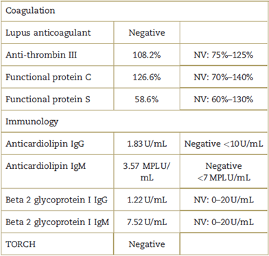
IgG=Immunoglobulin G; IgM=Immunoglobulin M; MPL=Immunoglobulin M units; NV=normal values; TORCH = Screening test for toxoplasmosis, rubeola, citomegalovirus, herpes and HIV.
Source: Authors.
The patient was scheduled for category 4 Caesarean section by Lucas et al. She has an obstetric history: 4 incomplete abortions, a gestation at term 4 years ago concluded by C-section (poor presentation), apparently without complications. No history of thromboembolic events. In normal general condition, accessible venous puncture areas, absence of varicose veins in the lower limbs, presence of edema +/+++, isochoric photorefractive pupils. Physical examination: Mallampati II score, thyromentonian distance of 6.5 cm, mouth opening greater than 4cm and mandibular subluxation greater than 0 degrees, adequate cervical movement, central trachea and denture without alterations, palpable spinous process, no scoliosis, and rest of apparently normal physical examination. American Society of Anesthesiologists (ASA) II physical fitness scale and pre-surgical laboratory ancillary examinations in the normal range (Table 2).
Table 2 Pre-surgical tests.
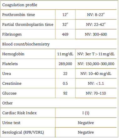
NV=normal values; RPR=rapid plasma reagin; VDRL=venereal disease research laboratory.
Source: Authors.
The patient is under pharmacological thromboprophylaxis from the 4th week of gestation, with enoxaparin 40 mg every 24 hours subcutaneously. Enoxaparin is suspended 24hours before surgery and mechanical thromboprophylaxis measures are recommended in the perioperative period. The patient is given the anesthetic options and the spinal technique is performed to provide greater security. Basic ASA monitoring was performed. Before antisepsis, the spinal technique was performed with spinal needle pencil tip No. 27 G and hyperbaric bupivacaine at 0.5% 10 mg plus fentanyl 20 mg in the intervertebral space L3 to L4, without any complication during the anesthetic act; we do not have ultrasound in the service to locate the intervertebral space for puncture.
The patient was hemodynamically stable before, during and after the surgical procedure. The birth occurred at 12:43 P.M., with a healthy newborn. Intraoperative bleeding of 500 mL was quantified. Tramadol 100 mg and metamizole 2 g were administered intravenously 30minutes before the end of surgery for the management of postoperative analgesia. The patient was transferred to the post-anesthetic recovery unit with stable vital functions, where she was monitored for 3hours; during the post-anesthetic evolution to discharge, according to the Aldrete scale, she had 10 points, and according to the Bromage scale, 0 points. She was then moved to the hospital for subsequent discharge from nosocomial on the 3rd day. There were no thromboembolic events in the 15 post-operative days.
Discussion
It is recommended that the risk of thromboembolism be stratified at the beginning of pregnancy, during pre-anesthetic evaluation, as well as in the immediate intra-and postpartum period (Table 3).
Table 3 Risk factors for venous thromboembolism.
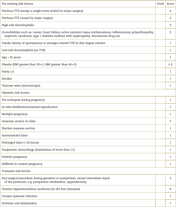
Score ≥ 4 antepartum, consider thromboprophylaxis in the first trimester. Score of 3, consider thromboprophylaxis from 28 weeks gestation. Score ≥ 2 postpartum, consider thromboprophylaxis 10 days later. Consider thromboprophylaxis in the antenatal period. Prolonged stay (≥3 days) or re-entry during puerperium, consider thromboprophylaxis. For patients at risk of bleeding, the balance between this risk and that of thrombosis should be evaluated by a hematologist with expertise in thrombosis and bleeding in pregnant women. BMI = Body Mass Index; VTE=venous thromboembolism.
Source: Adapted from Appendix III: "Risk assessment for venous thromboembolism" of the Royal College of Obstetricians & Gynecologists.5
In relation to low-risk thrombophilia, such as protein S deficiency, the recommendation for thromboprophylaxis is based on the presence of associated risk factors. In this patient the risk was stratified with a score of 3 (1 point for low-risk thrombophilia, 1 point for elective c-section, 1 point for pregnancy under 36 weeks).5 In relation to pharmacological prophylaxis, the agent of choice is low molecular weight heparin (LMWH) both antenatal and postnatal, and doses are based on maternal weight. LMWH is safe during breastfeeding; monitoring of factor Xa as follow-up is not necessary when used as prophylaxis. Unfractionated heparins may be useful in the peripartum, as they have a shorter action time, but their chronic use generates heparin-induced thrombocytopenia and osteoporosis.5
One of the advantages of LMWH in obstetrics is the extended half-life and increased bioavailability which is therefore used once a day as prophylaxis. Patients receiving heparin prophylaxis may benefit from neuro-axial techniques; however, their application should be personalized. In patients receiving LMWH by thromboprophylaxis, an interval of 12 hours after the last dose and 24hours after its use as treatment is recommended, before the start of a neuroaxial procedure; and restarting after 4hours of the use of a spinal technique or removal of the epidural catheter, in addition to the fact that it should not be removed within 12 hours after the last dose of LMWH.
Regardless of the anesthetic technique chosen, priority should be given to monitoring and avoiding decreased cardiac output, hypothermia, intraoperative hypovolemia, and bleeding, as this is associated with increased risk of thrombosis in patients with protein S deficiency. Postpartum prophylaxis should be maintained for at least 6 weeks with LMWH.5-7
It was decided to perform the spinal technique on this patient due to its simplicity, the anesthetic quality and the small caliber of the needle used. However, at discharge, consideration should have been given to continuing with thromboprophylaxis, given the risk of developing some thromboembolic event during the puerperal stage.1,5
Conclusion
Hemostasis changes occur in pregnancy, such as hyper-coagulability due to increased coagulation factors and reduced fibrinolytic activity. Neuroaxial techniques should be individualized during the surgical procedure and, according to the risk stratification of thromboembolism, antepartum, and postpartum thromboprophylaxis should be applied. Spinal anesthesia was effective and safe for this patient.
Recommendations
Pre-anesthetic evaluation is recommended days before surgery for anesthetic management in pregnant women with protein S deficiency, prior stratification of thromboembolic risk, and according to this, to perform the corresponding thromboprophylaxis, without forgetting the importance of strict hemodynamic monitoring during and after the surgical procedure, as well as the use of non-pharmacological thromboprophylaxis measures in the perioperative period, such as anti-thromboembolic stockings, effective analgesia, early ambulation and avoiding dehydration.
Ethical responsibilities
Protection of humans and animals. The authors state that no human or animal experiments have been carried out for this research.
Confidentiality of data. The authors state that they have followed their workplace protocols on patient data publication.
Right to privacy and informed consent. Authors have obtained informed consent from the patient and/or subjects referred to in the article. This document is held by the author of correspondence.

