


 text in
  English (pdf)
 Article in xml format
 Article references
 Send this article by e-mail
 Cited by SciELO
  Cited by Google
  Similars in
SciELO
Similars in
SciELO  Similars in Google
 Permalink
Introduction
Spiral embedded tubes, also known as reinforced or armored tubes, are commonly used in anesthetic practices especially during neurosurgeries, facial surgeries, and in patients with non-supine positioning during surgery.1,2
Although reinforced endotracheal tubes (ETTs) are less likely to get compressed because of having embedded metal coils, there are some reports of respiratory obstruction in the literature.(1 Using nitrous oxide is the main reason of obstruction which might cause dissection of armored tubes. Bubbles might appear in the tube wall developing with exposure to nitrous oxide. These bubbles might emerge due to the repeated usage or ethylene oxide sterilization. Obstruction of these tubes has also reported following prone positioning, however, it has never observed in critical care units.3
In this study, we report a rare case in which a reinforced ETT was collapsed due to the patient bite.
Case presentation
An 11-year-old girl, weighing 24 kg, with American Society of Anesthesiologists physical status grade II, was scheduled for an elective kyphoscoliosis correction surgery in Shahid Chamran hospital affiliated to Shiraz University of Medical Sciences, Shiraz, I.R. Iran. In pre-operative evaluation, she was on intermittent home oxygen therapy and had mild pulmonary hypertension. The baseline arterial blood gas showed a pCO2 of 69 mm Hg. The patient was under general anesthesia for a prolonged duration (7 hours) and she was intubated with a 6.0-mm-internal-diameter reinforced cuffed endotracheal tube (WELCARE®, Welford (UK) Ltd, London, UK; REF: ER-CF-R60). General anesthesia was induced by injection of propofol, vecuronium, and fentanyl, and it was maintained with N2O and isoflurane. This surgery had no complication for the patient and she was stable during the procedure. At the end of the surgery, the intubated patient was transferred to intensive care unit (ICU) for overnight monitoring under sedation and ventilation. The ETT was not removed due to high possibility of postoperative edematous airway as predicted by the anesthetist.
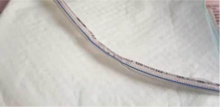
Five hours after admission to the ICU, when the agitated patient was under sedation (Midazolam infusion) and we were not following up The Richmond Agitation and Sedation scale (RASS scale), the high-pressure alarm of the ventilator started alerting and oxygen saturation dropped to 80%. The ventilation mode was on synchronized intermittent mandatory ventilation and the results of blood gas showed 100-mm Hg pCO2 and a PH of 7.1. At first, we tried to ventilate the patient with ambu bag. Then we tried to pass a suction catheter through the ETT's lumen, but we failed. We could not understand the reason and also, there was not any secretion. Then we attempted to replace the reinforced ETT with a standard (polyvinyl chloride) ETT doubting a probable obstruction in the patient's airway. After extubating the patient, we noticed that the kinked ETT was completely collapsed inside the inflated cuff because of the patient bite force (Fig. 1).
In the meantime, the patient was ventilated through high-flow O2 with an ambu bag (O2 = 100%) and flumazenil and naloxone were prescribed. Oxygen saturation was increased to 100% and blood gas showed pCO2 70mm Hg and a PH of 7.3. For maintaining the airway, head tilt and jaw thrust was done. Two hours later, blood gas showed pCO2 60 mm Hg that was exactly like the baseline value (before the surgery). At that time, she got completely continuous with breathing without need for tube replacement. After a 24-hour monitoring, we discharged her from ICU to the floor.
Discussion
The present case report describes a very rare complication after using a reinforced endotracheal tube in ICU. Although obstructions of reinforced ETTs were reported in some previous studies, there is just 1 case study that has reported this occlusion in ICU.1,3 These obstructions could be occurred because of mucus secretion, foreign bodies, tube-bites, or exposing to nitrous oxide. Since these tubes are not recommended in intensive care settings, they are routinely removed at the end of the procedure and replaced with standard endotracheal tubes (polyvinyl chloride) over a tube changer. However, the anesthesiologist in special situations (for instance, due to the probable occurrence of edematous airway) decides not to remove reinforced ETTs.1,2 To our knowledge, these special situations take place in almost 10% of ICU admissions in Shahid Chamran Hospital.
In this case, tube-biting was the probable reason of the ETT obstruction which led to permanent deformity of the lumen. Biting an ETT causes respiratory occlusions and might result in complications such as hypoxia, negative pressure pulmonary edema, increase of blood pressure, and rise in intracranial pressure. Early diagnosis of the problem might prevent these fatal complications.1,4 After this occurrence, we organized a root cause analysis team meeting with the ICU and anesthesia personnel and analyzed the leading causes of this issue to prevent or handle such situations. After ICU admission of intubated patients with endotracheal tubes in place, all the ICU staff have to be aware about the possibility of airway obstruction and the patient should be sedated and monitored with RASS scale.
Conclusion
To conclude, using of armored tube is not a guarantee for securing the patient's airway during ICU stay. Thus, it should be evaluated if there is a need for replacing reinforced ETTs with polyvinyl chloride ETTs before shifting from operating rooms to ICUs. Moreover, when such patients enter ICU, all the personnel must be aware of the possibility of obstruction and have to perform sedation monitoring with RASS scale.

