


 text in
  English (pdf)
 Article in xml format
 Article references
 Send this article by e-mail
 Cited by SciELO
  Cited by Google
  Similars in
SciELO
Similars in
SciELO  Similars in Google
 Permalink
What new knowledge does this study contribute?
About half (53%) of the anesthesiologist interviewed in Japan preferred the smaller display (4- or 5.5-inch) fixed at a distance of 30 to 40 cm. The rest preferred the larger display (7.9- or 9.7-inch) placed posterior to the probe with a visual distance of 45 to 60 cm.
Preferences should be considered when building a new system with in line positioning layout.
INTRODUCTION
Arterial cannulation is an important procedure performed in the operating room and intensive care unit to allow continuous blood pressure monitoring and repeated arterial blood sampling. The most common site for arterial cannulation is the radial artery because of its superficial location and relatively low rate of complications. However, sometimes complications such as arterial occlusion, pseudo-aneurysm, hematoma formation, and bleeding occur in radial artery cannulation, especially in difficult cases. 1 Recently, ultrasound-guided radial artery cannulation has become a common procedure, with a reportedly significant increase in first-attempt success rates. 2 The use of ultrasound, in-line layout of the equipment simplifies the procedure, and the success rate and time required for cannulation are improved compared to the conventional layout 3. Preferable size and distance of ultrasound display have not been previously discussed. Tsuchiya et al. 3 used a 20-cm ultrasound image display, with the needle puncture site and display in a straight line and a distance from the operator's eye to the display of about 60 cm. However, the most appropriate size and position of the display are unknown. In this study, a questionnaire was used to reveal the ideal ultrasound display size and distance to the operator's eye to facilitate radial artery puncture by the anesthesiologist.
METHODS
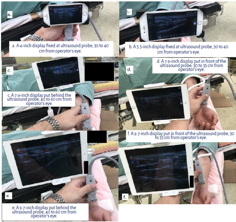
Wireless ultrasound image display system. A wireless display system similar to that used by Tsuchiya et al. 3 was designed. The Sonosite Edge IITM and L25X 13-6 MHz probe (Fujifilm Sonosite Inc., WA, USA) ultrasound equipment was used. Ultrasound image output was acquired from the DVI video outlet of the mini-doc of Sonosite Edge IITM. Resolution of the ultrasound image was up-scaled by an HDMI scaler HTCP-298HWTM (Cypress Technology Co., New Taipei City, Taiwan) from 640x480 to 1280x720. After that, a wifi HDMI transmitter, Accsoon CineeyeTM (Accsoon, Chengdu, China) was connected. Six display configurations were prepared. First, a 4-inch display (iPhone 5S, Apple Japan LLC, Tokyo, Japan) was fixed to an ultrasound probe. Second, a 5.5-inch display (iPhone 6S, Apple Japan LLC, Tokyo, Japan) was also fixed to an ultrasound probe. The distance of both displays distances from the operator's eye were 30 to 40 cm. Third, a 7.9-inch display (iPad mini, Apple Japan LLC, Tokyo, Japan) was placed on the front and back of the probe. Finally, a 9.7-inch display (iPad, Apple Japan LLC, Tokyo, Japan) was placed on the front and back of the probe. The distance from the operator's eye to the display placed on the front of the probe was 30 to 35 cm, and to the display placed on the back of the probe was 40 to 60 cm.
This study was conducted after receiving approval from the ethics committee of Ibaraki Prefectural Central Hospital (Res. Id. No. 793). No registration or informed consent was required since this was not considered a human clinical study.
Wireless ultrasound image display and probing were simulated using a demonstration patient (Figures 1a-f). Almost all anesthesiologists who are working in Ibaraki Prefecture, Japan, a total of 116 anesthesiologists, were asked which of the 6 display size and position configurations allowed the easiest radial artery puncture. They were also asked questions about levels of anesthesiology experience, proficiency in ultrasound-guided radial artery cannulation and nerve blocks, and certification by the Japanese Society of Anesthesiologists. The survey was answered by selecting one of the multiple choices, and all the responses obtained were analyzed.
RESULTS
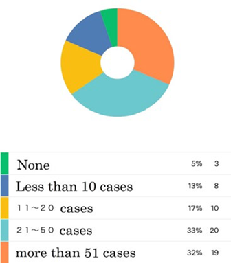
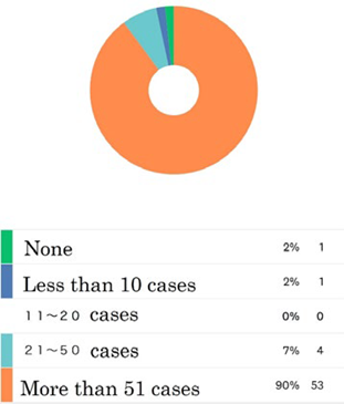
Of the 116 anesthesiologists to whom the questionnaire was administered, 60 (52%) completed the questionnaire, and one answered only half of the questions. Their levels of anesthesiology experience are shown in Figures 2,3a, and3b. In this study, 50% of the responders had more than 11 years of experience as anesthesiologists. In terms of in ultrasound-guided radial artery cannulation experience, 11 (18%) had performed less than 10 cases and 19 (32%) had performed more than 51 cases. However, most responders (90%) had performed more than 51 ultrasound-guided nerve blocks. About half of respondents (53%) preferred a smaller display (4- or 5.5-inch) fixed on the ultrasound probe, and most of the other respondents (44%) preferred a larger display (7.9- or 9.7-inch) positioned at the back of the probe (Figure 4). Nobody favored the 9.7-inch display configuration in front of the probe.


DISCUSSION
In this questionnaire, the preferred size of the display varied. About half of the respondents preferred a small display (4-or 5.5-inch), and about half preferred the tablet (larger display of 7.9 or 9.7 inches) behind the ultrasound probe.
When large wall-sized displays are used to read text, the display size does not affect individual performance if the visual angles are the same 4. The preferred distance for a large wall-sized display (cm) is reported as (2.73 x diagonal screen size + 75)/diagonal screen size 5, but preferred distances for small displays such as smart phones and tablets are not reported. Visual distance using a smart phone for reading text is 33.7 cm for Japanese young adults and 35.6 cm for young adults in the United States 6,7. In this investigation, visual distances for small displays are about 35 to 45 cm, and these distances are familiar for young doctors focusing on smart phones. The preferable visual distances for smart phones and tablets in middle-aged and elderly people are not known. Results may be affected by presbyopia. The anesthesiologists were asked about their levels of experience, but not about their ages; the influence of age is unknown.
Bababekova et al. also reported that font size for reading text using a smart phone is 1.6 mm 7. Mean radial artery diameter is reported as 2.8 mm (interquartile range, 2.4-3.1 mm) measured by ultrasound 8. When the image depth is set at 1.9 cm using the Edge II ultrasound equipment, a 2.8-mm diameter radial artery showed a 6.6-mm diameter on a 4-inch display. These dimensions are adequate to identify a radial artery for puncture when compared to a font size for reading text.
Placing a large display close to the person who is going to perform the puncture makes it easier to identify the artery, but interferes with the puncture process. The 7.9-inch display may have been a parting of the ways in terms of preference for either legibility or obstacles to puncture.
This investigation has several limitations. First, the questionnaire was not administered after the anesthesiologists performed radial artery puncture using this system. Second, a change in resolution was required to show the image on the mobile device. This change might influence the identification of the radial artery. Third, the probe weight, which increased when the display was affixed, was not considered. This weight change may affect the probe operability. Also, the anesthesiologists were asked about their level of experience but not their age; thus, the influence of presbyopia cannot be determined. Lastly, the anesthesiologists all worked in the small area of Ibaraki Prefecture, Japan, and most received their training at the Tsukuba University anesthesiology program. This narrow range of respondents may have affected the results. Moreover, this is the first survey of anesthesiologists working in Ibaraki Prefecture, and the responses were given anonymously over the Internet; there was no information about the age group of anesthesiologists who failed to answer the survey, making it difficult to conduct a sensitivity analysis with little information to examine bias.
CONCLUSION
Among anesthesiologists, there is variation in the display size and distance from the operator's eye needed to facilitate ultrasound-guided radial artery puncture, as long as radial artery is well delineated. These results can be used as a reference for anesthesiologists when building a new system with in line positioning layout in the future.

