


 text in
  English (pdf)
 Article in xml format
 Article references
 Send this article by e-mail
 Cited by SciELO
  Cited by Google
  Similars in
SciELO
Similars in
SciELO  Similars in Google
 Permalink
What do we know about this problem?
Neonatal airway pathology poses a challenge both for surgery as well as for anesthesia, with important variability in the published scientific literature as well as a paucity of protocols and guidelines for management and decision-making.
What new contribution is derived from this study?
This case report prompts a reflection about potential surgical approaches, with or without by-pass circulation, monitoring and anesthetic management, as well as potential complications that may arise in newborn patients. The importance of treating these cases in referral centers with multidisciplinary teams and consensus protocols is a key determinant of the probability of success in this complex condition.
INTRODUCTION
Airway pathology in neonates is one of the biggest challenges for the pediatric anesthetist. Creating multidisciplinary teams and inter-hospital referral committees is crucial in order to study each individual case and arrive at joint decision-making.
The case of a 28-day-old neonate with a diagnosis of severe tracheal stenosis is presented. The patient had been intervened previously to correct an esophageal atresia type IV, with two failed extubations and a torpid course which finally unmasked the diagnosis of tracheal stenosis. A slide tracheoplasty with extracorporeal circulation support was proposed. A series of complications occurred during the intervention, as described in the discussion section below.
CASE PRESENTATION
This case involves a 28-day-old neonate taken to slide tracheoplasty with by-pass circulation support at Hospital de la Fe in Valencia (Spain).
Previously, at a different center, the patient had been diagnosed with esophageal atresia type IV and distal tracheoesophageal fistula that required urgent surgery at 4 days of age. The postoperative course was torpid, with two failed extubations, which led to the diagnosis of severe mid-tracheal stenosis identified on fiberoptic bronchoscopy. Additional angiotomography studies (angio-CT) demonstrated a vascular ring component from the brachiocephalic trunk (Figure 1). When the diagnosis was made, the patient - intubated and under sedoanalgesia - was transferred to the referral Hospital specializing in these types of conditions, and scheduled for surgery.
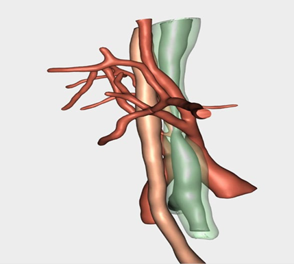
SOURCE: Authors.
FIGURE 1 The 3D reconstruction of the angio-CT image shows the tracheal stenosis in relation to the esophagus and the arterial component, as well as the vascular ring arising from the right brachiocephalic trunk.
After a few hours in the neonatal critical care unit, the patient was transferred to the operating room already intubated and under sedoanalgesia with fentanyl. Balanced anesthesia was initiated with sevoflurane, remifentanil and continuous rocuronium perfusion. Monitoring was established using the arterial pressure curve analytical recording technology based on the PRAM method (pressure recording analytical method) and regional cerebral oxygen saturation using NIRS (near infrared reflectance spectroscopy) as well as various metabolic modules for O2 and CO2, consumption and oxygen index (OI) for perioperative management, with goal-directed therapy.
During surgery, balanced inhaled anesthesia was maintained with sevoflurane at a minimum alveolar concentration (MAC) of 1.3% and remifentanil at 0.3 μg/kg/min, and train of four-adjusted (TOF) continuous rocuronium perfusion. OI values were maintained above 300 mL/min/m2, lactate remained normal with no variation over 20% of NIRS baseline values (INVOS®) throughout the surgery. Weaning from by-pass circulation proceeded uneventfully, with milrinone support. Importantly, an intraoperative ventilatory event occurred during recovery from anesthesia. After changing head position from hyperextension to neutral, total absence of ventilation and capnography loss were detected, with peak pressure elevation, suggesting tube dislodgement. Direct laryngoscopy and tube removal were performed although tube placement appeared to be correct under direct vision. With new intubation, the tube was found to be occluded by a remaining blood clot from the surgical dissection. Careful aspiration with a suction probe was carried out and damage control fiberoptic bronchoscopy allowed to determine that the suture was intact. Surgical time was 6 hours.
The patient was transferred to the neonatal critical care unit, intubated and under sedoanalgesia. On the second day of stay, the patient developed pneumonia, with positive bronchial aspiration culture for acinetobacter. The patient was extubated four days later and discharged after one week, with normal bronchoscopy findings and no complications.
DISCUSSION
In 40% of patients, the diagnosis of esophageal stenosis and tracheoesophageal fistula 1 is associated with tracheobronchial abnormalities. The presence of stridor or failed extubation may indicate association with bronchial stenosis. Some of these cases are often associated with vascular rings 2.
The airway stenosis protocol published by the task force of Hospital Universitario Politécnico de la Fe, in Valencia (Spain), was used to review the case. It is worth highlighting the importance of creating multidisciplinary teams specializing in pediatric airway pathology 3 in Level III hospitals that see these types of patients, because of their complexity. The slide tracheoplasty technique was selected because of stenosis greater than 50% of the tracheal thickness 4. It is the preferred intervention because it allows early extubation and results in less granulation tissue 5.
The recommended support system in these patients is peripheral venovenous extracorporeal membrane oxygenation (ECMO) because of the need for oxygenation during the repair maneuver. However, the surgical team preferred extracorporeal circulation with central cannulation because of familiarity with the technique and the difficulty of using peripheral cervical vessels without a secure airway 6. Other options such as intubation distal to the stenosis in the bronchus or close to the carina, or jet ventilation, were discarded due to the size of the stenosis (Figure 2). Other tracheal reconstruction procedures may be supported with tracheostomy distal to the stricture, but that option was not possible in this case because of the mid distal location of the stenosis.
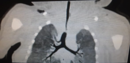
SOURCE: Authors.
FIGURE 2 Contrast CT image illustration the location of the tracheal stenosis and its relationship with the rest of the bronchial tree.
During recovery from anesthesia, the patient experienced an adverse event that could have been fatal. Tracheal suction at the time of weaning from by-pass circulation and ventilation verification is advisable in every surgery. In this case, in order to preserve the integrity of the surgery, careful suction under direct vision through fiberoptic bronchoscopy was required. However, the fiberoptic bronchoscope available at the hospital for these technical specifications does not have a working channel (Machida®). For small sizes, only bronchoscopes with no working channel are available, making it impossible to aspirate secretions under direct vision. The risk of suctioning without direct vision was high because of potential injury of sutured structures: however, there was no other option to solve the problem. Fiberoptic bronchoscopy was used to check for damage, and suture integrity was confirmed.
It is crucial to follow protocols in cases as complex as this. The results of the surveillance cultures made in the unit where the patient was hospitalized before the surgery were overlooked during preanesthesia stage. As determined during the immediate postoperative period, and for epidemiological reasons, the patient was infected with acinetobacter from the critical care unit in which the neonate remained prior to the intervention. Surveillance cultures made in the original hospital did not show any infectious agent, although the study was incomplete.
In conclusion, a complex airway pathology in a pediatric patient requires a multidisciplinary approach through the creation of inter-hospital referral units. Adherence to the protocol along every step of the process is mandatory. Likewise, readers are offered a series of recommendations derived from teamwork experience and meticulous familiarity with the technical devices so that they can be adapted to the surgical requirements:
Need for multi-disciplinary teams with expertise in complex pediatric airway pathology.
Reliance on assistance devices for patient oxygenation when needed during complex airway repair procedures.
Suction under fiberoptic bronchoscopic visualization after weaning from by-pass circulation in these types of surgery.
Importance of following protocols in complex cases: order surveillance cultures of all critical care units in which the patient has stayed, in particular in patients transferred to referral units.
ETHICAL RESPONSIBILITIES
Human and animal protection
The authors declare that no human or animal experiments were carried out as part of this research.
ACKNOWLEDGEMENTS
Authors' contributions
DMC: Study planning, Data collection and references, Initial manuscript drafting, Resident during surgery.
FJEA: Conception of the original project. Study planning. Data collection. Correction of the final manuscript. Assistant during surgery.
PAN: Service leader. Final manuscript approval.

