


 texto em
  Inglês (pdf)
 Artigo em XML
 Referências do artigo
 Enviar este artigo por email
 Citado por SciELO
  Citado por Google
  Similares em
SciELO
Similares em
SciELO  Similares em Google
 Permalink
INTRODUCTION
The use of advanced invasive techniques for the control of chronic pain is increasingly common. Anesthetists must be knowledgeable of the procedures and be aware of patient comorbidities in order to develop their anesthesia plans 1. The aim of this narrative review is to describe anesthetic management in pain control surgery, as well as the perioperative anesthetic considerations in patients with a history of pain control surgery undergoing other types of procedures.
Neuromodulation is a diverse burgeoning field that has revolutionized the management of chronic oncologic and non-oncology pain 2. The modern era of neuromodulation dawned in 1967 when Gol reported that repeated intracranial stimulation of the septal area resulted in effective pain control in several cancer patients 3. Neuromodulation is described as the electrical or chemical signal transmission alteration in the vicinity of nociceptive afferent fibers, interneurons and ascending spinal cord fibers using implantable devices or non-invasive techniques 2. This review will focus on invasive therapies, including implantable drug delivery systems, spinal stimulation and peripheral nerve stimulation.
IMPLANTABLE DRUG DELIVERY SYSTEMS
Infusion pumps
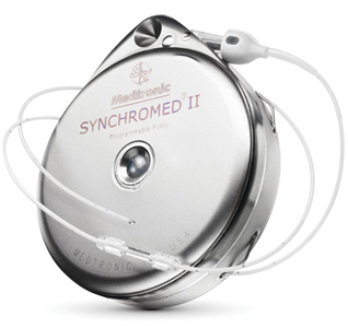
This form of neuromodulation consists of infusing one or more drugs into the epidural or intrathecal space using a fully implantable infusion pump. A small, battery-powered programmable pump (Figure 1) is implanted between the subcutaneous cellular tissue and the abdominal muscle wall connected to a small tunneled catheter at the spinal entry site 4. The epidural route is usually reserved for patients with short life expectancy of only days or weeks, because long-term epidural infusions have been associated with higher adverse event rates and catheter-related infections 5. These implantable devices are used mainly in oncologic patients with intractable pain that does not respond to conventional analgesic treatments, and in patients with insufficient pain relief or adverse effects from systemic drug therapy 2,3.
Different drugs have been used with these systems, including bupivacaine, clonidine, fentanyl, hydromorphone, sufentanil, morphine and ziconotide; the latter two are the only ones that have received FDA approval 2,6. Most pumps are designed to last 5-7 years, requiring drug reloading every 1 to 6 months according to pump size and infusion rate 7.
Regarding complications associated with these devices, battery failure, bleeding, kinking, breakage, catheter obstruction or disconnection, catheter tip granuloma, drug-related adverse events, infection, neurologic injury, post-puncture headache and pump malfunction have been described 2,8.
Spinal cord and peripheral nerve stimulation
Spinal stimulation is a minimally invasive technique involving placement of electrodes in the epidural space in order to deliver electrical stimuli to the myelinated fibers of the dorsal horn and, occasionally, stimulate lateral fibers. Electrodes are connected to a subcutaneously implanted pulse generator (Figure 2) 9. Before implanting the device, a trial lasting between 3 and 10 days is carried out in order to assess the effectiveness of the treatment 2.

There are several theories to explain the mechanism of action, the gate control theory being the most predominant. Stimulation by antidromic conduction of Aβ fibers in the dorsal columns reduces pain in the stimulated segment. Although the theory provides a partial explanation, it does not fully explain the mechanism. The second theory is the opioid theory, based on the fact that spinal neurostimulation increases endorphine levels mainly in the raphe nuclei and periaqueductal gray matter nuclei. The third theory explains the control of diffuse noxious inhibitory centers which begins in the nucleus reticularis dorsalis in the reticular formation of the medullary neurons and ends in the wide dynamic range neurons in the spinal cord. The GABAB system, substance P (protein) and CGRP (calcitonin gene-related peptide) are involved at least in part in the mechanism of action 10.
The most common indications for spinal cord stimulation include regional complex syndrome, failed back surgery syndrome and intractable angina pectoris 11. Different studies have shown that these patients improve of their symptoms with spinal stimulation, 12,13 with lower analgesic consumption demonstrated in patients with intractable spine or limb pain 14. In oncologic patients, it has been shown to be effective in the management of chemotherapy-associated neuropathic pain 15.
Adverse reactions to these stimulation devices include battery failure or malfunction, dural fibrosis, infection, electrode migration, breakage or failure, loss of analgesia over time, neurologic injury and post-puncture headache 16.
Peripheral nerve stimulation is an important area in neuromodulation. It consists of placing the electrode by the peripheral nerve, proximal to the injury site, added to an implanted or external pulse generator 17. Clinical applications are neuropathic pain due to peripheral nerve injury, nerve trapping or nerve plexus damage. This technique is considered a good management option 18.
ANESTHETIC CONSIDERATIONS FOR CHRONIC PAIN PROCEDURES
Knowledge of the different available techniques is required. This article describes the anesthetic considerations that have to be borne in mind with implantable drug delivery systems and spinal and peripheral nerve stimulation devices.
All of these procedures require a venous access for administering drugs, including perhaps antibiotics, apart from basic ASA (American Society of Anesthesiology) monitoring 1. They can usually be performed under sedation, with oxygen supplementation, patient collaboration and monitoring for signs of hemodynamic instability, anaphylaxis and vasovagal episodes. Although rare, these complications may occur during these procedures 1,6. If the patient is awake or under light sedation, constant verbal communication must be maintained between the anesthetist, the practitioner placing the implant and the patient, in order to minimize the risk of nerve injury 19.
Anesthesia for insertion of implantable drug delivery systems (IDDS) and intrathecal and epidural pumps tunneled to port
Implantation of a drug delivery system requires placement of an intrathecal catheter connected to a drug reservoir system used mainly for oncologic and chronic non-oncologic pain, and refractory spasticity 6. The system includes a rotor pump connected to a subcutaneously implanted battery. The most common implant site is subcostal, on the side less frequently used for sleeping 20. Some specialists implant always on the left side considering that the majority of surgical procedures will probably be performed on the right side (laparoscopic cholecistectomy, appendectomy). In oncologic patients, should a colostomy be required, the decision for the implant must be tailored in accordance with the prognosis 1. In patients with a survival prognosis of 2-3 months, and intrathecal or epidural catheter tunneled to a subcutaneous port is indicated for intermittent or continuous percutaneous medication administration 19. The catheter implantation procedure is similar to that of the intrathecal pump, except for the absence of a drug reservoir pump which is replaced by a port usually implanted subcutaneously in the chest wall, connected to an external pump/drug reservoir system.
All anesthetic modalities are allowed (general, regional/neuraxial or controlled local anesthesia) 19. General anesthesia is the most commonly used depending on the surgical skills, team speed, and patient comorbidities and tolerance of the procedure.
Preoperative period
The practice of pain surgery has grown exponentially, especially in patients with oncologic pain, who are usually very compromised by the time the decision is made to provide implantable intrathecal therapy 19. Therefore, during the preanesthetic assessment, the anesthetist must consider the patient's nutritional status, the location of the primary tumor, the presence or absence of metastases, and disease prognosis 1.
Intraoperative period
Patient positioning is very important in this procedure which is performed in lateral decubitus 4. The neuraxial catheter is usually advanced to the mid-thoracic region or one level higher under fluoroscopy. In lateral decubitus, the arms must be positioned at shoulder level or slightly higher, depending on patient tolerance, in order to ensure clear fluoroscopic visualization of the spine 1,20. A lateral arm rest for the "up" or non-dependent arm helps maximize the space, enabling the fluoroscopy machine to acquire anteroposterior thoracic views.
As for the choice of the anesthetic technique, it must be based on individual patient characteristics. If there is a history of lung compromise due to the underlying disease, with extensive resection or metastases, extubation may be difficult to accomplish and, consequently, a neuraxial technique should be considered. For the neuraxial approach, the patient is positioned in lateral decubitus and the intrathecal space is accessed to administer 0.5% isobaric bupivacaine 2.5-7.5 mg through a catheter placed at the T10 level; the final step is tunneling and fashioning of abdominal pocket 1. Anesthetic need will depend on the length of the procedure.
The procedure can also be performed under intravenous sedation with peri-incisional local anesthetic administered on the lumbar midline for catheter placement and fixation at the thoracolumbar junction, along the tunnel for the catheter and at the site of the pocket for the pump 19,21. This technique requires constant communication between the surgeon performing the procedure and the anesthetist in order to ensure adequate control of local anesthetic use and prevent drug toxicity.
Postoperative period
For analgesic management, the pain surgeon may administer an intrathecal bolus to initiate therapy immediately after surgery or for postoperative pain management 21. Care must be taken to avoid additional opioid or sedative doses and the staff of the postanesthetic recovery unit must be informed of the potential consequences of neuraxial doses of those medications, including respiratory depression, hypotension and skin reactions 22.
SPECIAL CONSIDERATIONS IN SPASTIC PATIENTS
There are special considerations for patients with spasticity-related pain requiring intrathecal baclofen therapy. The main indications for this intrathecal device include intractable spasticity due to cerebral palsy, cerebrovascular event or spinal cord injury from multiple sclerosis or trauma 1,23. These patients suffer from painful spasticity refractory to increasing doses of oral baclofen or other muscle relaxants.
Anesthetic planning must consider patient mobility and functional status. The patient must be advised to take the morning dose of baclofen or muscle relaxants to avoid perioperative spastic exacerbations 1.
Patient positioning can be challenging and, in spasticity cases, general anesthesia is the preferred choice 24. It is worth remembering that patients with cerebral palsy have a higher incidence of gastroesophageal reflux and care must be exercised when using a laryngeal mask 25,26. Muscle relaxants can be used for airway management an positioning; if the patient has been immobilized as a result of the spasticity or if there is any functional limitation of one or more limbs, succinylcholine may be contraindicated because of the probability of hypercalcemia even with only one atrophic limb due to lack of use 27.
ANESTHESIA FOR THE INSERTION OF SPINAL CORD, PERIPHERAL NERVE OR FIELD STIMULATORS AND IMPLANTS
Stimulator device trials
Spinal and peripheral nerve stimulator trials can be carried out in the hospital or on an outpatient basis 1,3. They are outpatient procedures performed under light sedation with no need for general anesthesia so that the patient can be alert and able to communicate. Trials enable to determine the optimum position for the device and ensure that the paresthesia area is placed on the painful region 28. It requires a venous access and a dose of prophylactic antibiotic 1. Occasionally, the stimuli may trigger anxiety attacks or vasovagal episodes. An airway team, a crash cart and oxygen supplementation need to be available at the site where the trial is carried out 13,29,30. Fluid restriction is required unless a urinary catheter is in place.
Permanent implantation of stimulator devices
Anesthetic planning for implantation of a spinal or peripheral nerve device depends on the skills and the technique of the practitioner performing the procedure, anesthetist satisfaction and patient preferences and comorbidities 31. Overnight stay is usually not required, unless warranted by comorbidities 30. Unlike with the intrathecal pump, neuraxial anesthesia is contraindicated because it requires active patient cooperation during the stimulation test 28. The incision is very small and not very painful and, therefore, a subcutaneous local anesthetic injection suffices; moreover, the battery is smaller than the intrathecal pump device, which is usually implanted in the external upper quadrant of the buttock or in the posterolateral flank 32. Some pain surgeons prefer placing the implant in the abdominal lower quadrant, which requires changing patient position from prone to lateral decubitus. In this situation, general anesthesia is preferred for the second part of the procedure.
Constant communication is recommended between the pain surgeon and the anesthetist in order to plan the different stages of the procedure. Superficial sedation is recommended for the first phase so as to ensure optimum placement and prevent excess drowsiness. Once the test is performed and adequate placement is confirmed, deep sedation is used in order to facilitate tunneling and fixation of the device. Local anesthetic infiltration is made for tunneling, paying close attention to the maximum dose in order to prevent toxicity from local anesthetics 9,33.
When general anesthesia is selected either because of patient intolerance of prone positioning while awake or because of surgical team preference, important confirmation of stimulator placement is lost and could lead to a failed procedure 34.
Patient positioning is critical to the success of the implant because the patient must be awake, comfortable and cooperative for the acquisition of the best fluoroscopic views 35-37. When the objective is to manage chronic lower limb or back pain, the stimulator must be inserted in upper lumbar levels and guided along the epidural space towards middle-low thoracic levels. For spinal stimulation, the patient is initially placed in prone position on the fluoroscopy table, with one or two pillows under the abdomen to diminish lumbar lordosis 38. Pressure zones must be avoided. In women, pressure on the breast must be avoided. Arms must be placed in an anatomical position to acquire lateral fluoroscopic views.
If the objective is to manage chronic neck or upper limb pain, the stimulator is inserted in upper thoracic levels and the pulse generator is placed in or under the axillary region or in the posterior flank/ upper gluteal area 32. The patient is placed in prone position on the fluoroscopy table. The head is placed in anatomic position with the neck slightly bent forward and supported by a protective gel pack. Too much head extension my impair approach to the epidural space. One or two pillows can be used to achieve slight cervical flexion and avoid pressure zones. Shoulders should preferably be relaxed, with the arms in anatomic position 38.
For permanent peripheral nerve stimulator implantation, placement depends on the anatomical area to be intervened 39-41. General anesthesia could be avoided by using an adequate dose of local anesthetic and sedation, which is sufficient for lower limb, abdominal and lumbar stimulators.
For occipital or craniofacial stimulation involving sensory areas of the head or the neck, patient comfort must be ensured, hence general anesthesia is recommended. The implant is placed by marking the precise site where the pain was elicited during the therapeutic test 42,43.
Checking and explanting implantable drug delivery systems and stimulation devices
With time, spinal and peripheral nerve stimulators tend to migrate or break, or individual electrodes simply stop working, requiring device exchange 44. Similarly, in implanted drug delivery systems, the catheter may dislodge from the pump/ reservoir, ending up in an adjacent position, where granulomas may develop 1.
For the pain surgeon, this procedure involves careful check of the electrode or catheter, intraoperative evaluation of all device components or exchange for a new device (and new performance tests) 44. This is a procedure that lasts between 2 and 4 hours. Checking of spinal cord, peripheral nerve or peripheral stimulation devices requires sedation, posing a challenge for the anesthetist because it is a lengthy procedure that involves alternating between light sedation and deep sedation in a patient lying in prone position. Communication with, and feedback from the patient is needed during the intraoperative period. Sedation titration without having secured the airway in a patient in prone position is always a challenge, particularly in patients with chronic pain, anxiety or conditions that obstruct the airway such as obstructive sleep apnea-hypopnea syndrome (OSAHS). Dexmedetomidine infusions can be effective in long procedures because of their anxiolytic effect and negligible respiratory depression 45,46. For revisions of implantable drug delivery systems, the choice is usually general anesthesia 1.
In cases of explantation due to infection or malfunction without planned replacement, general anesthesia must be chosen, if not contraindicated 1.
ANESTHETIC CONSIDERATIONS IN PATIENTS WITH NEUROMODULATION DEVICES
Preoperative period
A consult with the pain treating physician is needed whenever possible in order to have a clear idea of the device the patient utilizes, the time of use, the last time the pump was checked, the current medication in the pump and whether the dose is on demand or flexible; also, it is important to ascertain when the patient needs to reload the medication in the pump 22.
The anesthesia plan must consider regional anesthesia, whenever possible 47. In the event continuous infusion techniques through a peripheral or neuraxial catheter are considered, insertion must be performed using a strictly sterile technique, with the infusion lasting 48 hours. Neuraxial techniques should generally be avoided, although it is not an absolute contraindication 6; however, if an epidural lumbar technique is required, access to the epidural space must be accomplished under imaging guidance, avoiding the implanted components 11. For obstetric patients scheduled for cesarean section, a neuraxial technique may be considered, provided the exact location of the IDDS is known and the puncture is performed caudal to it 48,49; should that not be the case, general anesthesia is recommended 47.
Regarding spinal cord stimulators, each brand has specific recommendations for the device which can be found in the product manuals 50. In the preoperative area, the device must be brought down to its lowest setting using the patient's remote or with the help of the product representative, and then it must be turned off. Some types of devices can be set to "surgery mode," with no additional steps required 50.
Intraoperative period
Opioid infusions must be used cautiously in patients with IDDS, and multimodal analgesia with adjuncts including NSAID, acetaminophen, steroids and ketamine should be administered 47. In the event the IDDS is damaged during surgery and no immediate repair is possible, the pump must be stopped and the patient should be switched to oral or intravenous opioid postoperatively 47, bearing in mind that there is no reliable way to make the conversion from the equianalgesic opioid dose to an intravenous drug 22. Estimates of equianalgesic doses when rotating from oral administration to intrathecal morphine administration range between 12:1 and 300:1 (Table 1) 51; consequently, the dose calculation according to opioid equianalgesia with morphine must be taken into account (Table 2) and then adjust the equivalent dose to the route of administration used.
TABLE 1 Equivalent doses according to the route of administration.
| Route of administration | Equivalent dose |
|---|---|
| Oral | 300 mg |
| Intravenous | 100 mg |
| Epidural | 10 mg |
| Intrathecal | 1 mg |
SOURCE: Adapted from Sylvester RK, et al 50.
TABLE 2 Equianalgesic dose table.
| Drug | Equianalgesic dose | |
|---|---|---|
| Parenteral | Oral | |
| Morphine | 10 mg | 30 mg |
| Fentanyl | 0.1 mg | - |
| Hydromorphone | 1.5 mg | 7.5 mg |
| Codeine | 100 mg | 200 mg |
| Oxycodone | 10 mg | 20 mg |
| Tramadol | 100 mg | 120 mg |
SOURCE: Modified from Hernández-Ortiz A 54.
Intraoperative pain management varies because it is patient-dependent. Maintenance may require opioid doses up to 20% higher than the usual daily dose according to the type of surgical procedure performed 52. The dosing method for opioid analgesics towards the end of the procedure involves avoiding the use of muscle relaxants or reversing their effect before the end of the procedure and then adjusting analgesia according to respiratory rate; although any desired respiratory rate can be used, a target ofmore than 12-14 breaths per minute should be considered 53.
It is important to regulate the patient's body temperature and make sure that the IDDS is not subjected to marked temperature changes. If patient temperature rises, the temperature of the device could also rise, leading to increased drug delivery and potential overdosing. Infusion pump manufacturers recommend that temperature should not exceed 39°C if the risk of altering the infusion rate is to be minimized 22.
Temperature also affects the operation of the spinal stimulator: the electrocautery may overheat the electrodes and harm the spinal cord, or device settings may be altered, resulting in inadequate therapy 50. The surgical team should use a bipolar electrocautery whenever possible; if there is a need to use the monopolar electrocautery, the plate must be positioned contralateral, away from the device 50.
Postoperative period
If the patient has an IDDS, short-acting pain control medications should be administered, avoiding long-acting or continuous opioid infusions, and continue with multimodal analgesia depending on the pain level. If continuous infusions through a catheter are required, local anesthetics must be used. The use of patient controlled analgesia (PCA) pumps can be considered, but without a baseline infusion 54. In case the patient undergoes a very painful procedure, as in the case of total knee arthroplasty, increasing the dose of the intrathecal drug by 10% can be considered, with programmed tapering to the preoperative dose over a 4-6 week interval 47. No more than two weeks should elapse before the chronic pain physician assesses the patient postoperatively (Table 3).
TABLE 3 Perioperative management summary.
SOURCE. Authors.
CONCLUSIONS
Advanced invasive techniques for chronic pain control are increasingly being used as a treatment modality. In this context, the anesthetist must be aware of the anesthetic considerations at play when facing a surgical procedure for implanting pain management devices. Additionally, patients with electric neuromodulation or drug delivery devices may require anesthesia due to surgical indications unrelated with their chronic pain conditions. Consequently, it is important to be familiar with the basic components of these devices, how they work, what medications are used, and potential perioperative complications that may arise, in order to ensure adequate management and patient safety.

