


Servicios Personalizados
Revista
Articulo
 Inglés (pdf)
 Articulo en XML
 Referencias del artículo
 Enviar articulo por email
Indicadores
-
 Citado por SciELO
 -
 Accesos
Links relacionados
-
 Citado por Google
 -
 Similares en
SciELO
Similares en
SciELO -
 Similares en Google
Compartir

 Permalink
Biomédica
versión impresa ISSN 0120-4157
Biomédica vol.35 no.1 Bogotá ene./mar. 2015
https://doi.org/10.7705/biomedica.v35i1.2291
PRESENTACIÓN DE CASO
doi: http://dx.doi.org/10.7705/biomedica.v35i1.2291
1 Department of Internal Medicine, Mayo Clinic Arizona, Scottsdale, Arizona, United States of America
2 Division of Gastroenterology, Mayo Clinic Arizona, Scottsdale, Arizona, United States of America
Author´s contributions:
Diana L. Franco: study concept and design, acquisition of data and drafting of the manuscript
Sameer Islam: study concept and design and drafting of the manuscript
Kevin Ruff: critical revision of the manuscript for important intellectual content
Recibido: 21/04/14; aceptado: 21/10/14
A 79-year-old female with benign past medical history presented to the gastroenterology clinic complaining of long-standing symptoms of dyspepsia. Esophagogastroduodenoscopy showed nodular smooth mucosa in the second part of the duodenum. The morphologic and immunophenotypic findings were consistent with low-grade follicular lymphoma. The purpose of this manuscript is to educate the reader on this unusual finding that is pathognomonic for gastrointestinal lymphoma.
Key words: Lymphoma, duodenum, endoscopy, duodenal diseases, neoplasm.
doi: http://dx.doi.org/10.7705/biomedica.v35i1.2291
Hallazgo endoscópico de linfoma folicular primario
Una mujer de 79 años, sin antecedentes patológicos de importancia, consultó al servicio de gastro enterología por síntomas de dispepsia de larga data. Se practicó una esofagogastroduodenoscopia en la que se observó mucosa nodular en la segunda porción del duodeno. Esta morfología y los hallazgos inmunofenotípicos eran indicativos de linfoma folicular de bajo grado. El propósito de este manuscrito es ilustrar al lector sobre esta inusual condición en el duodeno, la cual es patognomónica de linfoma.
Palabras clave: linfoma, duodeno, endoscopia, enfermedades duodenales, neoplasia.
doi: http://dx.doi.org/10.7705/biomedica.v35i1.2291
A 79-year-old female with long-standing symptoms of cough, nighttime heartburn and mild intermittent right upper abdominal pain was referred to the gastroenterology clinic for evaluation of possible reflux disease. Family history was significant for leukemia in a sister and non-Hodgkin lymphoma in a brother. Past medical history was benign and she denied smoking, proton pump inhibitors use, weight loss, blood in the stool or dysphagia. Basic laboratory tests including complete blood count, comprehensive metabolic panel, and liver function tests were normal except for low leukocytes of 2,900/mm 3 .
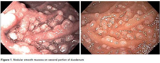
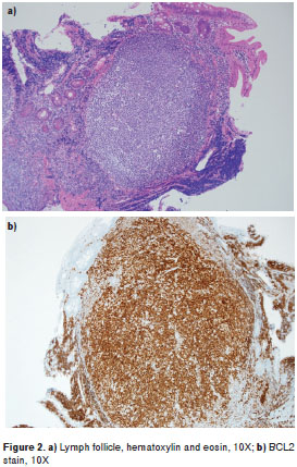
An esophagogastroduodenoscopy (EGD) revealed a 2 cm hiatal hernia, and grade A esophagitis with a nonobstructing Schatzki´s ring. In addition, nodular smooth mucosa was noted in the second part of the duodenum (figure 1). Biopsy showed a dense submucosal infiltrate of mature CD20-positive lymphocytes with disrupted and scattered lymphoid follicles (figure 2). Lymphocytes exhibited coexpression of CD10 and BCL2, but were negative for CD5 and CD43 (figure 2). This morphologic and immunophenotypic findings were consistent with duodenal low-grade follicular lymphoma that exhibited a follicular and diffuse growth pattern. The patient was started on proton-pump inhibitors therapy and was referred to the hemato/oncology clinic for further staging and management.
Primary gastrointestinal follicular lymphoma is a rare malignant disease, in the spectrum of non-Hodgkin B-cell lymphoma. It is commonly found in the duodenum, and is a frequent lymphoma in the gastrointestinal tract (1). The clinical course tends to be indolent with a continuous pattern of relapse (2). The median survival is 10 years after diagnosis, though in a recent multicenter, retrospective study male sex and the presence of abdominal symptoms were independently associated with worse clinical outcomes (1). Follicular lymphoma accounts for 1% to 3% of primary gastrointestinal lymphomas, and endoscopically appears in the form of small intestinal whitish smooth nodules as in our case (3). Follicular lymphoma can be part of a spectrum of disease called multiple lymphomatous polyposis, which is characterized by the formation of multiple mucosal nodules on various regions of the gastrointestinal tract including the esophagus, stomach, duodenum, and intestine. Histological findings tend to lead to the classification of most multiple lymphomatous polyposis as mantle cell lymphomas (4).
Immunohistochemical techniques positive for CD10 and bcl-2 markers establishes the diagnosis (3). Differential diagnosis includes pancreatic cancer, as this is the most frequent tumor in this area, and mantle cell lymphoma, the most common lymphoma in the gastrointestinal tract, which is characterized by CD10 negativity in immuno-histochemistry stains (2) (table 1). This case was chosen to highlight the macroscopic and pathology images, as diagnosis can be done by recognizing those typical patterns.
The authors are grateful for the support and assistance of Dr. Ryan Robetorye for the pathology images.
The authors declare that they have no conflict of interest.
The authors declare not receiving any type of finan cial aid for the creation of the present manuscript.
Corresponding author: Diana L. Franco, 13400 East Shea Blvd, Scottsdale, AZ, 85259, United States of America Phone: (480) 515 6296 franco.diana@mayo.edu
1. Takata K, Okada H, Ohmiya N, Nakamura S, Kitadai Y, Tari A, et al . Primary gastrointestinal follicular lymphoma involving the duodenal second portion is a distinct entity: A multicenter, retrospective analysis in Japan. Cancer Sci. 2011;102:1532- 6. http://dx.doi.org/10.1111/j.1349-7006.2011.01980.x [ Links ]
2. Higuchi K, Komatsu K, Wakamatsu H, Kawasaki H, Murata M, Miyazaki K, et al . Small intestinal follicular lymphoma with multiple tumor formations diagnosed by double-balloon enteroscopy. Intern Med. 2007;46:705-9. [ Links ]
3. Nadal E, Martínez A, Jiménez M, Ginés Á, Campo E, Piqué J, et al . Primary follicular lymphoma arising in the ampulla of Vater. Ann Hematol. 2002;81: 228-31. http://dx.doi.org/10.1007/s00277-002-0436-9 [ Links ]
4. Hirata N, Tominaga K, Ohta K, Kadouchi K, Okazaki H, Tanigawa T, et al . A case of mucosa-associated lymphoid tissue lymphoma forming multiple lymphomatous polyposis in the small intestine. World J Gastroenterol. 2007;13:1453-7. http://dx.doi.org/110.3748/wjg.v13.i9.1453 [ Links ]
