English (pdf)
 Article in xml format
 Article references
 Send this article by e-mail
 Cited by SciELO
  Cited by Google
  Similars in
SciELO
Similars in
SciELO  Similars in Google
 Permalink
Triatomines (Hemiptera : Reduviidae) are blood are blood - sucking insect vectors of the protozoan Trypanosoma cruzi which is the causative agent of Chagas' disease (American trypanosomiasis). Vector-borne transmission is the main infection route and occurs in many areas of the Americas (1). Despite significant progress towards the control of domestic vector infestation, Chagas' disease remains a major public health problem in Latin America due to its epidemiological and economic burden (2-4).
According to the eco-epidemiological studies carried out in Colombia, Rhodnius prolixus is the mo important vector of T. cruzi in the country and has successfully adapted to human domiciles (2,5,6). The preference for feeding on humans, the high susceptibility to infection with T. cruzi, the rapid development cycle and the short defecation time are factors favoring its vector capacity (5).
The first extensive studies on the blood-feeding of triatomine bugs were published in 1952 and 1953 by Barth, who detailed the internal anatomy of the head of Triatoma infestans and its feeding behavior on guinea pigs (7,8). Thereafter, Lavoipierre, et al., observed the mechanism of feeding of three triatomine species (R. prolixus, T. infestans and T. protracta) on a rabbit, a white rat, and a guinea pig, directing particular attention to the structure and function of the mouthparts of R. prolixus (9). An extensive morphological review on R. prolixus was published in 1969 by Ramírez-Pérez (10). Friend, et al., also made observations of mouthpart movements, salivation and ingestion in R. prolixus and correlated them with changes of electrical resistance (11). More recently, the functional anatomy of the hypopharynx and the salivary pump of R. prolixus has been elucidated (12).
The head, mouthparts and the probing process of R. prolixus are shown in figure 1. Triatomines are regarded to be vessel-feeders because they obtain blood from vertebrate hosts by directly inserting their mouthparts into vessels (solenophagy or capillary feeding) (9). Triatomines rely on the sense of CO2 to search for blood meals from sleeping hosts (13). Contact with the host occurs only during feeding, which lasts approximately 20 to 30 min (14). It has been described that these insects feed on a host swinging their proboscis forward and piercing the skin by rapid alternating movements of the resistant mandibles which penetrate only into the superficial tissues. Then, the maxillae (maxillary bundle) penetrate deeply into the dermis with restless twisting movements (9).
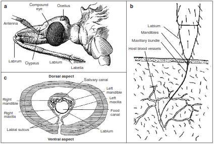
Figure 1 Schematic diagrams of the head and mouthparts of Rhodnius prolixus. a. Lateral view of the head and proboscis of R. prolixus. b. Closer look of the distal part of the proboscis during the end of probing. The mandibles pierce the host skin and the maxillary bundle is thrust into the tissues and enters the lumen of a vessel. c. Cross-sectional view of the proboscis close to the base of the labium Figure part a by courtesy of The Trustees of the Natural History Museum, London, UK; parts b and c adapted and modified from reference 9 with permission for reproduction from the publisher Taylor and Francis
Triatomines possess a wide variety of bioactive molecules in the saliva which facilitate the blood meal by reducing host physiological responses related to haemostasis, inflammation, and immune activation. In R. prolixus, saliva deposition into the host skin occurs throughout the probing and engorgement phases of the feeding (12,14). Feeding takes place from blood vessels rather than from small hemorrhages which are formed as a result of probing. When the maxillary bundle enters a blood vessel of suitable caliber, probing ceases and the insect gut begins to engorge (9). Blood ingestion through the food canal is aided by the cibarial pump, which is regulated by several strong muscles of the insect head (7). The bug continues to suck up its blood meal until it is full, but the feeding process is not always continuous and may be interrupted and repeated (9).
Microscopic techniques are useful for visualizing and describing the morphology of biological structures. Here, we show images of the blood-feeding of R. prolixus, including some histological features of the mouthpart structures implicated.
Third- and fourth-instar uninfected nymphs of R. prolixus were obtained from the insectary colonies of the Departamento de Microbiología of the Facultad de Salud of Universidad del Valle in Cali, Colombia. In the Laboratorio de Histología of the University, an anesthetized laboratory mouse (Mus musculus) was exposed to the triatomines for blood-feeding (figure 2 and figure 3). Triatomines were decapitated with a curved iris scissor during blood-feeding. We obtained biopsy samples of the skin pierced by the feeding apparatus or proboscis, and of the head of the insect, and processed them accordingly.
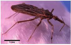
Figure 2 Interaction between Rhodnius prolixus and a laboratory mouse. The triatomine extended its proboscis to reach the skin of the mouse and performed several biting attempts
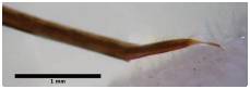
Figure 3 Proboscis of Rhodnius prolixus piercing the skin of the mouse. The terminal portion of the proboscis is thinner at the level in which the mandibular and the maxillary stylets are thrust into the tissues
Light microscopy (figure 4 and figure 5) was performed in the Laboratorio de Ingeniería de Materiales of Universidad del Valle and scanning electron microscopy procedures (JEOL microscope JSM6490LV) (figure 6, figure 7 and figure 8) were performed in the Laboratorio de Materiales de la Facultad de Ingeniería of Universidad del Valle. Animal experimentation was part of the practical component of the subjects "Basic histochemical techniques" and "Electron microscopy techniques" taken by one of the authors.
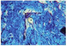
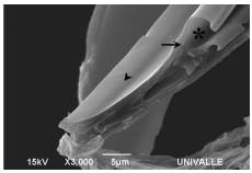
Figure 4 Blood vessel (arrow) collapsed likely due to active sucking up of blood through the maxillary bundle (asterisk). The connective tissue is stained blue. Masson's trichrome stain, 400X
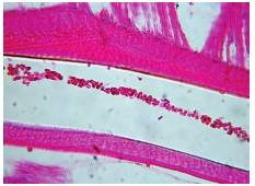
Figure 5 Blood flow throughout the lumen of the pharynx of Rhodnius prolixus during blood-feeding. Haemotoxylin and eosin stain, 400X
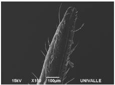
Figure 6 Scanning electron micrograph of the distal end of the labella of the labium of Rhodnius prolixus. The ventral lid is not visualized
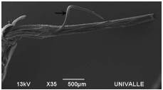
Figure 7 Scanning electron micrograph of the left half of the proboscis of Rhodnius prolixus. Note the retraction of the maxillary bundle (arrow) due to the great flexibility of its structure