


 Inglês (pdf)
 Artigo em XML
 Referências do artigo
 Enviar este artigo por email
 Citado por SciELO
  Citado por Google
  Similares em
SciELO
Similares em
SciELO  Similares em Google
 Permalink
INTRODUCTION
The zoo breeding of wild animals is one of the most useful methods for the conservation and repopulation of endangered species, especially in areas where they have been diminishing through different actions generated by the human being such as the indiscriminate felling of forests or the hunting of wild animals (Brusatin et al., 2011). In Colombia, the geographical distribution of caiman is located on the north coast and in the rivers of inter-Andean valleys, from the Sinú river to the Guajira peninsula. This shows the high adaptability of these species and, therefore, that the possibility of implementing zoo breeding sites in the area is feasible (Reina, 2007). In Colombia there were approximately 47 installations for a common caiman in 2008 (Brusatin, 2008), dedicated to breeding and raising and whose main objective is the commercial area (meat and skin) and a few companies dedicated to the biological conservations, that is, to repopulate.
One of the species studied in zoo breeding is the common caiman (Caiman crocodilus fuscus). This is one of the eight species of native caimans found in Colombia and is cataloged as a minor concern in Colombia with the classification of IUCN; globally, IUCN classified it like Low Risk and minor concern; besides, according the classification of CITES for Colombia, it is in Appendix II (Morales, 2013). For this reason, it is necessary to know its physiological and clinical parameters in order that these are available as a diagnostic tool for diseases in these animals, even more so when they are in captivity, where treatment is more feasible.
Among the aspects to be evaluated within the normal physiology of any animal are the hematological values, such as hematocrit, hemoglobin, average corpuscular volume, concentration of the average corpuscular hemoglobin, etc., which are very important because they are constituted for the biologist and/or the veterinarian as a diagnostic aid for a wide range of diseases, etiological agents such as viruses, bacteria or protozoa (Manzanilla et al., 2011).
Contrary to what happens with domestic animals, where the information of the hematological values for each species is well defined, in the case of caiman species, included Caiman crocodilus fuscus, the information is scarce (García and López, 2010). Therefore, the objective of this work was to determine the hematological values of Caiman crocodilus fuscus, reared in captivity in order to offer the typical reference values for further investigations.
MATERIALS AND METHODS
Site and Times of Sampling
The study was conducted in a zoo breeding located in the area of La Antigua, township Carmen de Apicala, Tolima department, Colombia, which has an altitude of328 m.a.s.l, an average temperature of 27.2 °C and an annual rainfall of 1630 mm. (IDEAM, 2018). The collection of the samples occurred during the period between March and May 2018.
Animals
We took 120 blood samples from a group of common caimans (Caiman crocodilus fuscus) with ages from one to two years old; following the instructions of CITES all the animals were between 121 cm and 180 cm, which means a degree of growth of semi-adult (Morales, 2013). None animal was captured in wildlife.
All the animals sampled for the research were subjected to a physical examination in order to sample only those who were in an optimal state of health, i.e., without skin damage (lacerations and flaking), or damage to the locomotor system (lameness, amputations of hoof or tail), and the sex of each of them was verified (Lane, 2002).
Diet
The diet of common caiman is based on discarded animals from nearby poultry incubators, as well as eggs, beef and horse meat, supplied every fifteen days, at 7:00 a.m. All the diet depends on the situation if the incubators have animals to give them, and sometimes supplemented with animals like cows and horses that are out of the production; for this reason, the diet has high variations.
Specimen Collection and Transportation
For sampling, the method described by Martínez et al. (2011) was used, where after physically evaluating the animal, it was placed in dorsal decubitus and the interscales portion between the second and third caudal scales was disinfected with ethyl alcohol. The needle (caliber number 16) was introduced at 90 degrees (angle) with the tip forward, and 4 mL of blood was drawn. Blood samples were collected with vacutainer tubes with Ethylene-diamine-tetra-acetic (EDTA) as anticoagulant (Carlos et al., 2017).
The samples were transported the same day of collection in a polyethylene container with ice to maintain the temperature cold and prevent their degradation, until their arrival at the Laboratory of Veterinary Hematology of the Universidad Pedagógica y Tecnológica de Colombia at Tunja, Boyacá, Colombia.
Laboratory Procedures
In the laboratory, the micro-hematocrit procedure was performed to fill the capillaries to three quarters with the blood and sealed with plasticine and put in a micro-centrifuge micro-spin 12 for 5 minutes at 12 500 rpm (Lara, 2013).
For the hemoglobin determination, the method of cyanmethemoglobin with Drabkin reagent was used (Manzanilla et al., 2011), which is based on the combination of a blood volume of 0.02 ml with a solution of Drabkin 0.5 ml, stirred for 5 minutes and put in a spectrophotometer at 540 nm to measure the absorbance of this solution. Besides, a standard solution was made to compare and thus have the result of the factor (concentration of the pattern on the absorbance), after which the hemoglobin levels that resulted from the absorbance factor were evaluated (González et al., 2012).
The mean corpuscular volume (MCV), red blood cells (RBC), concentration of the mean corpuscular hemoglobin (CMCH) and mean corpuscular hemoglobin (MCH) were studied with the Horiba® brand ESV60 hematological analyzer. The machine used is for mammals, but being the most accessible device, was decided to use it, with the clarification that all processes to find the results were done manually to avoid a mismatch of the machine and compared with the manual results. Later it was chosen the results which are presented in the tables.
Data Tabulation and Statistical Analyses
Once the hematological values were determined, they were transferred to the Excel 2013® program for Windows 10®, through which the mean, standard deviation, minimum and maximum were determined. An analysis of variance (ANOVA) was performed between each of the means of males and females for each parameter evaluated with the statistical program Statgraphics Centurion XVII® for windows. With a confidence level of 95 %, it was determined that one value was statistically different from the other when p was less than 0.05 (p < 0.05).
RESULTS
The results obtained from the 120 blood samples of the common crocodile are found in Table 1; Table 2 shows a comparison of the red line between Caiman crocodilus fuscus, Caiman crocodilus, and Caiman latirrostris; and Table 3 shows the comparison of the red line of Caiman crocodilus fuscus, between males and females.
Table 1 Hematological values in the species of common caiman (Caiman crocodilus fuscus), in captivity.
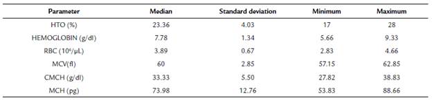
HTO: Hematocrit, RBC: Red blood cells, MCV: Median corpuscular volumen, CMCH: Median corpuscular hemoglobin concentration, MCH: Median corpuscular hemoglobin.
Table 2 Comparison of the red line between Caiman crocodilus fuscus, Caiman crocodilus and Caiman latirrostris.
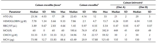
Table 3 Comparison of the red line to Caiman crocodilus fuscus, between males and females.
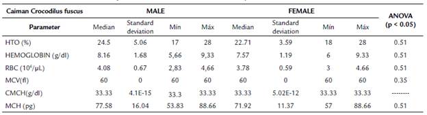
HTO: Hematocrit, RBC: Red blood cells, MCV: Median corpuscular volumen, CMCH: Median corpuscular hemoglobin concentration, MCH: Median corpuscular hemoglobin.
HTO: Hematocrit, RBC: Red blood cells, MCV: Median corpuscular volumen, CMCH: Median corpuscular hemoglobin concentration, MCH: Median corpuscular hemoglobin. Caiman crocodilus fuscus a (this study), Caiman crocodilus b (Carlos et al, 2017) Caiman latirrostris" (Barboza et al, 2011).
DISCUSSION
The results obtained in the present studies for the Caiman crocodilus fuscus are similar in some parameters to that reported by Koza et al. (2011) in their study on Caiman crocodilus yacare fed with two diets, the first of them with beef and the second with cassava. For the hematocrit, values were 22 % and 23 %, respectively. Table 1 shows the mean value obtained in this investigation for the hematocrit, which was 23.36 %. With respect to the first diet evaluated by Koza et al. (2011), this value varied by 1.36 %, while in the case of the second diet, it varied by 0.36 %, changing more in the case of the beef diet, even though in our study this was the only source of food. This comparison can be seen in Table 2, where a complete set of the data collected and provided by other authors and for other species of alligators is shown.
Comparing the values between of Caiman crocodilus fuscus males and females for hematocrit value, not statistical differences were found between them (p > 0.05), as shown in Table 3. With respect to the data reported by Peng et al. (2018) for this parameter, the average result of Caiman crocodilus fuscus was lower than in Alligator sinensis; both are of the same order, however, the average values of Alligator hematocrit was 28 %, which is the same as the maximum result of our study for males and females (Table 3). This was due to the difference between species; also, their reptiles were in the eastern territory of China, where the climatic conditions are different from those that are found in our Andean region (Peng et al., 2018).
These results are similar to that reported by Manzanilla et al. (2011), in which the hematocrit of Crocodylus intermedius kept in captivity was 24.76 ± 2.58 %. There is not a very marked difference concerning the average determined in our study, shown in Table 1 and mentioned previously. Likewise, the data of Manzanilla et al. (2011), had a lower standard deviation than our study, which was of 4.03 %.
In another investigation carried out by Carlos et al. (2017) with the wild spectacled caiman (Caiman crocodilus crocodilus), the hematocrit had a difference of 0.73 % of that reported in this study, despite the conditions of the different species and habitats (Table 2). The alligators of our study were in a zoo breeding and the spectacled caiman that was compared lived in wildlife (rivers and channels), which can change these values.
The typical hematological values for the Caiman latirostris were determined for Barboza et al. (2011), who found that the hematocrit was 21 %, with a balanced diet and crude protein of 38 %. This value was 2.36 % less than that reported in this study (Table 2). For the diet with 64 % of crude protein the hematocrit was 20 %, a 3.36 % lower than that the value reported in our study, as shown in Table 2.
Both previous reports can differ from our results possibly due to the influence of the diet, because our animals have a strict diet based on beef, horse and eggs, compared to a wild animal that lives in a natural environment, which feeds on other animals that live in its environment. This includes turtles, birds, picures, deers and some small reptiles, while the animals in zoo breeding could be feeding in captivity whit fish, chicken and all of tipes of food, incluid viscera or other tipes the waste (Morales, 2013). Thus, it is so hard to compare all these diets and include it' effect in the production.
In the case of hemoglobin, Koza et al. (2011) found for animals fed the beef diet, that this value was 6.10 g/dL, meanwhile for cassava diet, its value was of 6.38 g/dL. In our study, this value was 7.78 g/dL on average, which shows that there is a variation of1.77 g/dL for the first diet and 1.49 g/dL for the second. In addition, Koza and collaborators' results are closer to the minimum value reported in our study, which was 5.66 g/dL. The value of this parameter in Alligator sinensis (Peng et al., 2018), varied in 1.89 g/dL for males and 1.84 g/dL for females, very low in comparison with that found in the specimens of our study. Table 1 shows the maximum value obtained in the present study. Manzanilla et al. (2011) found 8.27 g/dL for Crocodylus intermedius, results that are in the average established in this investigation, which was 7.78 g/dL (Table 1).
The variation of hemoglobin in the spectacled caiman was 0.08 g/dL (Carlos et al., 2017) (Table 2), even though these animals were wild and in captivity. The variation (2.1 g/dL) was higher than that found in our study, which was 1.34 g/dL (Table 2). In the study carried out by Barboza et al. (2011) the hemoglobin of Caiman latirostris was 6.26 g/dL for diet 1 (balanced feed and crude protein 38 %), while for diet 2 (balanced feed and 64 % of crude protein) was 6.04 g/dL; those results are closer to the minimum value found in our study, which was 5.66 g/dL, despite being different species with different diet.
Regarding the CMCH, in the case of Caiman crocodilus yacare descript by Koza et al. (2011), in animals of diet 1 (beef) the value was 28 g/dL, while for diet 2 (Cassava) it was 29 g/dL. In Table 1, CMCH concentration was 33.33 g/ dL, which varied with respect to the first value in 5.33 % for the diet 1 and in 4.33 % for the diet 2, being the parameter of greater variation with respect to the issues evaluated by Koza et al. (2011). For the case of the CMCH in spectacled caiman, Carlos et al. (2011) found a value of 34.96 g/dL on average, that was different from the average of the present study (33.33 g/dL), being 1.63 g/dL above. However, it was among the range found in our study, which ranges from 27.82 g/dL to 38.83 g/dL (Table 2).
The CMCH reported by Barboza et al. (2011) for Caiman latirostris was 30 g/dL on average, for both diets, varying from what was reported in our study, which was 3.33 g/dL (Table 2). According to authors who worked with Alligator sinensis (Peng et al., 2018), the CMCH was 34.24 g/dL for males and 34.75 g/dL for females, results that were higher than found in Caiman crocodilus fuscus, in which the mean was 33.3 g/dL, without statistical differences between males and females (p > 0.05). Results of both studies were similar, even though the geographical and species conditions were totally different, as was previously explained (Peng et al., 2018).
In males of Crocodilus intermedius the MCH was 196.99 pg and in females was 193.67 pg, contrasting widely with reported in our study, which was 73.98 pg, and being 2.66 times lower than that reported for males and 2.61 times lower than that of females (Manzanilla et al., 2011). In the present study the values of MCH in males and females had no statistical differences (p > 0.05) (Table 3).
The parameter which differed to those reported in other studies was the MCV. For example, Carlos et al. (2017) found for spectacled caiman a value of 190.6 fl., which is three times higher than that of Caiman crocodilus fuscus (60 fl). On the other hand, for Alligator sinensisPeng et al. (2018) established that the MCV was 573.13 fl for males and 557.38 fl for females, resulting 9.5 points more than what was stated in our study for males, and 9.2 points more for females. However, comparing males with females we don't had statistical differences between sexes (p > 0.05).
The MCV of the Caiman latirostris reported by Barboza et al. (2011), was 449 fl (diet with 38 % of crude protein) and 439 fl (64 % of crude protein), while for Caiman crocodilus fuscus it was 60 fl. This means that the value of the first diet was 7.48, and that of the second was 7.31 times greater than that of common caiman found in our investigation.
Another value that presented the highest variance in this study was the MCH. The values reported by Manzanilla et al. (2011) for males of Crocodylus intermedius was 196.99 pg and for females 193.67 pg, contrasting widely with reported in our study, which was 73.98 pg, the latter being 2.66 times lower than that reported for males and 2.61 times lower in the case of females. As was mentioned, no statistical differences were found between sexes (p > 0.05) (Table 3). Similarly, Barboza et al. (2011) for the MCH found 135 pg (diet with 38 % of crude protein) and 130 pg (diet with 64 % of crude protein), results that are almost twice more than found in this study (Table 2). In the case of spectacled caiman, the MCH value of 63.49 pg (Carlos et al., 2017) was 10.49 pg lower than that found for Caiman crocodilus fuscus; nonetheless it is within the normal range that was determined for this species, which oscillates between 53.83 pg and 88.66 pg.
The value of the red blood cells found in this study was 3.89 106/µL, meanwhile, for Carlos et al. (2017) the value of red blood cells was 1.45 106/µL on average, which is far from reported in this study at 2.44 106/µL. Comparing the two sexes, no statistical differences were found between them (p > 0.05).
The studies carried out in the species of the Caiman yacare by Cañadilla (2015) validate the great importance of the feeding of this family of alligators, taking into account that in the family of Alligatoridae are the Melanosuchus niger, Paleosuchus trigonatus, Paleosuchus palpebrosus, Caiman crocodilus fuscus, Caiman crocodilus crocodilus and Caiman crocodilus apaporiensis, among others species (Morales, 2013). It is required an adequate management of the diets; for example, feeding schedules, which is done approximately every 10 to 15 days in the morning, could explained that the animals have a slow metabolism, so this feeding schedules provide a good alimentary conversion.
Feeding at will animals of C. latirostris and C.yacare species three times a week with meat meal supplemented with vitamins and minerals, generate doubts because this way to feed this kind of animals may cause lack of recognition of the prey or inadequate sizes of the food (Barboza et al., 2006), or that animals break their homeostasis (Cañadilla, 2015). This not only affects the red blood cells but the usual levels of minerals and vitamins that animals need to be in an optimal state of health, and their susceptibility to the contagion of diseases that could generates significant losses to the producer (Coppo et al., 2006).
Hemoparasites are another relevant aspect that can affect hematological values in the young crocodile. In the present study, the animals were one to two years old, and for this reason the hemoparasites are so important to evaluate, since different studies have found the presence of parasitic groups in ectotherms animals, such as Hemogregarins and Hepatozoon in Alligator mississippiensis (Enríquez et al., 2014); It is important to evaluate the ranges of hemoparasites in the zoo, because they could be changing the values of the hematological parameters. In our study the evaluation of hemoparasites was not taken into account, for which it is necessary to make an analysis on this factor that can affect the common crocodile.
It is recommended to directly assess the presence or absence of hemoparasites, cell morphology in general and white line evaluation in these animals, in addition to a more specific classification by sex and age in order to have greater security in the results obtained, as well as increase more information about these species that have been little studied. These studies could generate a great step for the development of this species' breeding. The diseases that can affect the alligators and could change the normal values in the blood are, for example, Mycobacterium szulgai and M. chelonae, which generate, in addition to apathy, cachexia and the rest of symptoms in these diseases, that the hematocrit and levels of hemoglobin and leukocytes decrease in the first day of action, which can subsequently lead to leukopenia (Slany et al., 2010).
CONCLUSIONS
There are no statistical differences between males and females for any hematological value of Caiman crocodilus fuscus, in captivity, with a degree of growth sub-adult.
Despite the differences between families and species of caiman compared in this study, biologically the results were not very different from each other. There was only one distinct difference in MCV and MCH between the Caiman crocodilus fuscus and the other species of caimans that were referred in the text, which could be due to the environmental conditions in which each of these species was studied, one in natural medium with a varied diet and that sampled in this study with different feeds and ambient temperatures.
It is necessary to analyze the typical hematological values from other ontogenetic stages of Caiman crocodilus fuscus and complement the results of this study.


