


 English (pdf)
 Article in xml format
 Article references
 Send this article by e-mail
 Cited by SciELO
  Cited by Google
  Similars in
SciELO
Similars in
SciELO  Similars in Google
 Permalink
INTRODUCTION
A cyst is defined as a pathological cavity lined on the inside by epithelium and covered on its outer side by connective tissue, which encloses a liquid, semi-liquid or gaseous content; it is not a cavity created by accumulation of pus, generally lacks internal pressure since the content enters the cavity by osmosis. They are asymptomatic and are discovered in routine radiographic studies 1. According to the classification given by the WHO in 1992, cysts are classified according to their pathogenesis, as lesions produced during development, odontogenic and non-odontogenic, which can be transformed into a pathology with signs of malignancy or radicular cysts of inflammatory origin 2,3. In 2005 the WHO classified cysts into three main categories, jaw cysts, cysts associated with cavities of the maxillaries and cysts associated with soft tissues of the mouth, face, neck and salivary glands. 4
Residual cysts are radicular cysts that remain or form after the responsible tooth has been extracted, this pathology may have been previously a lateral cyst or an apical cyst and after the tooth has been extracted, the cyst remains in the bone tissue without having been excised 5,6. This type of cyst usually does not present symptoms and goes unnoticed until it is discovered in a routine radiographic examination or indicated to evaluate another clinical situation; however, many times, when the cysts reach a larger size, they cause facial asymmetry accompanied by some degree of pain 1,7. Regarding its frequency, it occupies the second place among inflammatory cysts, and is usually of greater incidence in males, they occur mainly in non-vital teeth, in older individuals and in the mandibular region 8.
The objective of this publication is the presentation of a clinical case of a residual cyst with semi-solid content.
CLINICAL OBSERVATIONS
A 61-year-old female patient, consulted for anodontics in both posterior and anterior upper region, referring that she wants to get dentures for that area. In the anamnesis she reports not having any systemic diseases, nor consuming drugs, also states that in her youth many extractions were performed in her upper and lower jaw because of deep caries, the physical examination revealed a facial asymmetry in the middle third of the right hemi-face, secondary to a tumor. During the stomatological examination, swelling of 4 x 6 cm was observed, located in the right alveolar ridge, which produces effacement of the superior buccal sulcus, the mucosa that covers it has an appearance and color within normal parameters, of stony consistency, asymptomatic, non-depressible, non-mobile and with more than four years of evolution (Figure 1). Clinical diagnosis was established: residual cyst and as a differential diagnosis: development cyst, odontogenic and non-odontogenic tumor and mucocele.
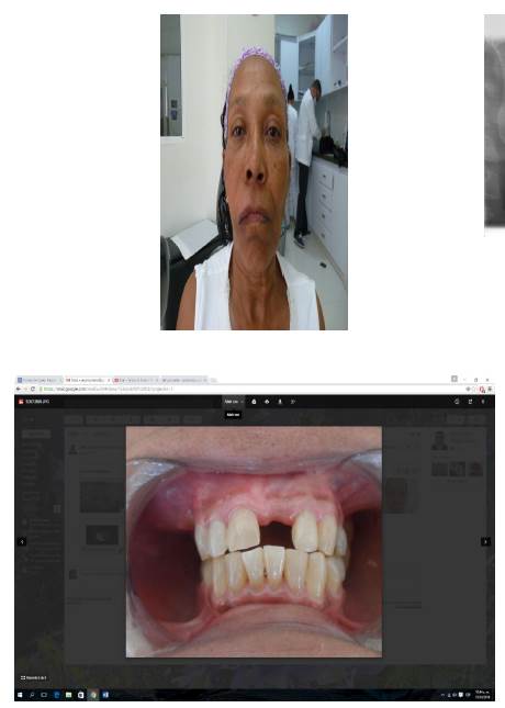
Figure 1 A. The facial asymmetry at the level of the cheekbones and above the right upper lip is observed. B. Intraoral photography before the procedure, there is swelling and effacement of the superior buccal sulcus, with a normal appearance on the mucosa that covers it, the dates indicate the comparison of the right and left cheeks.
On the radiographic examination, with panoramic x-rays (Figure 2), a radiolucent image with well-defined, smooth edges of 4x6 cm was observed, involving the edentulous area of the dental organs 14, 15, 16, 17 and without maxillary sinus involvement.
Figure 2 Panoramic x-rays, a radiolucent lesion was observed in the upper premolar area of the right side, with defined edges, without involvement of the maxillary sinus.
To corroborate the diagnosis and verify the real magnitude of the lesion, it was decided to perform a simple CT scan (computerized axial tomography) of the face (Figure 3), where an expansive image of 6 cm in diameter was observed, which presses the floor of the maxillary sinus, producing a reduction in its dimension. Taking into account the clinical history of the patient, clinical and radiographic characteristics, the diagnosis of residual cyst (RC) is presumed. Due to the patient's urgent need to perform the rehabilitation of the area, it was decided to perform the complete removal of the lesion as a definitive treatment, since it did not present malignant characteristics and an unknown content, it was decided to send the specimen to pathology in order to confirm the presumptive diagnosis.
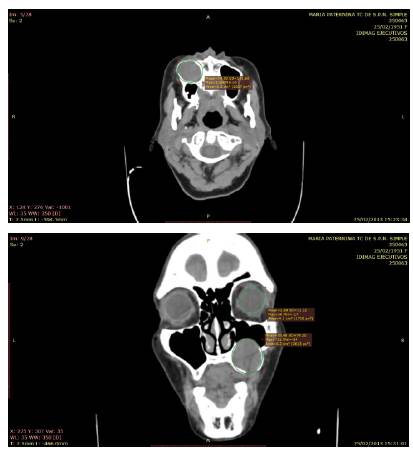
Figure 3 On the left side, in a coronal section, the well-defined lesion is observed, with a diameter of 6.2 mm. On the right side, the lesion was observed in a cross section and its diameter was compared with the diameter of the eyeball with a difference of 2.1 mm, with a larger diameter of the cyst and a close relationship with the maxillary sinus.
The procedure was initiated with local anesthesia, with a posterior superior alveolar nerve block, medial superior alveolar nerve block, infraorbital nerve block, anterior and posterior palatine nerves, with previous asepsis and antisepsis of the area; an intrasulcular incision was made from the distal of the dental organ #13 to the mesial of dental organ #18, with anterior and posterior relaxants, complete Neumann flap, then a mucoperiosteal flap was raised, an osteotomy was performed to obtain a bone window, exposing the cystic membrane and extracting its contents, which had a yellow semi-solid consistency (Figure 4), all its content was removed and proceeded to remove the cystic membrane, which was deposited in a container with 10% formaldehyde and it was hermetically sealed and referred to pathology, which showed the result of inflammatory cyst and, taking into account the 2005 WHO classification and the patient's history, it was diagnosed as a residual cyst.
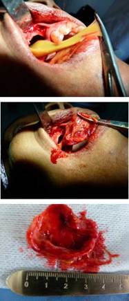
Figure 4 The semi-solid content of the cyst, its capsule and its diameter after its enucleation are shown.
The tissues were repositioned, sutured with 4/0 silk; the procedure was well tolerated and without complications. The pertinent recommendations were given and antibiotics and analgesics were prescribed. 1) Amoxicillin 500 mg. capsules every 8 hours for 5 days. 2) Nimesulide 100 mg. tablets every 12 hours for 3 days. Clinical and radiographic follow-ups were performed at three, seven, 15 and 21 days and then at one, three and six months period during which the patient did not report any discomfort, confirming in the radiographic studies the complete bone healing in the intervened area.
DISCUSSION
The authors presented a case of a slow-growing residual cyst in the upper right maxillary region, without noticeable macroscopic changes, without symptoms or discomfort in the patient. It was detected by means of a routine examination for the installation of prosthetic elements, which agrees with many authors who affirm the eventuality of the findings with this type of inflammatory pathologies 2,3,7,9,10; when performing the enucleation of the cyst, a semi-solid, viscous, yellowish-colored content was found, which is not usual in this type of cysts. There are few studies that report this type of contents in residual cysts, hence its importance. Sridevi et al report a clinical case of residual cyst which, after aspiration biopsy prior to surgical removal, reveals serosanguinous content with cholesterol crystals 10, Jamdade et al report a clinical case of a periapical residual cyst that, upon cytological examination of the aspirates, the extracted liquid seemed to be a chronic inflammatory fluid, at the moment of the enucleation a yellowish and solidified material surrounded by a thin capsule was observed.
In 2014, Rivero et al reported a case of a giant residual cyst, which was the result of trauma in the lower posterior area, for which the definitive treatment was its complete enucleation. The most coherent general reasons to treat these cystic lesions are: 1) the growth of the cyst, which destroys the surrounding bone and can cause the involvement of neighboring structures, such as the nasal cavity or the maxillary sinus and cause repeated nasal obstruction or sinusitis; 2) the possibility of infection attributable to the cyst, with its corresponding complications; 3) the need to perform a histopathological study to be certain of the true nature of the lesion 7.

