


Serviços Personalizados
Journal
Artigo
 Inglês (pdf)
 Artigo em XML
 Referências do artigo
 Enviar este artigo por email
Indicadores
-
 Citado por SciELO
 -
 Acessos
Links relacionados
-
 Citado por Google
 -
 Similares em
SciELO
Similares em
SciELO -
 Similares em Google
Compartilhar

 Permalink
Ingeniería e Investigación
versão impressa ISSN 0120-5609
Ing. Investig. v.31 n.3 Bogotá set./dez. 2011
Characterising structural, mechanical and cytotoxic properties of coral-based composite material intended for bone implant applications
Caracterización estructural, mecánica y citotóxica de un material compuesto a base de polvo de coral para posible uso en osteo-implantación
Angela Samper Gaitán1, Fabio Arturo Rojas Mora2, Diana María Narváez3, Luis Miguel Méndez Moreno4
1 Mechanical and industrial engineering, Universidad de los Andes. Business Analyst, Mckinsey & Company. angela_samper@mckinsey.com
2 Mechanical engineer, Master in Mechanical Engineering Universidad de los Andes, Ph.D. in Mechanical Engineering Universidad Federal de Santa Catarina, Brasil. Associate professor, Universidad de los Andes. farojas@uniandes.edu.co
3 Biology and Master, Universidad de los Andes. Research Assistant, Universidad de los Andes. dinarva@hotmail.com
4 Mechanical engineer, Master in Mechanical Engineering Universidad de los Andes. Professor, Universidad Nacional de Colombia.. lmmendezm@unal.edu.co
ABSTRACT
Studies concerning the application of Porites asteroides coral for bone implant purposes have demonstrated the biological viability of its use. As a complement to previous research regarding the development of bone-powder based composite materials which are useful for such applications, this study was aimed at developing a coral powder-based composite material which would be able to satisfy the appropriate structural, mechanical and cytotoxic properties required for its use. A composite material made of coral powder, calcium sulphate powder and water was therefore developed, and its properties were tested in different compositions. The results showed how the resulting composite material had properties which were comparable to those of human cortical bone (from both a structural and mechanical point of view), as well as being non-toxic below a 0.35 mg/ml critical composite material concentration.
Keywords: Porites asteroides coral, bone implant, composite material, calcium sulphate, biomaterial characterisation.
RESUMEN
Diferentes estudios referentes a la aplicación del coral tipo Porites asteroides en implantes óseos han demostrado su viabilidad biológica para este fin. Como complemento a investigaciones previas relacionadas con el desarrollo de un material compuesto a base de polvo de hueso susceptible de osteo-implantación, el propósito de este estudio es desarrollar un material compuesto a base de polvo de coral, que satisfaga las propiedades estructurales, mecánicas y citotóxicas requeridas para ser considerado como un material adecuado para este fin. A partir de esto se desarrolló un material a base de polvo de coral, polvo de sulfato de calcio y agua, y se realizaron los respectivos ensayos en diferentes composiciones del mismo. Los resultados generales muestran que el material compuesto desarrollado posee propiedades comparables a las del hueso cortical humano (desde un punto de vista tanto estructural como mecánico) y adicionalmente no muestra evidencias de citotoxicidad si se utiliza en concentraciones menores a 0.35 mg/ml.
Palabras clave: coral Porites asteroides , implantes óseos, material compuesto, sulfato de calcio, caracterización de biomateriales.
Received: May 25th 2010 Accepted: June 23th 2011
Introduction
This study was based on developing a Porites asteroides coral powder-based composite material for possible use in future bone implants. P. asteroides coral is made up of 99% calcium carbonate, known as aragonite mineral, which grows throughout the Pacific Coast coral reefs. This material has been studied for implant purposes for more than 15 years because of its biological properties (Vuola et al., 1996). Its structure is similar to that of human cortical bone and it is considered to be, "one of a limited number of materials that will form chemical bonds with bone and soft tissues in vivo" (Ben-Nissan, 2003, p. 285). A mineral known as porous hydroxyapatite (HA) can be obtained from P. asteroides coral by subjecting it to several heat treatments. This substance is structurally similar to P. asteroides coral and is also used for implant purposes. The difference between HA and coral is that HA is not biodegradable, making the implant last longer (Vuola et al ., 1996; Ben-Nissan, 2003). The application of this type of coral in bone implants to replace diseased bone has been widely studied and its features have been analysed (Khavari et al., 1993; Begley et al., 1995; Vuola et al., 1996; Ben-Nissan, 2003; Ripamonti et al., 2008). These studies have shown P. asteroides (and other coralline samples) to be biologically viable for this purpose, as it is a biocompatible, osteo-conductive and inert material. It has been stated that this type of coral has osteo-inductive properties when exposed to bone marrow cells, but not in itself (Braye et al., 1996; Vuola et al., 1996).
In spite of it having biological properties which are highly appropriate for implant purposes, P. asteroides coral has limited mechanical properties (Ben-Nissan, 2003) which are equally fundamental for developing bone grafts and implants that will assure a suitable level of in vivo resistance. The mechanical strength that the matrix material will need to provide in developing an appropriate composite material for bone implant purposes must be considered. Local research has focused on this concern, having mainly examined the development of human (Rojas, 2000) and bovine bone powder-based (Peñaloza, 2007; Quevedo, 2004; Reina, 2005; Salazar, 2009) composite materials satisfying implant requirements. Bone powder has been shown to be an appropriate alternative for developing bone implants, as it was easy to obtain at a minimum amount of lost material. This study describes two main phases: the development of a coral powder-based composite material and the characterisation of its structural, mechanical and cytotoxic properties to determine its level of appropriateness in bone implant applications.
Materials and methods
Producing coral powder particles
Tangential milling was chosen as the most appropriate machining mechanism for obtaining coral powder, given that the specimen's shape did not have to be modified or combined with any other substance. According to prior related studies (Lugo, 2009), the most influential cutting parameters affecting this material's surface roughness and cutting force are depth of cut (p) in mm and feed rate (f) in mm/min and tool nose radius (r) in mm, to a lesser extent. It was found that the rest of the observed parameters, (i.e. rake and clearance angles (α and β) in ° and cutting speed (v) in m/s) had little or no influence on the variables being analysed. While processing the coral specimen, a factorial experiment was designed to study the effect of varying the specimen's machining conditions on the type of powder obtained from each one, especially in terms of its size and shape. Two main factors were chosen for analysis: t feed rate (f) in mm/s and depth of cut (p) in mm. Both factors were varied according to three levels, thus giving six treatment tests (see Table 1).
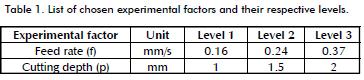
The feed rate (f) levels were set according to the milling cutter's available values; the depth of cut (p) was divided into three uniform levels, each one separated from the other by 0.5 mm. Factors kept constant during the experiment were cutting tool speed (v) and rake and clearance angles (α and β), along with tool nose radius (r) and tool diameter (d). Even though these factors remained constant throughout the experiment, they may nevertheless have affected the size of the obtained particles, according to theoretical relationships between this variable and cutting parameters during regular milling (Micheletti, 1980).
Characterising the coral powder so produced
Initial results showed that a mixture of different sized particles appeared in each sample. After separating each mixture by sieving, three main types of powder were obtained, differentiated by their size range and grouped into the following categories: Type
1 particles (see Figure 1) which were smaller than 250 µm, Type
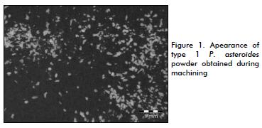
2 particles (see Figure 2) which were sized 250 µm to 500 µm and Type 3 particles (see Figure 3) 500-1,000 µm.
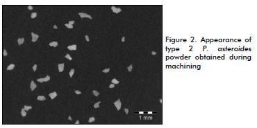
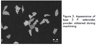
Particles larger than 1 mm were discarded as they had a much larger size and irregular shape due not to machining but to specimen tearing while machining its edges. These particles were therefore classified as Type 4.
After having completed the factorial experiment, each sample was statistically analysed to examine the influence of the chosen factors on the powder's main features. Given that three main types of powder were obtained per sample, three separate analyses were carried out for each one5. The response variables chosen for studying each type of powder were the following: particle size (s) in µm, particle shape factor (sf) and percentage weight (w) each type of powder represented within the complete sample. Particle size (s) was obtained through an optical analysis of each type of powder sample by calculating the smallest diameter upon which each particle could be fully surrounded. Likewise, particle shape factor (sf) was calculated by dividing the largest diameter that could be inscribed inside each particle, by its previously calculated size; 30 particle measurements were made for each test, to acquire a statistically-relevant sample of the total population. Each powder's percentage weight (w) was measured to examine its variability over the different tests carried out. As three types of powder were analysed per test, 18 samples were studied (counting the 6 tests originally made).
A two-way analysis of variance (ANOVA) was built for comparing the averages obtained for size (s), shape factor (sf) and percentage weight (w) in each sample to property averages of samples relative to the same type of powder.
Developing the composite material
Recent studies have suggested the use of calcium sulphate as being appropriate for implant purposes (Khavari et al., 1993; Cho et al., 2005). Commercially known as gypsum, this substance has been widely used for developing dental and orthopaedic implants (Pecora et al., 1997; Tay et al., 1999), demonstrating its in vivo biocompatible and osteo-conductive properties while implanted (De Long et al., 2007; Jung et al., 2010). An additional advantage of using calcium sulphate is its easy manipulation, given that it hardens through a chemical process at room temperature, without the need for adding any substance or applying any further treatment to it. The composite material was therefore made of coral powder, calcium sulphate powder and water. A factorial experiment was designed to analyse whether different amounts of coral powder and calcium sulphate added would influence the properties of the final composite material (chosen experimental factors are shown in Table 2).
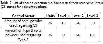
The amount of coral powder related to the amount of calcium sulphate was studied in order to examine the effects of adding more coral powder (regardless of its type) to the composite material, whereas the amount of Type 2 regarding Type 3 coral powder added was analysed to evaluate the difference between using different types of coral while developing the composite material.6
Mechanical properties
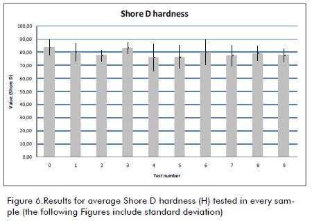
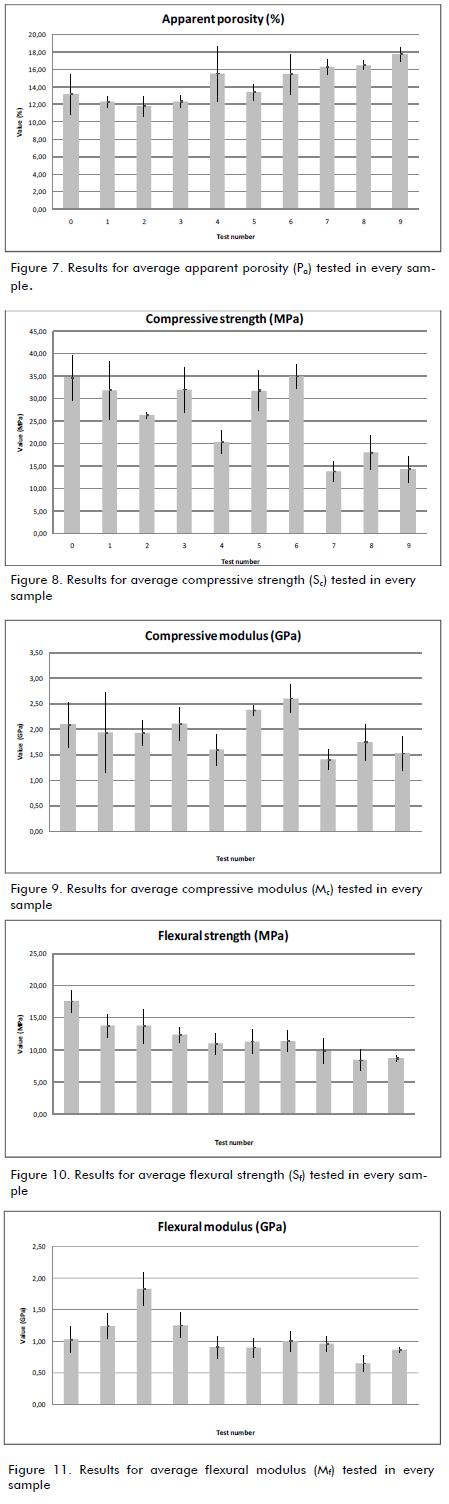
Tests for Shore D hardness (H), apparent porosity (Pa), compres-sive strength (Sc) and modulus (Mc) and flexural strength (Sf) and modulus (Mf) were carried out for each sample in the Universidad de los Andes' Mechanical Properties laboratory. The Shore D hardness test was carried out on 10 specimens from each sample in line with ASTM D695-08 while compressive and flexural tests were made upon five specimens per sample, as suggested in ASTM D790-02 and D2240-05, respectively. Porosity was tested upon the same specimens used for testing flexural properties, as suggested by Peñaloza (2007).
Cytotoxic properties
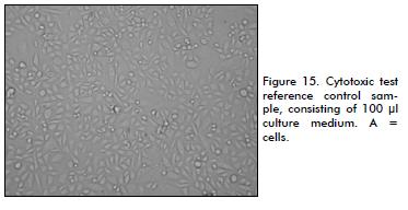
Chronic cytotoxicity tests were carried out in the Universidad de los Andes' Human Genetics Laboratory. They were measured by a colorimetric method using 3-[4,5-dimethylthiazol-2-yl]-2,5-diphenyltetrazolium bromide (MTT), according to Mosmann (1983). Each test involved Chinese hamster ovary K1 cells (CHO-K1) exposed to different concentrations of coral powder alone, calcium sulphate powder alone and three different samples of the composite material.7 The CHO-K1 cells were grown as monolayers in Roswell Park Memorial Institute (RPMI) 1640 medium (Sigma), supplemented with 10% foetal bovine serum (FBS), 1% penicillin/streptomycin (Gibco) and 2% glutamine (Gibco). All samples tested had been previously left in UV for 10 minutes to eliminate bacteria from manipulation, according to the laboratory standards for cell culture. After UV irradiation, the samples were pulverised and then prepared in RPMI medium, as follows: 2mg/ml of just coral powder, 2mg/ml of just calcium sulphate and 0.05, 0.5 and 2 mg/ml of the 10% 30% and 50% coral powder and calcium sulphate mixture, respectively. 3x10⁵ cells per sample were grown in 96-sample flat-bottomed plates, with four replicate samples per treatment. The fifth test of each treatment corresponded to a blank reference, consisting of just 100 µl culture medium. Cells were examined after 24 hours incubation and treated. The first sample corresponded to the reference control, consisting of 100 µl culture medium, and the remaining samples contained 100 µl of medium having a known concentration. The plate was then incubated at 37°C in a humidified 5% CO2 atmosphere for 72 hours. After this, MTT (5 mg/ml) was added to each sample. Cells were incubated for a 4 further hours. Afterwards, dimethyl sulfoxide was added to dissolve formazan crystals. Each sample was analysed on a Bio-Rad micro plate reader at 595 nm after 5 minutes' shaking using a 655 nm reference wavelength. The results were expressed as the percentage of living cells calculated from detected absorb-ance, assuming 100% as the absorbance reported by the reference control sample. The chronic cytotoxicity test was repeated twice and a two-sample t-test was performed to test differences between experiments, along with a Pearson's statistical correlation test to look for correlation between cell viability and the different concentrations used.
Results
Characterising the coral powder so produced
As the three different types of powder obtained had statistically equivalent analysed properties, they could be grouped into three categories, regardless of the sample they came from. Table 3 describes the average obtained as statistical results for these groups.
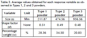
As can be seen, each type of powder differed considerably regarding size (s) but not shape, as all shape factor (sf) results were fairly similar. On the other hand, the weight proportion (w) represented by each type showed that Type 2 powder was more abundant than the other two types within each sample.
Structural properties
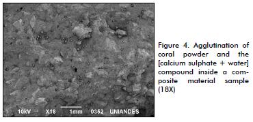
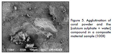
The samples' structural properties were inspected using optical and electronic microscopes to examine the way in which coral particles became agglutinated with the [calcium sulphate + water] compound. Structural imperfections such as pores, impurities or particular defects were also examined. The different samples showed a high level of agglutination between the different substances forming the composite material. Coral powder was effectively surrounded by the [calcium sulphate + water] compound, thereby preserving the composite's integrity (see Figures 4 and 5). However, samples were shown to have a large amount of pores throughout their structure, mainly due to calcium sulphate becoming solidified inside the different moulds used for constructing specimens.
Mechanical properties
Average values for each sample's mechanical properties were recorded (see Figures 6 through 11), samples being tested in their dry form.8 ANOVA test results showed that variations in apparent porosity (Pa), compressive strength (Sc) and flexural strength (Sf) values could be explained due to variations in the amount of coral powder used in relation to calcium sulphate in each sample, given that their corresponding p-value was below the 5% reference level. Other properties' variations proved not to be caused by any of the treatments analysed here, as the difference between the averages in each sample did not prove statistically different.
Cytotoxic properties
After 72-hour exposure to pure coral powder (2 mg/ml), CHO-K1 cells exhibited a dose-dependent increase in viability (see Figure 12). Pearson's correlation value suggested that the increase in pure coral powder concentration was significantly correlated with increased cell viability. Exposure to pure calcium sulphate powder produced a decrease in dose-dependent viability, as an increase in calcium sulphate concentration was significantly correlated with decreased cell viability, according to the corresponding Pearson's correlation value. The lethal concentration at which 50% of the cells died (LC50) was around 1.3 mg / ml. Images according to these results are shown in Figures 13 to 14 compared to the control in Figure 15.
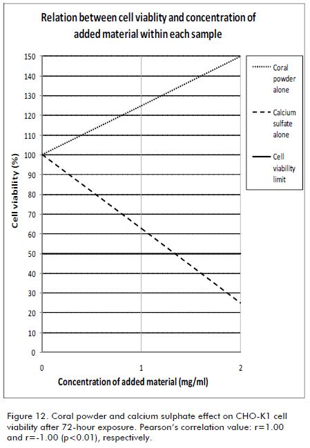
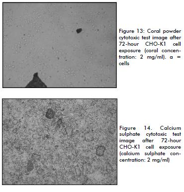
It can be observed how coral powder represented no danger for cell viability at 2 mg/ml concentration whereas a 2 mg/ml calcium sulphate concentration was harmful for living cells (Figure 12). Only about 20% of them were able to survive in these conditions.
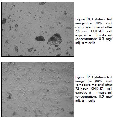
Cell exposure to the three composite material samples did not have significant correlation with cell viability (see Figure 16). LC50 levels were found to be 0.5 mg/ml for the 10% coral powder sample and 0.35 mg/ml for the 30% and 50% coral powder samples. Images of these results (0.5 mg/ml composite material concentration) are shown in Figures 17 to 19.
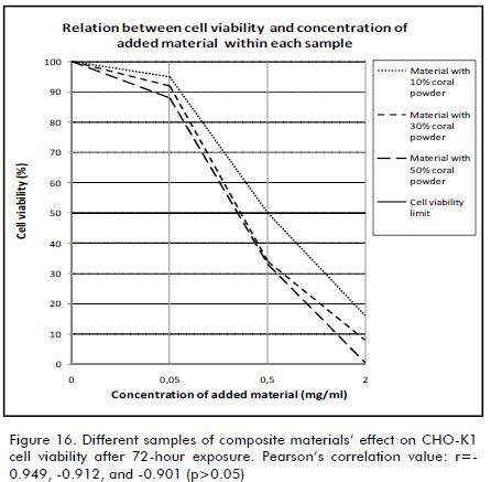
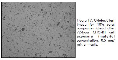
The results showed that a 0.5 mg/ml concentration of composite material was only suitable when using a 10% coral powder sample (Figure 17). Higher coral powder percentage within the composite material resulted in decreased cell viability at 0.5 mg/ml concentration, as seen in Figures 18 and 19.
Conclusions
A coral powder-based composite material was produced by a reproducible process involving a minimum level of material waste. Its structure was analysed and tests were made for its apparent porosity (Pa), Shore D hardness (H), compressive strength (Sc) and modulus (Mc), flexural strength (Sf) and modulus (Mf) and cytotoxic levels. The results showed how the properties tested varied with a greater amount of coral powder being incorporated within the compound (regardless of the type of powder present). As this factor increased, the compound's compressive and flexural properties decreased, while its apparent porosity showed a slight increase and Shore D hardness remained stable. Such result has also been seen in similar studies regarding the testing of coralline + calcium sulphate materials for future implant purposes (Khavari F. et al., 1993).
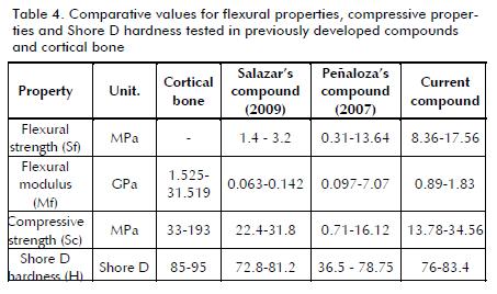
Compared to previously developed bone-implant compounds (Peñaloza, 2007; Salazar, 2009) and with lyophilised cortical bone, the compound had appropriate values for most of the properties tested (see Table 4).9 However, efforts should be made to improve its compressive strength results.
Future studies should focus on testing this material when exposed to human cells, as well as determining whether its mechanical properties change during exposure to living cells for a determined amount of time (i.e. the specimen's lifetime value). For the moment, the composite can be used in up to 0.5 mg/mL concentrations without affecting cell viability. This type of material's level of machinability could be tested to determine whether it can be developed in different forms for replacing different kinds and shapes of diseased human bone.
Nomenclature
p: Depth of cut (mm)
f: Feed rate (mm/min)
r: Cutting tool nose radius (mm)
α: Rake angle (°)
β: Clearance angle (°)
v: Cutting speed (m/s)
d: Cutting tool diameter (mm)
s: particle size (µm)
sf: particle shape factor (µm/µm)
w: percentage weight (%)
H: Shore D hardness (Shore D)
Pa: Apparent porosity (%)
Sc: Compressive strength (MPa)
Mc: Compressive modulus (GPa)
Sf: Flexural strength (MPa)
Mf: Flexural modulus (GPa)
NOTAS AL PIE
5Due to Type 4 particles' observed irregularities regarding shape and size these were not submitted to further characterisation or analysis.
6Type 1 coral powder was not used to build the composite material as its small size could have potentially affected its biocompatibility.
7These included the three different levels of amount of coral added regarding the amount of calcium sulphate, regardless of coral powder type.
8Samples labelled "0" in the graphs represent the reference sample consisting of [calcium sulphate + water] compound only
9Cortical bone data was collected on November 25th, 2009 from Salazar (2009) and through the link: www.bonesim.com/. Missing properties were those not found for all specimens.
References
Begley, C.T., Doherty, M.J., Mollan, R.A.B., Wilson, D.J., Comparative study of the osteoinductive properties of bioceramic, coral and processed bone graft substitutes., Biomaterials, Vol. 16, 1995, pp. 1181-1185. [ Links ]
Ben-Nissan, B., Natural bioceramics: From coral to bone and beyond. Current opinion in solid state and materials science 7(4-5), 2003,. pp. 283-288. [ Links ]
Braye, F., Irigaray, J.L., Jallot, E., Oudadesse, H., Weber, G., Deschamps, N., Deschamps, C., Frayssinet, P., Tourenne, P., Tixier, H., Terver, S., Lefaivre, J., Amirabaldi, A., Resorption kinetics of osseous substitute: natural coral and synthetic hydroxyapatite., Biomaterials 17, 1996., pp. 1345 -1350. [ Links ]
Cho, B.C., Chung, H.Y., Lee, D.G., Yang, J.D., Park, J.W., Roh, K.H., Kim, G.U., Lee, D.S., Kwon, I.C., Bae, E.H., Jang, K.H., Park, R.W., Kim, I.S., The effect of chitosan bead encapsulating Calcium sulfate as an injectable bone substitute on consolidation in the mandibular distraction osteogenesis of a dog model., Journal of oral and maxillofacial surgery, Vol. 63:12, 2005., pp. 1753-1764. [ Links ]
De Long Jr., W.G., Einhorn, T.A., Koval, K., McKee, M., Smith, W., Sanders, R., Watson, T., Bone grafts and bone graft substitutes in orthopaedic trauma surgery. A critical analysis., The Journal of Bone and Joint Surgery, 2007;89, pp. 649-658. [ Links ]
Jung, H., Song, G., Lee, Y., Baek, J., Ryoo, H., Kim, G., Choung, P., Woo, K.M., Modulation of the resorption and osteoconductivity of a-Calcium sulfate by histone deacetylase inhibitors., Biomaterials 31, 2010., pp. 29-37. [ Links ]
Khavari F, Bajpai PK. Coralline-sulfate bone substitutes. Biomed Sci Instrum. 1993;29:65-9. [ Links ]
Lugo S., H., Estudio de la mecánica de corte en materiales celulares: coral tipo Porites Asteroides., Tesis presentada a la Universidad de los Andes, para optar al título de Magister en Ingeniería Mecánica, 2009. [ Links ]
Micheletti, G.F., Tecnología mecánica: Mecanizado por arranque de viruta., Ed. Blume, Barcelona, España, 1980. [ Links ]
Mosmann, T. Rapid colorimetric test for cellular growth and survival - application to proliferation and cytotoxicity tests., J. Immunol, 1983., Métodos 65, 55, 63. [ Links ]
Pecora, G., Andreana, S., Margarone, J.E., Covani, U., Sotto-santi, J.S., Bone regeneration with a Calcium sulfate barrier., Oral surgery, Oral medicine, Oral pathology, Oral radiology, and Endodontology, Vol. 84;4, 1997., pp. 424-429. [ Links ]
Peñaloza, J., Diseño de un material compuesto para implantes óseos.,Tesis presentada a la Universidad de los Andes como requisito de grado del programa de pregado en Ingeniería Mecánica, 2007. [ Links ]
Quevedo, S. M., Desarrollo de una metodología para la fabricación de injertos compuestos de polvo de hueso y biopolíme-ro. Tesis presentada a la Universidad de los Andes como requisito de grado del programa de pregado en Ingeniería Mecánica, 2004. [ Links ]
Reina, D. Y.,Implementación de un sistema de manufactura de injertos de polvo de hueso por inyección., Tesis presentada a la Universidad de los Andes como requisito de grado del programa de pregado en Ingeniería Mecánica, 2005. [ Links ]
Ripamonti, U., Crooks, J., Khoali, L., Roden, L., The induction of bone formation by coral-derived Calcium carbonate/ hydroxyapatite constructs., Biomaterials, Vol. 30, 2009, pp. 1428-1439. [ Links ]
Rojas, F.A., Fabricación de implantes ortopédicos a partir de hueso humano., Tesis presentada a la Universidad Federal de Santa Catarina para optar al título de Doctor of Philosophy, 2000. [ Links ]
Salazar, A., Diseño de un proceso de fabricación de implantes dentales y ortopédicos de polvo de hueso y sulfato de calcio con aplicación industrial., Tesis presentada a la Universidad de los Andes como requisito de grado del programa de pregado en Ingeniería Mecánica, 2009. [ Links ]
Tay, B.K., Patel, V.V., Bradford, D.S. Calcium sulfate - and Calcium phosphate based bone substitutes: Mimicry of the mineral phase of bone., Orthopedic Clinics of North America, Vol. 30;4, 1999., pp. 615-632. [ Links ]
Vuola, J., Göransson, H., Böhling, T., Asko-Seljavaara, S., Bone marrow induced osteogenesis in hydroxyapatite and Calcium carbonate implants., Biomaterials 17, 1996., pp. 1761-1766. [ Links ]

