


 Inglés (pdf)
 Articulo en XML
 Referencias del artículo
 Enviar articulo por email
 Citado por SciELO
  Citado por Google
  Similares en
SciELO
Similares en
SciELO  Similares en Google
 Permalink
A 77 year-old woman with STEMI, who underwent thrombolysis and coronary angiography, severe three-vessel coronary artery disease and clinical features of heart failure. The surgery was postponed. At six weeks a trans-thoracic echocardiography (TTE) showed a ventricular septal defect (VSD). She was referred to our Institution. The patient was in severe cardiogenic shock, requiring inotropic support and mechanical assistance. A RT3D transesophageal echocardiogram (TEE) showed a VSD (Fig A) and severe shunt by color Doppler. Schematic figure of the VSD (E). The decisión was made to proceed with percutaneous closure of VSD. An occluder device was placed guided by TEE (Fig C), which confirmed adequate positioning with minimal residual shunt (Fig D). The clinical course of the patient was initially good, but a week ago she deteriorated. TEE showed a significant leakage (Fig F). The patient was taken to emergency surgery. Inspection of the occluder device showed extension of the VSD at the inferior and superior border (Fig G). The VSD was closed with a Dacron single patch (Fig HD). The clinical course was excellent.
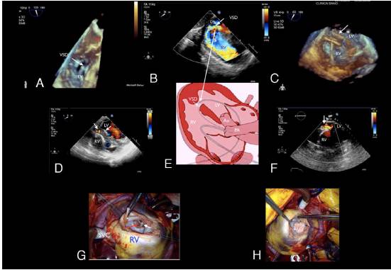
Figure 1

