


 English (pdf)
 Article in xml format
 Article references
 Send this article by e-mail
 Cited by SciELO
  Cited by Google
  Similars in
SciELO
Similars in
SciELO  Similars in Google
 Permalink
Introduction
Atherosclerosis continues to be the leading cause of coronary artery disease, which can cause the full spectrum of acute coronary syndrome as a consequence of the rupture or erosion of an atherosclerotic plaque. The etiology of acute myocardial infarction is of non-atherosclerotic origin in 1-7% of cases in all age groups. Non-atherosclerotic causes of coronary artery disease include coronary embolism, vasospasm, spontaneous coronary dissection, vasculitis, and congenital anomalies of the coronary arteries. The prevalence of coronary embolism in patients with acute ST-segment elevation myocardial infarction ranges from 4% to 13% based on angiographic and autopsy studies1,2. This article presents the case of a young patient with no medical history, who presented an acute myocardial infarction caused by coronary embolism.
Clinical case
A 20-year-old man with no previous medical history entered the emergency room for the first episode of oppressive retrosternal chest pain radiating to the left arm and mandibular region associated with diaphoresis which started 3 h ago.
In the initial assessment, vital sings of 140/90 mmHg blood pressure were documented. The other vital signs were within the normal range without pathological findings in the cardiopulmonary examination and in the rest of the physical examination. An electrocardiogram was taken (Fig. 1) showing ST segment elevation of 3 mm in the inferior leads associated with ST-segment depression in V1-V2 and aVL.
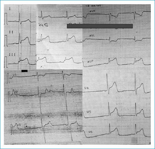
Figure 1 Electrocardiogram on admission: Sinus bradycardia rhythm 55 beats/min, without delay AV conduction, QT: 400 ms, ST elevation of 3 mm in inferior leads, V4-V6, and ST segment depression in V1-V2 and aVL.
Laboratory test showed an initial ultrasensitive troponin I greater than 10 times the 99th Percentile, 5390 ng/L (reference range 0-25 ng/L). The patient received loading doses of acetylsalicylic acid 300 mg, clopidogre 600 mg, anticoagulation with enoxaparin, and high-intensity statin therapy.
A transesophageal echocardiogram showed a 52% left ventricular ejection fraction, hypokinesia of the middle, and apical anterolateral segment; the rest of the segments with preserved contractility without valvular alterations; and with agitated saline solution test image compatible with patent foramen ovale with significant passage of bubbles to the left atrium with valsalva maneuver. Doppler ultrasound of the lower extremities showed no deep or superficial vein thrombosis. Test for thrombophilias was performed and the result was negative (Table 1).
Table 1 Laboratory data
| Variable | Results | Reference range |
|---|---|---|
| Factor V Leyden mutation | Negative | Negative |
| ANAs | Negative | Negative |
| Lupus anticoagulant | 42.6 s | 31-44 s |
| Antithrombin III | 96.1% | 79.4-112% |
| Anti-cardiolipins IgG | 1.07 UGPL/ml | < 20 UGPL/ml |
| Anti-cardiolipins IgM | 4.45 UMPL/ml | < 13 UMPL/ml |
| Homocysteine | 9.9 umol/L | 5-12 umol/L |
| B2 glycoprotein IgG | 1.35 U | 0-20 U |
| B2 glycoprotein IgM | 1.24 U | 0-20 U |
| Creatinine | 0.95 mg/dl | 0.6-1.2 mg/dl |
| Ureic nitrogen | 18.8 mg/dl | 6-20 mg/dl |
| Total cholesterol | 146 mg/dl | |
| HDL cholesterol | 33 mg/dl | |
| LDL cholesterol | 90 mg/dl | |
| Triglycerides | 113 mg/dl | |
| Thyroxine | 4.3 uUI/ml | 0.4-4.0 uUI/ml |
| Glycosylated hemoglobin | 5.4% | |
| VDRL | Negative | Negative |
Emergent coronary angiography performed (Fig. 2) and showed the left anterior descending artery with 85% thrombotic occlusion in its apical portion, TIMI I flow, and the second diagonal artery with thrombus in the distal portion without evidence of significant atherosclerotic lesions. The other coronary arteries were without significant lesions. Intracoronary thrombolysis was performed with tirofiban infusion with improvement of the thrombus at the apical level. The patient was admitted to the Coronary Care Unit for medical surveillance.
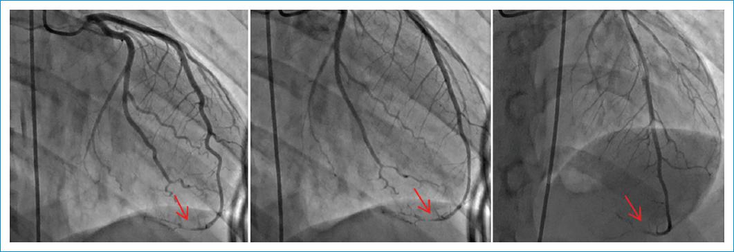
Figure 2 Coronary angiogram: anterior descending artery (apical portion), compatible image with thrombi that compromise flow (TIMI flow I at this level). Note: Optical coherence tomography (OCT) was not performed, because it is located in the apical portion and the diameter of the vessel at this level is 1.6 mm.
The patient had an adequate clinical evolution, resolution of chest pain without symptoms of acute heart failure. Antiplatelets were suspended and Warfarin was indicated until a goal of INR 2-3 was achieved. He was discharged in good condition with anticoagulation goals for cardiology outpatient follow-up.
Discussion
There are several risk factors that should raise suspicion for coronary embolism in a patient with acute chest pain, such as the presence of valve prostheses, atrial fibrillation, cardiac myxoma, intracardiac thrombus, rheumatic valve disease, intracardiac shunts (patent foramen ovale), dilated cardiomyopathy, infective endocarditis, congenital anomalies of the coronary arteries, and the presence of a hypercoagulable state3.
Coronary embolism frequently affects anterior descending artery especially its distal epicardial portion and its intramural branches. This is because the proximal portion of the anterior descending artery is straighter than the circumflex artery. In the systematic review of coronary embolism cases by Lacey et al., involvement of the anterior descending artery accounted for 45.3% of cases followed by right coronary artery 15.3%, circumflex artery 14.7%, and left main trunk 5.3%4. In the study by Shibata et al., which included a population of 1776 patients diagnosed with acute myocardial infarction, the patients with coronary embolism had fewer cardiovascular risk factors (hypertension, diabetes, dyslipidemia, and smoking) compared with the group without coronary embolism5.
Three types of coronary embolism have been described: direct, paradoxical, and iatrogenic. The criteria used for the diagnosis are described in the table 2.
Table 2 Diagnostic criteria for coronary embolism
| Major criteria |
| 1. Angiographic evidence of coronary artery thrombosis without atherosclerotic components. |
| 2. Concomitant multisite coronary embolism |
| 3. Concomitant systemic embolization, excluding intraventricular thrombi attributable to ST-segment elevation myocardial infarction |
| Minor criteria |
| 1. < 25% stenosis on coronary angiography, except for the culprit vessel |
| 2. Evidence of an embolic source detected by any imaging modality |
| 3. Coexistence of a potential condition for thromboembolic disease such as intracardiac tumors, infective endocarditis, prosthetic heart valves, atrial fibrillation, hypercoagulable states, patent foramen ovale, and atrial septum defects |
Definitive diagnosis: ≥ 2 major criteria, 1 major criterion plus≥2 minor or 3 minor criteria.
Probable case: 1 major criterion plus 1 minor criterion or 2 minor criteria. The diagnosis of coronary embolism is excluded in cases of pathological evidence of atherosclerotic thrombus, history of coronary revascularization, presence of coronary ectasias or disruption/erosion of plaque detected by intravascular ultrasound (IVUS) or optical oherence tomography (OCT) in the proximal portion of the guilty vessel7.
Direct coronary embolism is the most common mechanism and occurs in conditions that favor cardioembolism, such as: atrial fibrillation, presence of valve prostheses, infective endocarditis (mainly of the aortic valve), mitral stenosis, post-surgical cardiac surgery, and in rare cases by atrial myxoma6,7.
The paradox occurs in the presence of a patent foramen ovale where an embolus of venous origin crosses the foramen and lodges in an arterial bed. Iatrogenic coronary embolism adduces post-procedure development such as cardiac catheterization1,4.
The treatment of coronary embolism is subject to the underlying cause, and a case-by-case approach to management must be made. There are several therapeutic options that have been described without reaching a consensus including catheter thrombus aspiration, thrombolysis, balloon angioplasty, stent implantation, anticoagulation, antiplatelet therapy, and use of antibiotics in cases of infective endocarditis8,9. Lacey et al. found that thrombectomy was the most used technique in 47.6% of the cases out of the 147 analyzed patients, balloon angioplasty was used in 44 cases (49.9%), 17 out of 147 patients had a stent implant, and thrombolysis was used in 14.9% of the cases.
At discharge, 49 patients (33.3%) were prescribed anticoagulation and/or antiplatelet agents. Oral anticoagulation as monotherapy was prescribed in 25 cases. A combination of oral anticoagulation with a single antiplatelet agent (acetylsalicylic acid or clopidogrel) was used in 16 cases. Triple therapy (oral anticoagulation, acetylsalicylic acid, and clopidogrel) was indicated in six patients, and monotherapy with acetylsalicylic acid or clopidogrel was used in two patients5.
Paradoxical embolism has been described for several decades as a potential mechanism of coronary embolism. This occurs in individuals with the presence of patent foramen ovale when an embolus from the venous circulation crosses the cardiac shunt and lodges in the arterial circulation, thus producing ischemic stroke or systemic embolism, including coronary artery. The management of these patients continues being a topic of debate especially in the coronary embolism scenario10.
Conclusion
Coronary embolism is probably underdiagnosed given the difficulty in its diagnosis and is potentially fatal. At present, there are no management guidelines, which leave the treatment to the discretion of the treating physician. A high level of suspicion is required since the clinical presentation may be indistinguishable from an acute coronary syndrome due to coronary atherosclerosis. In our case, a patent foramen ovale (PFO) with a right-to-left shunt was found only during the valsalva maneuver. It was not possible to identify an embolic source from the venous circulation that could explain a possible paradoxical embolism or a frequent coagulation disorder. The management consisted of thrombolysis with tirofiban infusion and later anticoagulation with warfarin until reaching INR goals of 2-3, which was also continued at discharge. The need to close the PFO will be defined during the follow-up.
Ethical disclosures
Protection of human and animal subjects. The authors declare that no experiments were performed on humans or animals for this study.
Confidentiality of data. The authors declare that they have followed the protocols of their work center on the publication of patient data.
Right to privacy and informed consent. TThe authors have obtained the written informed consent of the patients or subjects mentioned in the article. The corresponding author is in possession of this document.

