


Serviços Personalizados
Journal
Artigo
 Inglês (pdf)
 Artigo em XML
 Referências do artigo
 Enviar este artigo por email
Indicadores
-
 Citado por SciELO
 -
 Acessos
Links relacionados
-
 Citado por Google
 -
 Similares em
SciELO
Similares em
SciELO -
 Similares em Google
Compartilhar

 Permalink
Revista Facultad de Ingeniería Universidad de Antioquia
versão impressa ISSN 0120-6230
Rev.fac.ing.univ. Antioquia no.64 Medellín jul./set. 2012
ARTÍCULO ORIGINAL
System of heart and lung sounds separation for store-and-forward telemedicine applications
Sistema de separación de sonidos cardiacos y pulmonares para aplicaciones de telemedicina de almacenamiento y envío
Antonio José Salazar*1, Catalina Alvarado1, Fernando Enrique Lozano22
1Biomedical Engineering Group (GIB). University of Los Andes. Edificio Mario Laserna Cra 1 Este No. 19A-40. Bogotá, Colombia.
2Microelectronic center (CMUA). University of Los Andes. Edificio Mario Laserna Cra 1 Este No. 19A-40. Bogotá, Colombia.
*Autor de correspondencia: teléfono: + 57 1 339 49 49 (3360), fax: (571) 332 4316, correo electrónico: ant-sala@uniandes.edu.co (A. Salazar)
(Recibido el 11 julio de 2011. Aceptado el 31 de agosto de 2012)
Abstract
Auscultation is a medical procedure that provides a general idea of heart and lung behavior as the physician listens to the breath sound. Due to the fact that the sounds from these organs overlap in time and frequency domains, important affections in one of them could be discarded. For instance, the objective of this work is to implement two different methods of cardiac and pulmonary sound separation. First, we apply modulation filters to the time- frequency representation of the original signal, recorded on the chest. Second, we apply an iterative algorithm of wavelet decomposition and reconstruction filters. Results show that they both separate signals appropriately. Taking lung signals as noise, we determine that signal to noise (SNR) ratio is 10.21 for the first method and for 6.61 the second. Applications in telemedicine are encourager since the bandwidth of the signal transmission could be reduced by sending it separately.
Keywords: Auscultation, cardiac sounds, pulmonary sounds, modulation filters, wavelet filters
Resumen
La auscultación es un procedimiento médico que proporciona una idea general del comportamiento del corazón y los pulmones, a partir de los sonidos producidos por estos dos órganos. Debido a que estos sonidos se sobreponen en sus componentes de tiempo y frecuencia, afecciones importantes en uno de los órganos podrían no ser detectadas, dando lugar a un falso negativo. Por lo tanto, el objetivo de este trabajo es implementar dos métodos diferentes de separación de sonidos cardiacos y pulmonares. Primero, se aplica filtros de modulación a la representación en tiempo-frecuencia de la señal original, tomada sobre el pecho del paciente. Segundo, se aplica un algoritmo iterativo con filtros wavelet de descomposición y reconstrucción. Los resultados muestran que ambos separan las señales correctamente. Tomando el sonido pulmonar como ruido, se determina que la relación señal a ruido (SNR) es 10.21dB para el primer método y 6.61dB para el segundo. Aplicaciones en telemedicina son interesantes al poder reducir el ancho de banda de la señal transmitida, enviando las dos señales por separado.
Palabras clave: Auscultación, sonidos cardiacos, sonidos pulmonares, filtros de modulación, filtros wavelet
Introduction
Auscultation is a primary procedure that examines the thoracic and abdominal cavity where it is possible to hear to the heart sounds corresponding to the blood flux along the heart valves (mitral, tricuspid, aortic and pulmonary), and the lung sounds, corresponding to air flux along the pulmonary conducts (trachea, bronchi and alveoli) during inspiration and expiration phases [1]. Nevertheless, heart and lung sounds overlap in their time and frequency components, which could hide different affections in one of these organs. For this reason, the question of how to separate heart and lung sounds, keeping the integrity of both signals has motivated scientists work in the last 3 decades.
The purpose of this work is to provide a system of heart and lung sounds separation for telemedicine store-and-forward applications (reducing the file size and improving sound quality) to make an easy remote auscultation. At present time we are working also to extend the system to real time applications using implanting faster algorithms in systems-on-chips.A first approximation to the problem is the use of conventional filters, which filter heart signal (20-150Hz) from breath signal. However some components of pulmonary sound (25-1600Hz) are included in that frequency band and are incorrectly discarded from lung signal [2]. In order to better separate the components that belong to heart and lung sounds, it has been proposed the use of adaptive filters [3], whose coefficients are fitted at each iteration depending on a reference signal, commonly the ECG [3,4]. To avoid the problem of synchronizing reference and signal, coefficients are computed from the fourth order statistics [5] or with the multi-resolution wavelet transform [6-8]. Recent studies propose a statistical method, the independent components analysis (ICA) [9,10].
In this work, we show the implementation and validation of two different separation methods. We first implement a method based on modulation frequency filters, applied over the short time Fourier transform (STFT) representation of the signal [11]. Secondly, we present a separation algorithm based on discrete wavelet transform (DWT) with a multi-resolution analysis, which separates stationary component (lung sound) from non-stationary component (heart sound) [6] by a threshold applied to the coefficients of the wavelet representation [8].
Materials
Methods are validated with signals from four healthy subjects, two women and two men aged from 20 to 40 years. We ask them to breathe normally, to hold the respiration and to breathe deeply during 10 seconds. For each case, one sensor is located in the apex and another in the second intercostals space, so that 24 segments are analyzed. Data acquisition module (figure 1) records the signals using an omnidirectional microphone. After that, sound is converted into a digital voltage signal and acquired by an ADuC7020 microcontroller. We use 8-bit resolution and sampling rate of 3500Hz, since Nyquist frequency is 3200Hz. The software used to acquire and process data is Matlab2008b.

Methods
Two different methods are implemented and validated over the recorded signals:
Modulation filters
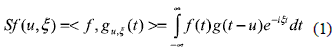
Original signal is represented as a summation of elementary functions well concentrated in time and frequency domains, known as STFT. In consequence, variations of ''instantaneous'' frequency can be measured. The general expression for STFT is defined by the equation 1:
Where each atom gu,ξ(t) is constructed with a window g translated u units in time and modulated by the frequency ξ as shown in equation 2:
For any value of the parameters (u, ξ), the window must be normalized , ||gu,ξ||= 1, so that the energy of the signal segment concentrated around this point can be measured with the spectrogram Psf(u,ξ)= |Sf(u,ξ)|2. Each element of this matrix corresponds to the energy density of the signal in the neighborhood of u and ξ.
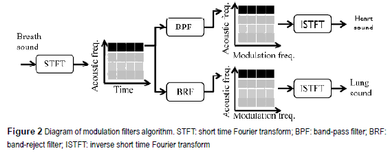
Once the spectrogram is obtained, it is evident that stationary components (lung sounds) have less frequency variance than non-stationary components (heart sounds). Consequently, when band-pass and band reject modulation filters, are applied to the spectrogram it is possible to differentiate the components with more frequency variations over the time, non-stationary and the components with less frequency variation, stationary (figure 2) [11].
Wavelet filters
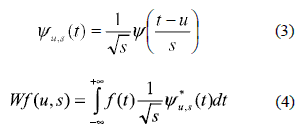
Wavelet transform also decomposes the signal in time-frequency domain. But instead of complex exponential atoms, the family of functions used to represent the signals corresponds to dilated (s) and translated (u) versions of a mother function ψ of zero mean value and normalized. The transform Wf (u,s) is defined by the equations 3 and 4:
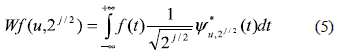
In order to minimize the number of scales we use a multi-resolution approach and we select the discrete set,  , that forms an orthogonal basis family [12]. The new expression for the discrete transform is defined by the equation 5:
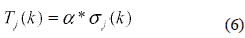
It is possible to create different levels of signal decomposition according to the nature of j. If j is negative, signal details are obtained, contrarily, if j is positive, signal approximation is obtained. Furthermore a decomposition-reconstruction iterative algorithm is implemented so that, at each iteration k, the coefficients are compared with a statistical threshold —defined by the equation 6—proportional to the standard deviation of the transform coefficients for each decomposition level.
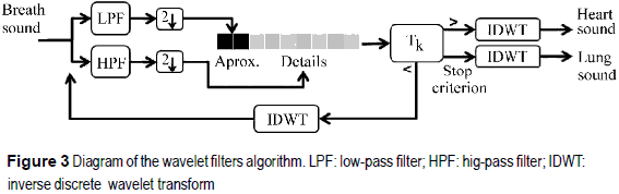
Coefficients of magnitude under the threshold represent non-stationary heart components and those over the threshold represent stationary lung components (figure 3) [6].
Results
Modulation filters
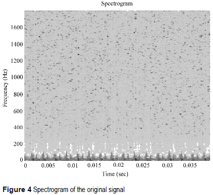
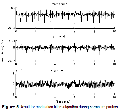
Modulation filters separate heart and lung sounds applying a band pass filter for heart signal, and a band reject filter for lung signal over the time- frequency representation. For the computation of STFT we use a Hamming window of 512 samples, with overlap of 50%, in order to avoid border effects. Once spectrogram is obtained (figure 4), for a fixed frequency the temporal trajectory is filtered with Butterworth coefficients. Furthermore, cardiac non-stationary signals change its frequency content across the time, so that they have bigger components in modulation frequency domain. Contrarily, pulmonary stationary signals have frequency components more uniform over the time, corresponding to lower components.
Heart sounds are separated with a band pass filter of 5-35Hz, while lung sounds are separated with a band reject filter at the same frequency range (figure 5). SNR in average for all patients is 6.61 dB.
Wavelet filters
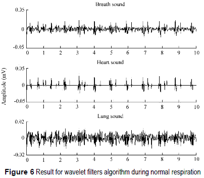
Wavelet filters separates cardiac and pulmonary signals when a fixed threshold is applied to the wavelet coefficients. An iterative decomposition- reconstruction algorithm is implemented using mother wavelet Daubechies-8. We realize that 5 iterations are enough to a correct separation. Besides, decomposition is obtained by filtering the signal at the fourth level, which means that, one low-pass and four high-pass filters generate one approximation and four detail levels, respectively. Those coefficients that are above the threshold belong to the cardiac signal, and those below the threshold are reconstructed and decomposed again, until the fifth iteration, when the remaining signal belongs to the pulmonary sound. For each level, the threshold is calculated with an adjust factor of 150± 15 (figure 6). Value of SNR for all patients is 10.21dB.
As the reconstructed signal (respiratory or heart signal) contained less information—due to the separation—better compression rates could be achieved than if the original signal were compressed without separation. For example, after applying MPEG-1 Audio Layer III compression (better known as MP3) to the original and separated signals, a 30% increase in the compression rate was achieved for the separated signal.
Conclusions
The most difficult aspect in separating cardiac and pulmonary sounds is the fact that time and frequency components are overlapped, such that conventional filters are not convenient. For this reason, we present two different advanced methods of separation.
With modulation filters it is possible to analyze the variations over each frequency component over the time. The result largely depends on the cut frequency for the modulation filters.
Wavelet filters also represent a good strategy of separation. The threshold determines whether signal components are stationary or not. In the first case, wavelet coefficients have lower values and components belong to the lung sound. In the second case, coefficients are higher and they belong to the heart sound.
The results are convenient for store-and- forward telemedicine applications, in which real-time process hardware and high-bandwidth communication channels are not required. Future work includes hardware implementation of the algorithms for real time telemedicine applications with low-bandwidth communication channels.
References
1. L. Bickley, P. Szilagyi, B. Bates. Bates' guide to physical examination and history taking. 8 ed. Ed. Lippincott Williams & Wilkins. Philadelphia, USA. 2003. pp 233-243, 277-295. [ Links ]
2. Z. Kazem, M. Zahra. Fundamentals of respiratory sounds and analysis. Ed. Morgan & Claypool Publishers, series Synthesis lectures on biomedical engineering #8. San Rafael, CA, USA. 2006. pp. 19–21. [ Links ]
3. V. Iyer, P. Ramamoorthy, F. Hong, Y. Plolysonsang. ''Reduction of heart sounds from lung sounds by adaptive filtering''. IEEE Trans. Biomed. Eng. Vol. 33. 1986. pp. 1141-1148. [ Links ]
4. S. Charleston, M. Azimi-Sadjadi. ''Reduced order Kalman filtering for the enhancement of respiratory sounds''. IEEE Trans. Biomed. Eng. Vol. 43. 1996. pp. 421-424. [ Links ]
5. L. Hadjileontiadis, S. Panas. ''Adaptive reduction of heart sounds from lung sounds using fourth-order statistics''. IEEE Trans. Biomed. Eng. Vol. 44. 1997. pp. 642-648. [ Links ]
6. L. Hadjileontiadis, S. Panas. ''A wavelet-based reduction of heart sound noise from lung sounds''. Int. J. Med. Inf. Vol. 52. 1998. pp. 183-190. [ Links ]
7. M. Pourazad, Z. Moussavi, G. Thomas. ''Heart sound cancellation from lung sound recordings using time- frequency filtering''. Med. Biol. Eng. Comput. Vol. 44. 2006. pp. 216-225. [ Links ]
8. I. Hossain, Z. Moussavi. An overview of heart-noise reduction of lung sound using wavelet transform based filter. in Proc. Ann. Int. Conf. IEEE EMBS. Cancún, México. 2003. pp. 458-461. [ Links ]
9. M. Pourazad, Z. Moussavi, F. Farahmand, R. Ward. Heart Sounds Separation From Lung Sounds Using Independent Component Analysis. in Proc. Ann. Int. Conf. IEEE EMBS. 2005. pp. 2736-2739. [ Links ]
10. C. Jen-Chien, H. Ming-Chuan, L. Yue-Der, C. Fok- ching. A Study of Heart Sound and Lung Sound Separation by Independent Component Analysis Technique. in Proc. Ann. Int. Conf. IEEE EMBS. New York City, USA. 2006. pp. 5708-5711. [ Links ]
11. T. Falk, C. Wai. Modulation filtering for heart and lung sound separation from breath sound recordings. in Proc. Ann. Int. Conf. IEEE EMBS. Vancouvert, Canadá. 2008. pp. 1859-1862. [ Links ]
12. S. Mallat. A Wavelet Tour of Signal Processing. 3 ed. Ed. Academic Press. San Diego, California, USA. 2009. pp. 299-371. [ Links ]
