


Services on Demand
Journal
Article
 English (pdf)
 Article in xml format
 Article references
 Send this article by e-mail
Indicators
-
 Cited by SciELO
 -
 Access statistics
Related links
-
 Cited by Google
 -
 Similars in
SciELO
Similars in
SciELO -
 Similars in Google
Share

 Permalink
Revista Facultad de Ingeniería Universidad de Antioquia
Print version ISSN 0120-6230
Rev.fac.ing.univ. Antioquia no.78 Medellín Mar. 2016
https://doi.org/10.17533/udea.redin.n78a10
DOI: 10.17533/udea.redin.n78a10
ARTÍCULO ORIGINAL
Biomechanical analysis of damaged intervertebral disc using reflective photoelasticity
Análisis biomecánico de un disco intervertebral con lesión empleando fotoelasticidad reflectiva
Ricardo Gustavo Rodríguez-Cañizo1*, Luis Héctor Hernández-Gomez2, Ariel Fuerte-Hernández3, Emmanuel Alejandro Merchán-Cruz1, Alejandro González-Rebatu4, Pedro Alejandro Tamayo-Meza1
1Escuela Superior de Ingeniería Mecánica y Eléctrica, Instituto Politécnico Nacional. Unidad Azcapotzalco, Av. de las Granjas 682, Col. Sta. Catarina. C.P. 02550. Azcapotzalco, México.
2Escuela Superior de Ingeniería Mecánica y Eléctrica, Instituto Politécnico Nacional. Unidad Profesional Adolfo López Mateos, Edificio 5, Tercer Piso. C. P. 07320. Gustavo A. Madero, México.
3División de Mecatrónica, Universidad Politécnica del Valle de México. Av. Mexiquense s/n, esq. Universidad Politécnica, Col. Villa Esmeralda. C. P. 54910. Tultitlán, México.
4 Área de ortopedia, Hospital Regional 1° de Octubre ISSSTE. Av. Instituto Politécnico Nacional 1669, Col. Lindavista. C. P. 07300. Gustavo A. Madero, México.
* Corresponding author: Ricardo Gustavo Rodríguez Cañizo, e-mail: ricname@gmail.com
(Received June 12, 2015; accepted December 1, 2015)
ABSTRACT
This paper presents an experimental evaluation of the structural integrity of the lumbar section (L2-L3-L4) considering a damaged intervertebral disc. In this study, porcine specimens were used due to the similarity of the mechanical properties of those of the human spine. The lumbar section L2-L3-L4 was tested under compression. Five cases were analyzed; in the first one, the lumbar section consisted of healthy intervertebral discs. For the other four cases, the disc located between L2 and L3 was divided into four quadrants: front, back, left and right. For each of these cases, a damage condition was induced by making an incision from the annular fibers to the pulpous nucleus, covering each quadrant; the back elements (pedicles and facet joint) were removed and only the vertebral bodies and discs were tested. As a damaged intervertebral disc is unable to properly perform its mechanical function, the load transferred from L2 to L3 through the disc is no longer optimal. The actual stress field on L3, considering the damaged disc, was obtained using reflective photoelasticity for each one of the previously mentioned study cases. The results show that the induced damage in the intervertebral discs increases the stresses on L3 considerably when compared to the case of an undamaged disc, being the most critical when the damage is located in the back quadrant of the disc. In the other three cases, the damaged disc does not reduce the structural integrity of the vertebral body significantly. However, the inter-vertebrae space is reduced as a result of the damage, thus compromising the structural integrity of the studied lumbar section.
Keywords: Photoelastic analysis, stress distribution, lumbar section, damaged intervertebral disc
RESUMEN
Este artículo presenta un análisis biomecánico de una sección de columna lumbar (L2-L3-L4) considerando que existe un daño inducido en uno de los discos intervertebrales. En este estudio, se utilizaron especímenes de columna lumbar de cerdo debido a su gran similitud biomecánica con la del ser humano. Las secciones lumbares fueron ensayadas bajo carga axial de compresión. Se analizaron cinco casos de estudio, el primero de ellos fue la sección lumbar de columna con el disco intervertebral sano. Para los casos restantes, se indujo una lesión en el disco intervertebral ubicado entre L2 y L3 dividiéndolo en cuatro cuadrantes: anterior, posterior, derecho e izquierdo. Para cada uno de estos casos, la lesión en el disco se indujo haciendo una incisión con bisturí desde el anillo fibroso hasta llegar al núcleo pulposo abarcando todo el cuadrante. Los elementos posteriores (pedículos y facetas articulares) fueron retirados manteniendo así solo los cuerpos vertebrales y los discos intervertebrales (unidad de carga); lo que simula una fusión vertebral. El campo de esfuerzos completo en L3, se observó utilizando fotoelasticidad reflectiva. Los resultados muestran que la lesión del disco intervertebral propicia un aumento en los esfuerzos observados en L3 a través del polariscopio, en comparación con el caso de un disco sano, siendo el caso más crítico cuando el daño está situado en el cuadrante posterior. En los otros tres casos (anterior, derecho e izquierdo), la lesión en el disco intervertebral no produce un aumento significativo del campo de esfuerzos observado en L3; sin embargo, el espacio interdiscal se reduce considerablemente, lo que compromete la integridad estructural de la columna lumbar.
Palabras clave: Análisis fotoelástico, campo de esfuerzos, columna lumbar, lesión de disco intervertebral
1. Introduction
A damaged intervertebral disc can be the result of trauma where the adjacent vertebra suffers a fracture, or due to a degenerative condition where the disc loses its capacity to transfer load between vertebrae. Whichever case, it is necessary to determine whether non-invasive conservative treatment or surgery is required. If the course of action is surgery, the surgical technique that should be followed has to be chosen taking into consideration the amount of damage suffered by the disc. In order to solve this issue, a detailed biomechanical analysis of the vertebral section is required. Although the current trend in the treatment of some spine injuries is to avoid surgery due to the complexity that these procedures involve, conservative treatments are often recommended [1-4]. However, from a biomechanical perspective, an important question arises: whether it is convenient or not to keep an injured disc untreated, as it has lost its mechanical capacity to maintain the structural integrity of the system.
In general, an injury on a lumbar intervertebral disc is closely related to lower back pain, which ranges from moderate to severe, even disabling, depending on the degree of the disc degeneration. Nowadays, approximately 80% of the world's population suffers from lower back pain (LBP) that requires medical treatment, constituting a health problem in developed countries as workers are often on sick leave due to LBP, representing important economic loses [5-7].
The aim of this interdisciplinary work is to provide scientific and relevant data to the medical community, guiding the assessment of an injury in intervertebral discs of the lumbar section. Thus, this paper focuses on the biomechanical study on the effect of annular fiber injuries in intervertebral discs as a result of trauma, evaluating the structural integrity of the whole system.
2. Materials and methods
The tested specimens are fresh porcine vertebrae sections of the lumbar region (L2-L3-L4). Young pigs were used with an approximate weight of 80 kg. All specimens are fresh (less than 24 hours after slaughter). The porcine vertebrae were chosen not only because of the biological similarity with the human spine; but, as established in [8], these specimens can be used as test subjects to simulate the human spine in certain mechanical tests.
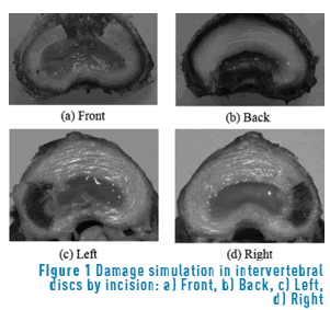
The damage simulation in the intervertebral disc was made by making a window shaped incision with a scalpel. The disc was divided into four quadrants (back, front, right and left side) were the damage condition was cut, as it can by appreciated in Figure 1. These cases reproduce the intervertebral disc injury and intervertebral fusion of those patients who have suffered an injury in L3 due to trauma. As this study focuses on the effect of the stress distribution, due to the load transfer, and not on the functionality of the lumbar section as a joint, the back vertebrae elements (pedicles and facet joint) were eliminated, using only the vertebral bodies and intervertebral discs. The depth of the incisions is such that reproduces the total damage of the intervertebral disc, as the fibrous ring is cut. The functional unit considered in this study is that constituted by L2, L3 and the intervertebral disc between them (Figure 2(a)).
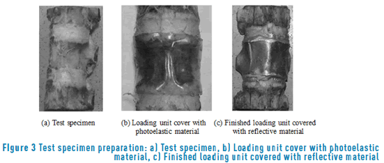
Surgically, this sort of injury can be treated using a "fixation device" that transfers the load from L2 to L4 while the fracture of L3 heals by consolidation (Figure 2(b)). For this study, it was necessary to determine the loading unit to be analyzed experimentally, e.g. the part of the lumbar section that is covered with photoelastic material (Figure 2(c)). Figure 3 shows the preparation of the test specimen for the undamaged intervertebral disc case.
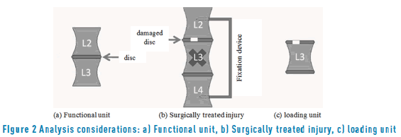
The difference between the functional unit and the loading unit is its lack of mobility. Its role is limited to be subjected to compression load only, as it happens in spinal fusion. This situation takes place when an internal spinal fixer is used; as a result, the mobility of the study section is restricted.
The in-vitro analysis results reported in [9], where the spine bends when it is loaded with two or three times the body weight, and considering that the average weight of a Mexican individual is 80 kg; the axial compression loads applied to each specimen were 1471.5N, 1962N and 2452.5N (150, 200 and 250Kgf respectively). The latest represent the most critical event that can take place where the external load exceeds the critical load of the spine and fails. The biomechanical tests were conducted on a universal test machine MTS 858 with a capacity of 5 tons.
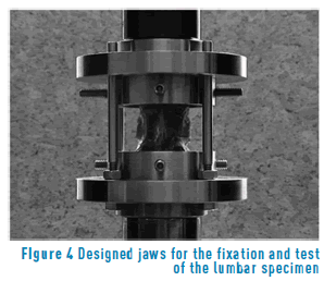
In order to ensure that the loads were fully applied along the axial direction, it was necessary to design an appropriate set of jaws to properly apply the compressive load to the spinal section, as shown in Figure 4.
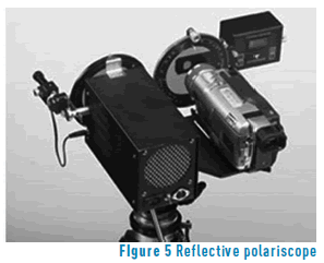
The mechanical behavior of the vertebral body L3 was analyzed using a reflective polariscope (Figure 5), as the reflective photoelestacity technique has proven to be reliable for the analysis of the complete stress field. The lacquer used in this case was PL1 with a PLH1 catalyzer. The reflecting adhesive used was PC1. The thickness of the coating lacquer was 1.5x10⁻³m. It was obtained from 0.15x0.10m plates which were previously prepared.
3. Results
Once the load was applied to the test specimen, the isochromatic pattern shows whether or not the disc damage caused some stress concentration. In addition to the mechanical tests, a load-displacement diagram was obtained. With this information, the stiffness change, due to the damage introduced in the disc, was assessed. This showed the reduction in the load carrying capacity of the damaged specimens when a comparison is made with the healthy case. The loading rate was set at 2x10⁻³m per minute.
After the loads were applied, the isochromatic patterns were photographed for each of the studied cases. Figure 6 shows the isochromatic patterns as seen with the reflective polariscope in order to determine the stress magnitude on L3 for each of the before mentioned cases under a compression load of 1471.5N.
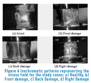
The intact case is taken as reference to establish in what order the stress magnitude increased during the tests for the damaged cases. It can be seen that, for the intact case the stress magnitude through the whole vertebra is very low (gray isochromatic shade). This is a clear indicator that the intervertebral disc is properly working as a load dissipater. The blue isochromatic shade in this case is present due to a structural irregularity in the test specimen, which does not represent a risk on the structural stability of the functional unit.
For the front and back damages, the isochromatic pattern indicates an increment on the stresses on L3 (orange isochromatic shade). Using an electronic compensator with the reflective polariscope to avoid human error on the interpretation of the isochromatic shades, it can be established that the stress magnitude is higher for the back damage case compared with the front damage case.
Finally, also from Figure 6, the right damage case which is analogous to the left damage case, presents an isochromatic pattern that reflects an intermediate magnitude stress field (pale yellow isochromatic shade).
It is important to point out that the zone of interest in this study is the central zone of the functional unit, as from the structural point of view, is the region where the load is transferred and the stresses are distributed. It can be appreciated that, for all cases, the stress distribution is homogeneous (Figure 6); implying that even with a damaged disc the load transmission is uniform. However, the magnitudes of these stresses are different. Quantitatively, the magnitudes of the stresses for each case are compared in Figure 7. The numeric value derived from the isochromatic patterns of the stress fields were processed using the PC-CALC code in conjunction with the reflective polariscope; model LF/Z-2.
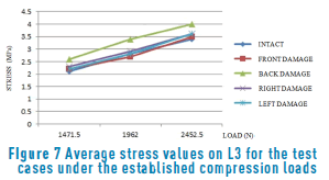
It can be seen that the stress values for the lateral and frontal damage do not vary significantly between one to another. Conversely, for the case where the damage is located in the back quadrant, a clear increment on the stress values can be appreciated for the three loading conditions.
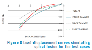
Additionally, in order to determine the stress value due to compression loads for the functional unit, an axial compression test with a controlled displacement of 4x10⁻³m applied at a 2x10⁻³m/min rate was performed. The 4mm limit is established taking into account that this is the height of porcine intervertebral discs, simulating the condition medically known as spinal fusion that takes place when the disc collapses. This test allowed the characterization of the biomechanical behavior of a loading unit. Figure 8 shows the load-displacement curves for the different test cases; it is clear that when the intervertebral disc does no present any damage (solid line) the loading unit is able to bear a load of 1964.27N to achieve a 4mm displacement. For the case where the damage is in the front quadrant (dashed line), the capacity to bear load of the loading unit reflects a reduction of approximately a 9% at 1785.71N with respect to the intact case, whereas for the loading unit with a lateral damage (dot-dashed line) this reduction is approximately a 20% with a load of 1642.85N. Finally, for the case that simulates a damage in the back quadrant, an important reduction is observed compared to the intact case; the total load where the system simulates an equivalent condition to the spinal fusion is approximately 43% less than the intact case with 1125N.
The displacement caused by the injury in the intervertebral discs was measured with a Vernier Calibrator, and presents a fluctuation of 0.30x10⁻³ to 1.0x10⁻³m, which represents up to 30% of the normal height of pork intervertebral discs (3x10⁻³m). These values are consistent with the results reported in [10]. They found a maximum displacement of 1.05x10⁻³ m. in an ovine intervertebral disc under loading conditions similar to those in this work.
4. Discussions
The mechanical characterization of the spine has been the subject of several studies. In recent years, the mechanical characterization of intervertebral discs has been widely reported. However, the available data vary considerably due to different animal specimens tested in each study as well as the diverse mechanical testing conditions that were followed such as those reported in [11-14]. Moreover, it is not common to find data related with the mechanical behavior of intervertebral discs under axial compression loading which simulates the upright position of the human being. In general terms, all these cases are analyzed from a medical perspective. Thus, in order to propose a design of appropriate engineered solutions of biomechanical components, that are expected to be used as a replacement or reinforcement of any part of the human body, it is necessary to produce and provide more consistent data on the characterization of the biomechanics of particular cases where the analyzed system fails.
The tests conducted in this work show that stresses in the middle region of the vertebral body L3 increases moderately when the intervertebral disc presents damage in any of its quadrants (Figure 6). The calculated stress range goes from 2.1 MPa, in the case of the healthy specimen under a compression load of 1471.5N, up to 4.0. MPa for the case of the intervertebral discs with damage in the back quadrant, with a 2452.5 N compression load. In all cases, the stress values are below the 4.6MPa fracture conditions of the bone under compression loading, as reported by [15]. Therefore, there is no risk that an injury of the intervertebral discs can fracture the adjacent vertebral body. However, attention must be paid when the damage is in the back quadrant, as it proves to be the most critical at 4.0 MPa, not far from failure conditions.
The reduction in the height of the damaged intervertebral discs can be up to 1x10⁻³ m. This may be a risk factor if it is considered that the average value at the height of the pork intervertebral discs is around 3x10⁻³m. It represents a loss of height of about 30%. In fact, this phenomenon causes that functional units physically located above the injured intervertebral disc collapse, causing all the elements involved (tendons, facets, discs, muscles, etc.) to react in order to compensate for the displacement, generating strain conditions that result in what is commonly known as back pain. Another factor inherent to this situation is the spine misalignment produced by the inclination of the functional units towards the quadrant where there the damage is located.
It can be established that the magnitude of the necessary load to cause structural instability in a healthy spine, is quantitatively different to that required to cause the same damage in the spine with an injured disc. In fact, the results obtained in this work opposes those reported in [16], as their results suggest that a unit with a damaged disc supports twice the axial load of a healthy disc; justifying this behavior, due to disruption in the stress distribution resulting from the damage in the intervertebral discs. But, as demonstrated using the photoelestacity technique and the data analysis, there is an important stress concentration when the studied specimen is axially loaded under compression for the specimens with an injury, proving that damaged units loose structural integrity, thus diminishing their mechanical capacity to bear compressive loads. This reduction is approximately 43% (Figure 8), which corresponds to the case in which damage is located in the back quadrant of the intervertebral disc.
The obtained data indicates that the structural stability of the analyzed segment is reduced. From the biomechanical perspective, due to the damage in the intervertebral disc, it is no longer able to perform its function as a load dissipater. Consequently, the adjacent vertebral bodies are subjected to increased loading conditions. This results in an increment of the peak stresses. Some authors suggest that the core of the disc is highly stressed, while others have reported that high levels of stresses are located in the annular fibers [17-19].
5. Conclusions
The results reported in this paper indicate that the damage in intervertebral discs do not represent an immediate risk factor. Therefore, failure in the adjacent vertebral bodies is not expected. In other words, the damage of the disc does not generate high stresses in the central zone of L3. This situation takes place in most of the analyzed cases in this work. Nonetheless, when the damage is in the back quadrant, the stresses in L3 are close to failure conditions. This situation has to be considered when the health conditions of a patient are assessed.
Moreover, the resulting vertebrae space reduction due to injured disc is also significant, as the injury induces a full collapse in all functional units above it. This may be a risk factor for other spine conditions that might present further complications in the patient's health. Therefore, it is necessary to evaluate other areas on which overloading takes place as a direct result of this condition.
The aim of the results presented in this paper is to provide orthopedists with additional information to decide the best course of treatment, whether it implies the possible removal of damaged intervertebral discs, instead of recommending only dry off of the extruded portion of the pulpous nucleus and maintaining the rest of the disc, which is a regular practice in surgical procedures as depicted in [5]. Derived from the results of this paper, the immediate recommendation would be the use of a prosthetic implement or bone graft in the inter-vertebrae space; nonetheless, that decision has to be taken considering the particular conditions of the patient. Nowadays, prosthetic disc spacers or bone grafts and space boxes are used to perform the biomechanical function of damaged intervertebral discs, however, its benefits in human cases are still being evaluated [20-25]. As a matter of fact, the problem becomes increasingly more complex, as the mechanical response of the biomechanical system is completely different to that of the natural uninjured system; the introduction of materials with entirely different mechanical characteristics to those of the human bone, such as titanium, steel and polymers; imply that the stiffness of the system is increased and must be compensated, as lower back pain may be induced.
6. Acknowledgements
The authors would like to acknowledge the support given to carry out this research, by the Instituto Politécnico Nacional, the Consejo Nacional de Ciencia y Tecnología (CONACYT), as well as the facilities offered by the Hospital General de la Villa, and the Instituto de Seguridad y Servicios Sociales de los Trabajadores del Estado (ISSSTE): Hospital 1° de Octubre.
7. References
1. L. Moreno, G. Gonzalez, L. Galan and S. Espinosa, "Cirugía fallida del disco intervertebral lumbar: Etiología", Rev. Mex. Ortop. Traum., vol. 9, no. 1, pp. 21-23, 1995. [ Links ]
2. J. McCulloch, "Focus issue on lumbar disc herniation: Macro and microdiscectomy", Spine, vol. 21, no. 24, pp. 45S-56S, 1996. [ Links ]
3. W. Caspar, B. Campbell, D. Barbier, R. Kretschmmer and Y. Gotfried, "The Caspar microsurgical discectomy and comparison with a conventional standard lumbar disc procedure", Neurosurgery, vol. 28, no. 1, pp. 78-86, 1991. [ Links ]
4. M. Wenger, L. Mariani, A. Kalbarckzyk and U. Gröger, "Long Term outcome of 104 patients after lumbar sequestrectomy according to Williams", Neurosurgery, vol. 49, no. 2, pp. 329-334, 2001. [ Links ]
5. M. Savitz, "Lumbar disk disease: Controversies in Neurosurgery", Mt. Sinai J. Med., vol. 58, no. 2, pp. 95-96, 1991. [ Links ]
6. F. Nykvist, M. Hurme, H. Alaranta and M. Kaitsaari, "Severe Sciatica: a 13-year follow-up of 342 patients", Eur. Spine J., vol. 4, no. 6, pp. 335-338, 1995. [ Links ]
7. P. Donceel and M. Du Bois, "Fitness for work after surgery for lumbar disc herniation: a retrospective study", Eur. Spine J., vol. 7, no. 1, pp. 29-35, 1998. [ Links ]
8. J. Dickey, G. Dumas and D. Bednar, "Comparison of porcine and human lumbar spine flexion mechanics", Veterinary and Comparative Orthopaedics and Traumatology, vol. 16, no. 1, pp. 44-49, 2003. [ Links ]
9. A. Nachemson and J. Evans, "Some mechanical properties of the third human lumbar interlaminar ligament (ligamentum flavum)", J. Biomech., vol. 1, no. 3, pp. 211-220, 1968. [ Links ]
10. D. Espino, J. Meakin, D. Hukins and J. Reid, "Stochastic finite element analysis of biological systems: Comparison of a simple intervertebral disc model with experimental result", Comput. Method. Biomec., vol. 6, no. 4, pp. 243-248, 2003. [ Links ]
11. F. Phillips, J. Reuben and F. Wetzel, "Intervertebral disc degeneration adjacent to a lumbar fusion. An experimental rabbit model", J. Bone Joint Surg. Br., vol. 84, no. 2, pp.289-294, 2002. [ Links ]
12. M. Naohisa et al., "Outcome of one-level posterior lumbar interbody fusion for spondylolisthesis and postoperative intervertebral disc degeneration adjacent to the fusion", Spine, vol. 25, no. 14, pp. 1837-1842, 2000. [ Links ]
13. G. Ghiselli, J. Wang, N. Bhatia, W. Hsu and E. Dawson, "Adjacent segment degeneration in the lumbar spine", J. Bone Joint Surg., vol. 86, no. 7, pp. 1497-1503, 2004. [ Links ]
14. C. Aultman, J. Drake, J. Callaghan and S. McGill, "The effect of static torsion on the compressive strength of the spine: an in vitro analysis using a porcine spine model", Spine, vol. 29, no. 15, pp. E304-E309, 2004. [ Links ]
15. A. White y M. Panjabi, Clinical Biomechanics of the Spine, 2nd ed. Philadelphia, USA: Lippincott Williams & Wilkins, 1990. [ Links ]
16. A. Baranto et al., "Vertebral fractures and separations of endplates after traumatic loading of adolescent porcine spines with experimental-induced disc degeneration", Clin. Biomech., vol. 20, no. 10, pp. 1046-1054, 2005. [ Links ]
17. M. Adams, D. McMillan, T. Green and P. Dolan, "Sustained loading generates stress concentrations in lumbar intervertebral discs", Spine, vol. 21, no. 4, pp. 434-438, 1996. [ Links ]
18. H. Ranu, "Measurement of pressures in the nucleus and within the annulus of the human spinal disc: due to extreme loading", Proc. Inst. Mech. Eng. H., vol. 204, no. 3, pp. 141-146, 1990. [ Links ]
19. K. Sato, S. Kikuchi and T. Yonezawa, "In vivo intradiscal pressure measurement in healthy individuals and in patients with ongoing back problems", Spine, vol. 24, no. 23, pp. 2468-2474, 1999. [ Links ]
20. X. Wang and G. Dumas, "Evaluation of effects of selected factors on inter-vertebral fusion – a simulation study", Med. Eng. Phys., vol. 27, no. 3, pp. 197-207, 2005. [ Links ]
21. H. Mathews, J. Lehuec, T. Friesem, T. Zdeblick and L. Eisermann, "Design rationale and biomechanics of Maverick Total Disc arthroplasty with early clinical results", Spine J., vol. 4, no. 6, pp. 268S-275S, 2004. [ Links ]
22. J. Zigler, "Lumbar spine arthroplasty using the ProDisc II", Spine J., vol. 4, no. 6, pp. 260S-267S, 2004. [ Links ]
23. A. Fantigrossi, F. Galbusera, M. Raimondi, M. Sassi and M. Fornari, "Biomechanical analysis of cages for posterior lumbar interbody fusion", Med. Eng. Phys., vol. 29, no. 1, pp. 101-109, 2006. [ Links ]
24. S. Trincat, G. Edgard-Rosa, G. Geneste and T. Marnay, "Two-level lumbar total disc replacement: Functional outcomes and segmental motion after 4 years", Orthopaedics & Traumatology: Surgery & Research, vol. 101, no. 1, pp. 17-21, 2015. [ Links ]
25. A. Meir, B. Freeman, R. Fraser and S. Fowler, "Ten-year survival and clinical outcome of the AcroFlex lumbar disc replacement for the treatment of symptomatic disc degeneration", Spine J., vol. 13, no. 1, pp. 13-21, 2013. [ Links ]
