Introduction
Vascular leiomyoma (VL) is a smooth muscle benign tumor. It frequently occurs in the female genital tract and lower extremities, with skin and stomach being less commonly affected. The head and neck region lack smooth muscle; therefore the occurrence of VL in this area is extremely rare, accounting for less than 1% of all lesions 1-5. The most frequent locations in the oral cavity are the lips and the tongue 4-7.
Vascular leiomyoma of the lip (VLL) can appear at any age; however, there is a peak of incidence between the 5th and 6th decades of life, with over 76 percent being found in patients older than 40 years. It shows a predilection for males. Clinically, lip lesions present as a submucosal, firm, single, slow-growing, painless nodule. The color may vary from pale red to purplish, and the size rarely exceeds two centimeters 7-10. Due to the clinical presentation, VLL may mimic other mesenchymal or salivary gland tumor-like lesions, as well as traumatic lesions 3-7.
Histopathological analysis is essential for the definitive diagnosis of these lesions, which may present the following subtypes: solid, vascular and/or epithelioid. Even with these well-defined variables, immunohistochemical analysis is necessary in some cases to define the diagnosis and conduct the treatment. Conservative surgical excision is the treatment of choice, and the prognosis is excellent 2-7-11.
Case Report
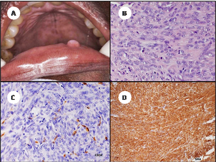
A 27-year-old black woman was referred for evaluation at the School of Dentistry of the Federal University of Maranhão complaining of a small enlargement in the lower lip mucosa. The lesion appeared as a non-ulcerated circumscribed exophytic mass measuring approximately 8 mm in diameter, which has been present for one year. The painful white nodule had a smooth surface, resilient consistency, well-defined borders, and was slightly raised from the surrounding oral mucosa (Figure 1). The patient did not describe any episode of ruptures or release of any fluid content. The rest of the oral cavity was healthy. The medical history was unremarkable. Despite this, a clinical diagnosis of mucocele versus benign tumor was made, and the lesion was surgically excised under local anesthesia. The postoperative course of the patient was uneventful after a 10 days follow-up. The tissue was completely healed, and there was no sign of a scar.
Gross examination of the formalin-fixed specimen revealed one piece of smooth brown tissue measuring on the aggregate 0.9 × 0.6 × 0.4 cm. Histological examination with hematoxylin and eosin stain showed a well-circumscribed, encapsulated mass formed by small and fusiform cells, with uniform, monochromatic, spindles nucleus, and blunt ends. There was neither nuclear atypia nor mitotic activity.
Due to the similarity of the histopathological profile with other neoplasms such as those with a fibroblastic and neural lineage, immunohistochemistry was necessary to confirm the diagnosis. The immunohistochemical study revealed intense and diffuse expression of smooth muscle actin (SMA) within the tumor cells and CD34 immunoreactivity of the endothelial cells lining the vascular spaces, indicating the presence of blood vessels (Figure 1). Based on these findings, a diagnosis of oral vascular leiomyoma was made. Nowadays, five years after the excision of the lesion, no signs of recurrence are observed.

A. clinical characteristics of the lesion, B. spindle cells with pale stained elongated nuclei and blunt termination, C. immunoreactive positivity of endothelial cells lining the vascular spaces, indicating the presence of blood vessels, D. Immunohistochemical reactivity for diffuse and strong SDMA for tumor cells, indicating the large amount of smooth muscle cells
Figure 1 Clinical and histological features of the lesion
Discussion
Oral leiomyoma is a rare tumor first described by Blanc in 1884 4-5. It is characterized by a neoplastic proliferation of mature smooth muscle cells associated with a variable amount of blood vessels 2-4-11. Its etiology is uncertain; however, some authors have associated its development with the medial tunic of smooth muscle blood vessel walls, and therefore it is called vascular leiomyoma or angioleiomyoma 12. In addition, the literature suggests other possible sources, such as heterotopic embryonic tissues, circumvallated papillae, and lingual ducts; origins that cannot be associated with the lesion presented in this report due to its location 3-13-14.
According to the literature, the majority of oral leiomyomas are observed in the lips, tongue, hard and soft palate, and with less frequency in the cheek. 15-16. Some authors report the lip as the most affected area 17, while others report the tongue 16-18-20. The small number of case series reported can explain this difference 7-10-16-21-22.
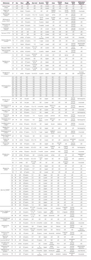
The involvement of the labial region is extremely uncommon, with only 78 biopsy-proven cases of VLL reported in the literature (Table 1). In 1959, Duhig & Ayer described the first two cases affecting the lip in a series of 61 skin cases, and the last case was reported by Matiakis et al. in 2018 23.
Although this case presented in the third decade of life, the review of the cases of VLL evidenced that 58,7% of them (n=37) occur in the fifth and sixth decades of life (Table 1), similarly to what has been reported in previous studies 7-9-10-16-24-25. Considering only the cases of lip leiomyomas, other authors reported a higher prevalence in the third and fourth decades of life 8-22-26. The age of 63 out of 78 patients could be retrieved from the literature, and their mean age was 47.7 years. Vascular leiomyomas are particularly rare in children and adolescents 27-28. In the present review, the youngest patient was 10 years old and the oldest was 83 years old, and only three cases were observed in adolescents (Table 1).
Regarding the gender, 41 cases were seen in males and 25 in females (Table 1), yielding a male to female ratio of 1.64:1. A similar gender distribution has been previously reported in the literature 7-9-29. However, some authors reported a slight predilection for females 4,5.
In the presented study, similarly to almost all previous reports of VLL, the tumor was less than 2 cm in diameter (Table 1). The size of the lesion in 55 out of 79 patients could be retrieved from the literature, and only one case reported a bigger size than 2 cm 30. According to Wang et al. (2004), the characteristic small tumor size in most vascular leiomyomas may be due to their superficial location and slowgrowing nature 9.
Pain was not a characteristic finding in VLL, and only 3 patients had this complaint besides the case described in this report (Table 1). The pain has been reported as intermittent or severe on palpation 30-32. There are three theories that try to explain the pain in leiomyomas. The first relates the pain to contraction of smooth muscle vessels, which may cause local ischemia, particularly in solid-type tumors (33). The second connect the pain to the compression of nerves accompanying the blood vessels in the lesion 34, and the third is related to a secondary mild to moderate inflammation of the tumor 1,21,34. None of these theories was definitive to explain our case.
Clinically, VLL are characterized by a small, well-delimited, superficial, and slowgrowing unspecific mass, ranging from 0.5 to 6 cm in size. The color of the lesion is varied, depending on its vascularization and its location 36. This diagnosis is hardly considered when a slow-growing, non-ulcerated asymptomatic mass is observed on the lip. Moreover, the labial lesions may present a varied clinical appearance with no specific diagnostic aspects (Table 1), which lead to a wide range of misdiagnosis, including mucocele 7-8-16-26-35-37-42, hemangioma 7-8-25-35-43-44, pleomorphic adenoma, 7-26-45-46, canalicular adenoma 7, neurofibroma 26, oral fibroma 23-26, fibrous hyperplasia 7, giant cell granuloma, pyogenic granuloma 23, hyperkeratosis, and papilloma 8.
On the lower lip, mucocele was the most common differential diagnosis observed among the VLL cases reported in the literature (78.6%), as well as one of the differential diagnoses proposed in the case described. The location of the lesions can justify this. Moreover, the rounded, spherical, oval, raised, or dome-shaped aspect of the lesion located in the submucosal layer of the lip is the characteristic presentation of this traumatic glandular lesion. On the upper lip, hemangioma (41.7%) was the most commonly reported differential diagnosis. The treatment of choice for VLL is surgical excision, and recurrences are not expected like it was observed in the present case. Despite the prominent vascular component, bleeding is rarely observed when removing the lesion 4-7-8.
Based on the histopathologic findings, the World Health Organization classified leiomyoma into three groups: solid leiomyoma, vascular leiomyoma (angioleiomyoma), and epithelioid leiomyoma (leiomyoblastoma), being the vascular leiomyomas the most prevalent in the oral cavity, as it was the case in our report 2-4-11. Histologically, the solid-type is characterized by bundles of smooth muscle cells and thin-walled vessels; the vascular type shows thick vascular channels’ walls and an arrangement of vascular musculature within the intervascular muscle bundles. The cavernous-type is composed of large vascular channels with delicate muscular walls. Some lesions may present mixed patterns, leading to a biphasic aspect 7.
Many other morphological variations have been described like areas of hemorrhage and deposits of hemosiderin, dense hyalinization of collagen, mature fat cells, and globlet-shaped endothelial cells 7-38. Foci of ossification are rare and indicates tissue degeneration, which may be due to an inadequate blood supply. Considering the histopathological aspects of leiomyoma, the abundance of spindle-shaped cells adds other benign lesions to its differential diagnoses, such as myofibroma, hemangiopericytoma, neurofibroma, neurilemmoma and schwannoma.
Hematoxylin and eosin stain is routinely used to define the diagnosis of vascular leiomyoma. Special stains such as Masson’s trichrome, Van Gieson’s stain, or Mallory’s phosphotungstic acid (PTAH) are specific for muscle cells and collagen fibers, and can also contribute in achieving the diagnosis 30-32-44-47-48. Nowadays, the confirmation of smooth muscle origin can be achieved immunohistochemically with smooth muscle markers such as SMA, desmin, HHF-35, calponin and h-caldesmon 7-23-30-32-47-53. For vascular endothelium the use of CD31, CD34, and factor VII are indicated 22. Our case demonstrated intense and diffuse expression of SMA within the tumor cells and CD34 immunoreactivity of the endothelial cell lining in the vascular spaces, indicating the presence of blood vessels and confirming the smooth muscle origin.
It is very important to differentiate vascular leiomyomas from its malignant counterpart leiomyosarcoma. The malignant lesions present myofibroblast-like cells and undifferentiated mesenchymal or fibroblast-like cells. Some immunohistochemical and molecular markers like Ki-67, p53, p16, p21, PCNA, B-cell lymphoma 2, cyclin-dependent kinase 4, and mouse double minute 2 homolog, can be used for identifying leiomyosarcoma 50-54-55.
The diagnosis of leiomyoma in the oral cavity is difficulty because the lack of suspicion by the stomatologist and the oral pathologist concerning a tumor that it is rarely encountered. Knowing the clinical, microscopical, and immunohistochemical features of VLL is essential to achieve a correct diagnosis and to perform an adequate treatment.


