


 English (pdf)
 Article in xml format
 Article references
 Send this article by e-mail
 Cited by SciELO
  Cited by Google
  Similars in
SciELO
Similars in
SciELO  Similars in Google
 Permalink
Introduction
Erosive tooth wear is a chemical-mechanical process characterized by the irreversible loss of dental hard tissue resulting from the chronic, localized action of acids and mechanical abrasive forces 1. The acids may be of intrinsic origin, arising from gastroesophageal reflux and frequent vomiting or extrinsic origin, which is related to the types of dietary habits and hygiene practices, without bacterial involvement 2,3. The complexity of this set of possible causative agents explains the fact that some individuals have a larger number of erosive lesions or of greater severity in comparison with others, in spite of having some similar risk factors 4.
People’s lifestyles have changed significantly, especially those of children and adolescents, with a much higher consumption of acidic foods and beverages being observed (2. Although preventing the consumption of acidic foods is a problem in oral health education, it is very difficult to prevent these potentially erosive substances from coming into contact with the teeth throughout a lifetime 5. Other factors that deserve emphasis are the gastroesophageal reflux diseases, as their complications also lead to the development of erosive lesions. Moreover, these tend to be aggravated in primary teeth due to the anatomic particularities, when compared with permanent teeth 6,7. Therefore, if dentists make correct diagnoses of erosive tooth wear and the probable etiological factors were well established, it would be possible to create preventive strategies. This is because erosive factors may occur in a similar manner in children’s permanent dentition (3, and their prevalence increases with age 8.
In this context, the aim of this study was to report two clinical cases of children diagnosed with erosive tooth wear, but these had different etiologies, unknown to the families, and that were associated with health problems that were initially identified by the dentist. Both cases were related to situations that caused concern and involve the lifestyle of the children and their families.
Case reports
Case 1
Patient O.L.F., a 9-year-old boy, presented to the Pediatric Dentistry Clinic of the Araraquara Dental School - UNESP, with indication for extraction of a supernumerary tooth for orthodontic purposes. On clinical oral exam no caries lesions were observed, however, various lesions characteristic of erosive tooth wear were observed in the patient’s primary teeth. The lesions were restricted to enamel, but their proximity to the dentin was evident. The mandibular teeth were more severely affected than the maxillary teeth (BEWE index scores 2 and 1, respectively). The occlusal surface had a polished, velvety aspect, and no anatomic details could be observed as a result of dental dissolution. A whitened halo could be observed around the occlusal surface delimiting enamel and dentin, in addition to “cupping lesions”, mainly in the cusp region (Figure 1).
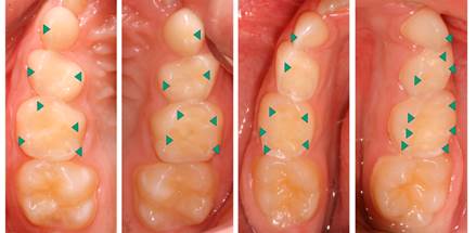
Figure 1 Occlusal surfaces with polished/velvety aspect and loss of microanatomy. Arrows indicate concave lesions with “cupping” aspect, especially in cusp regions.
The patient did not complain of hypersensitivity and no lesions were observed in the anterior teeth. During anamnesis, the mother was asked whether her son had frequent bouts of vomiting and/or belching. According to the mother’s report, the child vomited every morning, and this also caused disturbances at school, because he was frequently sent home. In addition, he had recurrent episodes that made him miss school. The school personnel and family believed that the child was creating a situation that would allow him to avoid attending classes, because in a certain medical consultation, no diagnosis of systemic change was established. Another impressive report was that the patient routinely vomited, right from the first months of life. No reports of frequent consumption of acidic foods and beverages, among other extrinsic agents, were recorded during the dental consultation. The data obtained in the anamnesis lead the dentist to believe that the erosive tooth wear was being caused by undiagnosed gastroesophageal reflux disease. However, to confirm this hypothesis the child was referred to a specialized doctor for evaluation, who confirmed our suspicion, by providing the diagnosis in a detailed medical report. Medical therapy was instituted and no further episode of vomiting was reported by the patient or guardian. In the dental clinic, the affected teeth received the application of remineralizing agents. Immediately after the first follow-up visits, a change in the child’s quality of life was observed. He told the dentist, “Doctor, I am now able to stay at school”.
Case 2
Patient R.S.S., a 10-year-old boy, presented to the Pediatric Dentistry Clinic of the Araraquara Dental School - UNESP, for a routine dental evaluation, without any reports of complaints. On clinical exam, we also noted no caries lesions, but observed lesions characteristic of erosive tooth wear in his primary and permanent teeth (BEWE index scores 1,2 and 3, respectively). Occlusal surfaces with a polished, velvety aspect and loss of microanatomy were observed, in addition to concave lesions that had a “cupping” aspect, especially in the regions of the cusps (Figure 2).
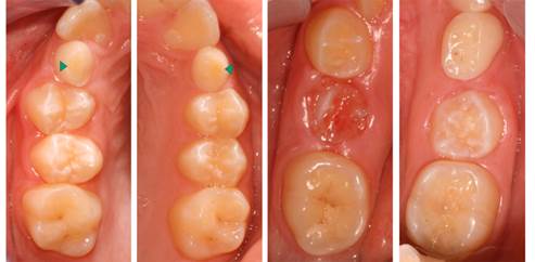
Figure 2 Occlusal surfaces with polished/velvety aspect and loss of microanatomy. Arrows in primary canines indicate the wear also attained dentin.
In the primary teeth, flat wear associated with erosive depressions were observed. In the primary canines, wear also attained the dentin (BEWE index score 3) (Figure 2). The patient did not complain of dentin hypersensitivity. No lesions were observed in the anterior teeth. During anamnesis, the patient’s mother was asked about dietary habits and gastric problems. The only report was that the child frequently consumed acidic beverages, especially sodas, without any control over the quantity ingested. This was the family’s daily routine, proved by the diet diary. At the same time, nocturnal tooth grinding and habits of playing games on tablets, smartphones and video games close to bedtime were reported, compatible with a pattern of anxiety.
Discussion
There has been growing prevalence of erosive tooth wear in the last few decades (5, which had been considered a public health problem, frequently involving the dentition of children 9,10. This has led to increased attention being paid to dental damage resulting from this process, particularly because it concerns a condition with a multifactorial etiology 7 that is difficult to diagnose in the initial stages 5.
Dentists must be alert during anamnesis and clinical exam, considering that the oral manifestations frequently are the indications that will make it possible to diagnose some of the etiological factors of the condition, such as gastroesophageal reflux disease (GERD) 11. In the clinical case of the patient who had erosive tooth wear related to intrinsic factors (GERD right from the time of early childhood), early diagnosis of erosive tooth wear was of extreme importance in minimizing the damage caused. Seeing that the patient did not report any type of dental hypersensitivity, it could have led to some delay in seeking clinical attendance. Another factor to consider was that the child had frequent episodes of vomiting, but had no proven diagnosis of GERD. During anamnesis, the health problems the child had were found, which made it possible to refer him for specialized medical attention and final diagnosis of gastroesophageal reflux disease. It must be taken into consideration that reflux may occur without symptoms, therefore, patients with erosive tooth wear must be evaluated relative to the possibility of having gastroesophageal reflux disease 5.
Gastroesophageal reflux disease is characterized by an involuntary movement of the esophageal sphincter, allowing acid reflux from the stomach through the esophagus into the oral cavity 11. This being so, the stomach acid can come into contact with the teeth as a consequence of chronic vomiting or reflux. In the present case, when the patient with intrinsic erosive tooth wear was evaluated by the doctor, he was found to have frequent episodes of vomiting, and was instructed to take medication to prevent repetition of this condition. The severity of erosive tooth wear is related to the frequency of reflux or vomiting, pH level, type of acid and protective capacity of saliva 12.
The acid coming into contact with the tooth surface causes dental demineralization. In addition to being of intrinsic origin, this acid could also arise from extrinsic sources, coming from the foods consumed. Eating habits represent an important factor in the development of tooth tissue loss 13,14. When evaluating the eating habits of the patient with extrinsic tooth erosion, a high frequency of consuming sodas and acidic juices could be observed. This high consumption of acidic foods, such as sodas, teas, sports drinks, salad dressings, among other foods, are considered potentially contributory factors to the development of and increase in erosive tooth wear 15.
Tooth surface wear may be accelerated by the presence of acids in the oral cavity associated with some stimulatory factors such as attrition between the teeth and abrasion 16. As seen in the present clinical case, the patient with erosive tooth wear related to extrinsic factors also had the habit of tooth grinding during the night.
In spite of the fact that neither of the two children were evaluated by a specialist to define the diagnosis of anxiety disorder, evidences have pointed out that the association of different lifestyles had a great influence on the occurrence and aggravation of the erosive lesions 17,18. As observed in this clinical case report, the patient with extrinsic erosive lesions consumed an excessive quantity of acidic beverages, had a habit of tooth grinding in the nocturnal period, as well as the practice of playing games on electronic devices, at all times, before going to sleep. There is evidence that anxiety is an outstanding contemporary disturbance as a result of the agitated world we live in, full of tensions that end up being reflected in the routine of children. This disturbance has affected the population in a general way, irrespective of age. However, increasing numbers of children have been affected by psychic disturbances of this nature. Anxiety in children is a reflection of the inconsistencies in the environment, both their predictable and unpredictable variables, and are also associated with their parents’ anxieties. In this sense, there may also be both a genetic and environmental influence 19,20.
The absence of early diagnosis of erosive tooth wear and changes in etiological factors result in a sever loss of dental structure 7. In the cases presented, the routine exam performed enabled the identification of erosive lesions in the initial stages, recognition of the risk factors, as well as institution of preventive measures and healthy habits in patients. Therefore, this case report showed evidence of the important role of dentists, not only relative to the oral cavity, but also in improving patients’ general state of health.
Conclusion
It was concluded that dentists play an important role, in helping with the diagnosis of health problems related to erosive tooth wear. Therefore, meticulous clinical exams must be performed and information collected in anamnesis to establish the correct diagnosis, because swift intervention in the clinical cases presented had a positive impact on halting the erosive process.

