


Services on Demand
Journal
Article
 text in
  English (pdf)
 Article in xml format
 Article references
 Send this article by e-mail
Indicators
-
 Cited by SciELO
 -
 Access statistics
Related links
-
 Cited by Google
 -
 Similars in
SciELO
Similars in
SciELO -
 Similars in Google
Share

 Permalink
Revista colombiana de Gastroenterología
Print version ISSN 0120-9957On-line version ISSN 2500-7440
Rev Col Gastroenterol vol.25 no.1 Bogotá Jan./Mar. 2010
Biliary fistula and Bouverets Syndrome. The same but different
Two case descriptions and a literature review
Luis Fernando Álvarez Chica, MD (1), Walter Bejarano Cuéllar, MD (2), Olga Lucía Rojas Cardozo, MD (3)
(1) General Surgeon. Mario Correa Rengifo Departmental Hospital and Imbanaco Medical Center. Cali, Colombia.
(2) Gastrointestinal Surgeon. Imbanaco Medical Center. Cali, Colombia.
(3) General Surgeon. Mario Correa Rengifo Departmental Hospital and Imbanaco Medical Center. Cali, Colombia.
Received: 26-02-10 Accepted: 02-03-10
Summary
Biliary fistula is an infrequently occurring disease in which gall stones pass from the gall bladder to the duodenum causing intestinal obstruction. In most cases the stones impact is at the end of the small intestine. When the stones impact in the duodenum it is called Bouverets syndrome. We present a case study of a patient with acute cholecystitis and another case study of Bouverets syndrome, together with a summary symptoms, diagnostic techniques, and treatment.
Key words
Laparoscopic cholecystectomy, cholecytitis acute, biliary fistula, intestinal obstruction, Bouveret syndrome.
CASE 1: BILIARY FISTULA
An 86 year old male patient with a one month history of the development of colicky pain accompanied by vomiting and diarrhea consulted a private physician who initially treated patients symptoms and requested ultrasound of patients liver and gallbladder. Sonograms showed acute cholecystitis and a hepatized gallbladder with multiple calculi in its interior. Surgery was recommended but the patient did not consult the doctor out of fear. When he entered the Imbanaco medical center 10 days later he had abdominal pain and vomiting. The patient was dehydrated, apparently acutely ill, and had sharp pain upon deep palpation of the right hypochondrium. He was positive for Murphys sign. Blood tests showed leukocytosis and neutrophilia. Diagnosed with acute cholecystitis caused by cholelithiasis, a laparoscopic cholecystectomy was performed. During surgery a peritoneal mass in the gall bladder and cholecystoduodenal fistula with a 4 cm gallstone were found near the ileocecal valve. The gallbladder mass was removed, the cholecystoduodenal fistula was resected, the duodenum was sutured, and a cholecystectomy and an enterotomy were performed in the distal ileum to extract the biliar calculi, all through one stage laparoscopy (Clinical video of the National Congress of Surgery). The patient had no postoperative complications.
CASE 2: BOUVERETS SYNDROME
A 67 year old male with a 3 month history of poor appetite, nausea without vomiting, sensation of fullness, weight loss, epigastralgia, heartburn and melenas on three occasions. Upon physical examination patient was pale, thin and in general not in good condition. His abdomen had no masses or organomegaly, but a gastric succussion was felt.
The patient gave the impression of having gastric cancer. An initial endoscopy showed grade C esophagitis, hiatal hernia, changes from chronic gastritis, and a duodenal lesion compatible with advanced neoplasias. Biopsies showed an acute ulcer. An abdominal CAT scan showed a lesion located in a topographic area of the gallbladder with images suggesting calculi and air content in its interior. These symptoms suggested cholecystitis with cholecystoduodenal fistula.
The presence of a tumor-like lesion eroding both the gallbladder and the second portion of the duodenum was not ruled out. There was no dilatation of the bile duct, and the liver was normal. A new upper endoscopy was performed which showed a giant calculus in the duodenum which impeded passage towards the distal part of the instrument. This obstruction was initially interpreted as a neoplastic lesion. A laparotomy was performed in which a large inflammatory peritoneal mass was found in the subhepatic area. It involved both the gallbladder and duodenum. Cholecystoduodenal fistula secondary to severe cholecystitis with two mayor 5 cm calculi was found. An exploratory laparotomy, a wide Kocher maneuver, a duodenotomy distal to the calculi, and a cholecystectomy were performed. The giant calculus and the other calculi were extracted and removed, and a T tube was placed in the gallbladder. (Neither cholecystectomy nor resection of the cholecystoduodenal fistula were performed). There were no post operative complications. 60 days later, the T tube was removed by cholecystectomy following the cholangiography.
INTRODUCTION
Gallstone ileus, also known as biliary ileus, is an uncommon cause of intestinal obstruction that affects older people. It is caused after one or more gallstones pass through a biliary-enteric fistula and impact in the small intestine. On very rare occasions the calculi can enter the intestine through fistulous communication between a choledocal cyst and the digestive tract (1). The term "biliary ileus" was first coined by Bartholin in 1654 (2). The diagnosis is normally late because symptoms tend to be intermittent. Treatment is either surgical or endoscopic removal.
The most common places for calculi to impact are the terminal ileus and the ileocecal valve. The least common place for the calculi to impact are the jejunum, the ligament of Treitz and the stomach, although it is also rare for them to impact the duodenum and colon (3).
When the gastric outlet is obstructed by one or more calculi impacted in the duodenum or the pylorus, this condition is called Bouverets Syndrome. This is in tribute to the French internist Leon Bouverets who described this syndrome in which the patient has epigastric pain, nausea, persistent vomiting, weight loss and general discomfort (4). A particular type of gallstone ileus occurs when the obstruction is in the duodenal bulb. This occurs in 3% of all cases in which a calculus impacts in the digestive tract. This figure represents 1% to 3% of all causes of intestinal obstruction. It was first described in 1770 by Beaussier (5-8). Until the year 2000, 175 cases had been reported in the literature for Bouverets syndrome (9).
INCIDENCE
Biliary fistula is a rare complication of the cholelithiasis which occurs in between 0.3% and 0.4% of patients. It is seven times more frequent in people who are over 70 years old. It is responsible for from1% to 4% of cases for mechanical obstruction, and it is responsible for 25% of intestinal obstructions without strangulation in patients older than 65 years.
The average age in patients with gallstone ileus is 70 years, and its incidence is greater in females. In a review of 128 cases of Bouverets syndrome the average patient was 74 years old. There were twice as many cases among women as among men (2:1 ratio) (10).
PATHOGENESIS
The way in which the calculus enters the intestine is through a biliary enteric fistula causing complications of cholecystitis associated episodes for 2% to 3% of patients with cholelithiasis. Between 53% and 68% of the fistulas are cholecystoduodenal. Least frequent are cholecystocolonic (5%) and cholecystogastric. The factors that favor formation of fistulas are size of the calculi 2cm to 8 cm, a long history of biliary disease, repeated episodes of acute cholecystitis, female gender and advanced age. In classic gallstone ileus, the calculi impact in the distal ileus in 73% to 90% of all cases. They impacts in the colon in 3% to 25% of cases, and only in 3% to 10% do they impact in the duodenum . These last instances are classified as Bouverets Syndrome (11).
Pericholecystic inflammation after cholecystitis allows formation of adhesions between the gallbladder and the intestine and necrosis due to the pressing of the calculus against the gallbladder. This results in the formation of the fistula (gangrene of the gallbladder). In other rare cases, the biliary ileus occurs after an endoscopic sphincterotomy (12) (Figure 1).
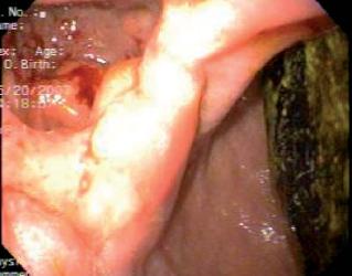
Figure 1. Patient with Bouverets Syndrome. Endoscopic vision of the calculus in the duodenum and the cholecystoduodenal fistula.
Once in the digestive tract, the calculus can be vomited, it can spontaneously pass through the rectum or it can impact and cause an intestinal obstruction. While the calculus advances in the intestine, its diameter increases due to the accumulation of debris and intestinal sediment on its surface. The vast majority of the calculi are more than 25 mm wide. 50% to 70% impact in the distal ileus which is the narrowest segment of the small intestine, and the place where there is less peristalsis. Obstruction of the colon happens when there is a preexisting pathology such as stenosis due to diverticulitis (13). The physician must take into account that several calculi may be found throughout the intestine.
Once in the intestinal lumen, the calculus can manifest in many ways depending on its size, the digestive tract segment involved upon whether or not there is any preexisting intestinal pathology. Hence, the calculus can be symptomatic or it can be eliminated by feces of vomit. The onset of symptoms may be acute, intermittent or episodic chronic pain. Occasionally, a calculus can enter the intestine through a fistulus route between the bile duct and the digestive tract (14).
CLINICAL MANIFESTATIONS
The classic presentation of gallstone ileus is in an adult woman with episodes of sub-acute intestinal obstruction. The transitory impact of the calculus produces abdominal pain and vomiting which alleviate the patients pain by moving the calculus and allowing it to continue its distal advance. This results in vague and intermittent symptoms that can persist several days before there is an assessment in the emergency room. In Bouverets syndrome, the main symptom is vomiting accompanied by epigastric or right hypochondrium pain. Other symptoms appear infrequently. They include digestive hemorrhaging secondary to duodenal erosion, the expulsion of a bile calculus while vomiting and/or the presence of esophageal lesions associated with an intense emesis. The biliary ileus rarely triggers perforation of the jejunum (15).
Upon physical examination the patient may have fever, dehydration, distension and abdominal pain, and increased peristalsis. Jaundice is rare, but occurs in at least 15% of patients. Less than 20% of patients have acute cholecystitis.
Laboratory study can show leukocytosis and altered acid-base, hydroelectrolitic and renal functions. The magnitude of these will depend on associated diseases, the degree of inflammatory response and each individuals compensatory mechanisms. Less frequently, hepatic tests may show alterations and plasma amylase elevation (16).
DIAGNOSIS
Generally, diagnosis is late as more than half of the patients have no history of vesicular disease and biochemical alterations resulting from ileus are not specific. A patient can live with leukocytosis, hydro-electric disequilibrium and elevated aminotransferases. Diagnosis for gallstone ileus tends to be difficult, generally depending on radiological findings. In 50% of the cases, the diagnosis is made during laparotomy (17).
A simple abdominal x-ray is of great value. 35% of patients with biliary fistula have Riglers triad: partial or complete intestinal obstruction, air in the bile duct and calculus visible in the x-ray (18). Other radiological signs are changes in a previously located calculus and two adjacent hydro-aerial levels in the right hypochondrium.
Two out of these first three findings are encountered in more than 50% of all patients with gallstone ileus. Pneumobilia occurs in up to 60% of patients but, it is not specific since it may occur in patients with Sphincter of Oddi or patients who have had an endoscopic sphincterotomy.
Less than 15% of calculi are visible in a simple abdominal x-ray because most of them are radiolucent. Studies by means of contrast are useful for identifying the degree of intestinal obstruction and for finding the biliary enteric fistula.
Abdominal CT scans are the most important diagnostic tests due to their superior resolution and ability to identify a calculus in 78% of patients. In addition they can show pneumobilia, intestinal obstruction and the thickness of the bile gallbladders wall. Nonetheless, it is not possible to observe the calculus with a CT scan in 15% to 25% of patients (19). Lassandro et al. compared the clinical values of simple abdominal x-rays, abdominal ultrasound and the abdominal CT scans in the diagnosis of 27 gallstone ileus cases. They found that Riglers triad is observable on 14.81% of simple abdominal x-rays, in 11.11% of abdominal sonograms and in 77.78% of abdominal CT scans (20-23). As noted, other imaging studies such as ultrasound have limitations when faced with anatomical alterations, abdominal distension, air in the gallbladder, or collapse of the gallbladder. Likewise, scintigraphy is rarely useful due to its multiple technical limitations (Figure 2).
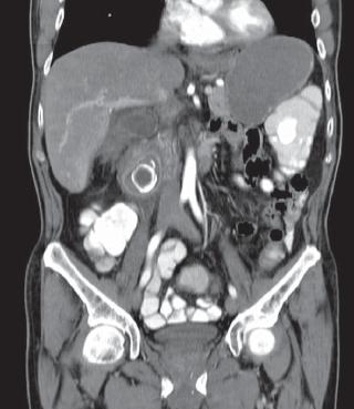
Figure 2. Patient with Bouveret’s syndrome. Tomographic image of the calculus in the duodenum.
Endoscopy also has a relative value because it rarely shows an impacted calculus in the duodenum, although Endoscopic Retrograde Cholangiopancreatography (ERCP) can show a fistula if the gallbladder can be filled with a contrast medium during examination.
TREATMENT
The patients hydro-electrolytic compensation is of vital importance. In addition to the patients hemodynamic resuscitation, it is very important to take her/his general state of health, including associated diseases, into account. Sometimes it is preferable to first perform the enterotomy and the extraction of the calculus, and later perform the cholecystostomy and the resection of the bile-digestive fistula in a second surgical stage.
The main objective of treatment is the extraction of the calculus to overcome the intestinal obstruction. This can be done with endoscopy in the case of an impacted calculus in the duodenum (Bouverets syndrome), surgically or through laparoscopy. Endoscopic extraction has an important role in this pathology because it is less invasive and is associated with fewer complications. Nevertheless, it is difficult and requires endoscopists trained in therapeutic procedures. Endoscopy is more likely to be successful in selected patients with intermediate size and relatively mobile calculi. Patients with big or firmly impacted calculi are probably better candidates for surgical resolution (24, 25).
Generally, the choice of therapeutic strategy takes into account the patients age, comorbidity, the effect of the obstruction on the patients general health, the size of the calculi and of the fistula, local inflammatory changes and the obstructions location. It has been proven that the length of the surgical intervention does not influence the subsequent evolution, but that evolution can be influenced by delay in establishing the correct diagnosis and by unjustified delay of intervention (26) (Figure 3).
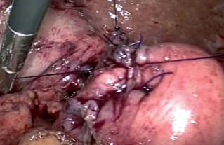
Figure 3. Laparoscopic vision of the patient with biliary ileus. Suture of the fistula in the duodenum.
One-stage surgery (complete lithotomy, fistula closure and cholecystomy) is very controversial. One-stage surgery proponents believe it prevents future complications such as more episodes of acute cholecystitis, cholangitis and recurrent biliary ileus (19). Some authors report good experiences with this procedure and suggest that complete lithotomies performed alone should be reserved for unstable patients and difficult cases. Nonetheless, other studies have shown that complications occur in up to 66% in patients who receive one-stage surgeries. These studies have suggested that this procedure should be reserved for selected low risk patients (27).
It has been argued that one-stage surgery significantly decreases morbidity and mortality since the removal of the gallbladder and the closure of the bile-enteric fistula prevents future recurrence of gallstone ileuses, other recurrent biliary symptoms, the associated morbidity and mortality, and the obvious necessity of a second operation (15).
The preference of some for performing only complete lithotomies to extract calculi should not be generalized. This procedure should be reserved for patients in critical conditions and those cases where local inflammatory changes make the intervention extremely difficult and predispose patient to intraoperative or perioperative complications. Those who defend performing complete lithotomies alone justify their decision by the resulting decreased mortality rate of 12% compared to the 20% to 30% range of mortality rates in cholecystomies combined with fistula closures. They also point to the fact that for a cholecystoduodenal fistula it can work as a biliary-digestive anastomosis if the cystic conduct is permeable. Aside from being safer in low and high risk patients, an complete lithotomy alone requires less surgical time than one-stage surgery, it is technically less demanding and it can be combined with an elective cholecystomy if biliary symptoms persist. Moreover, in most cases complete lithotomy alone is adequate treatment for patients of advanced age for whom subsequent cholecystomy is not mandatory (28, 29).
Complete lithotomy
By means of laparoscopy or laparotomy, a longitudinal lithotomy is performed along the entire proximal antimesenteric edge (10 to 15cm) to the point of impact of the calculus, but not over the impact area. It is then drained and removed. A careful transversal closure of the enterotomy is required to avoid residual intestinal stenosis. Manipulation of the calculus in the struggle to make it advance towards the cecum has been associated with mucosal lesions and/or the tearing and rupture of the serous. For this reason it should not be performed routinely unless it is an easy and expeditious maneuver. Intestinal resection can be required when there is perforation, significant ischemia or when a calculus cannot be removed (30) (Figure 4).
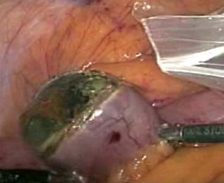
Figure 4. Laparoscopic vision of the patient with biliary ileus. Enterotomy and extraction of the calculus in the distal ileus.
The entire small intestine should be checked for more calculi since multiple calculi are found in 16% of all cases. In case of Bouverets syndrome, when it is possible calculi in the duodenum should be displaced towards the stomach for later extraction through gastronomy. Closure or repair should be preformed over healthy tissue. In patients in whom this maneuver is not possible, a duodenotomy should be performed, placing special emphasis on closure to avoid stenosis (31).
Biliary Surgery
Definitive biliar surgery combining cholecystomy and resection of the biliary-digestive fistula decreases recurrent gallstone ileus which is present in up to 17% of those patients treated with complete lithotomy alone. It also prevents the emergence of acute cholecystitis, cholangitis and gallbladder carcinoma. The last occurs in up to 15% of patients with biliary-enteric fistulas although it occurs in only 0.3% of patients whose gallbladders have been removed for other reasons (32, 33). Persistent fistulas that can cause weight loss and deficient absorption can also be prevented with one-stage surgery (Figure 5).
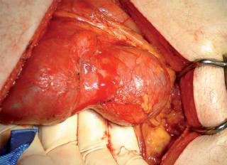
Figure 5. Patient with Bouverets syndrome. Mobilization of the distal duodenum to the cholecystoduodenal fistula and the exposure of the calculi.
In the largest review of biliary ileus to date, covering 1001 cases, Reisner et al. reported that one-stage surgery leads to a 16.9% mortality rate compared with an 11.7% rate for complete lithotomy alone. Their study also showed a recurrence for gallstone ileus of 4.7% with only 10% of patients requiring reoperations for symptoms related to the biliary tract. 57% of the recurrences occurred within six months of the initial surgery (Figure 6).
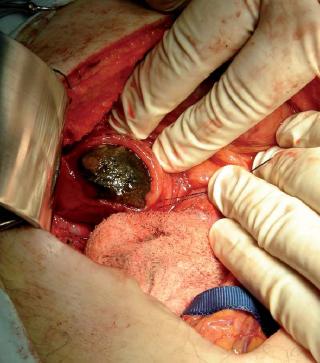
Figure 6. Patient with Bouveret’s syndrome. Duodenotomy and extraction of the calculi.
The following observations were also noted. Cholecystomy alone does not protect against all recurrences in biliary patients for two reasons: choledocal calculi can migrate distally and produce new intestinal obstructions and, most importantly, calculi within the intestine that are not detected during surgery can produce a recurrence of biliary ileus.
Choledocal lithiasis occurs in 15% of patients whose gallbladders were not removed.
Biliary enteric fistulas can spontaneously close, especially if the cystic duct is permeable and when there are no residual calculi.
It is very important to take into account that extensive dissection and the greater anesthesia time required for one-stage surgery can further compromise a critically ill patient (34).
Mortality rates range between 4.5% and 25%. Mortality is generally due to delayed diagnosis and concomitant diseases including cardiopulmonary complications, obesity, and diabetes mellitus. These rates are ten times higher than for any other cause of intestinal obstruction of none-malignant etiology. Hence, the one-stage procedure should be performed only in very specific patients whose risk is low (Figure 7).
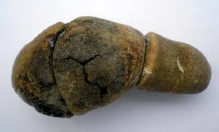
Figure 7. Patient with Bouveret’s syndrome. The calculi that molded the gallbladder. The two on the left were found in the duodenum and the one on the right was found in the gallbladders’ bottom.
Similar mortality rates were reported in a recent study which concluded that urgent repair of the fistula is associated with a high percentage of complications. That study found that the mortality rate for the one-stage procedure is two times greater than for complete lithotomy alone (35-40).
Despite large breakthroughs in perioperative care, gallstone ileus mortality remains high (15% to 18%). This implies that the population with this pathology is on the borderline of life suffering from multiple medical problems, and it means that the vast majority of these patients are classified from ASA III to ASA IV (41, 42).
Reports have recently appeared of totally laparoscopic management of gallstone ileus, complete lithotomy through laparoscopy and one-stage laparoscopic resolution through laparoscopy as in the case described by us (43-47).
Nonsurgical treatment
Non- surgical treatment is required for patients at very high risk from surgery. Extracorporeal lithotripsy, electro-hydraulic lithotripsy and lasers for impacted calculi in the stomach and duodenum have all been reported. The endoscopic removal of calculi in the colon and the duodenum has also been reported, nonetheless, surgery continues to be the primary recommended therapy (48-50).
CONCLUSIONS
Biliary ileus is an important, though infrequent, cause of mechanical obstructions of the intestines that affects elderly patients with associated diseases. It is caused by the impact of a calculus in the distal ileus after it passes through a biliary-enteric fistula.
Bouverets syndrome is a gallstone ileus that occurs in the duodenum or the pylorus.
The clinical chart for biliary ileus is characterized by sub-acute intestinal obstruction in an elderly woman. The transitory impact of the calculus produces abdominal pain and vomiting that is relieved when the calculus becomes disconnected, only to reoccur when it reimpacts more distally. This results in vague and non-specific symptoms that can be present for several days before definitive diagnosis.
The diagnosis can be suggested by a simple abdominal x-ray, though a CT scan can visualize the impacted calculus better.
Diagnosis and resolution of the intestinal obstruction after adequate repletion of liquids in the patient are essential for the treatment of gallstone ileus.
If the patients condition allows it, one-stage surgery (cholecystomy, cholecystoduodenal resolution of the fistula and complete lithotomy) should be performed.
If the patient is in a critical state, complete lithotomy alone should be performed.
Currently, either of the two pathologies described are susceptible to treatment with laparoscopy.
References
1. Keaveany AP, Afdhal NH. Gallstone ileus. UptoDate. Mayo 31 2008.
2. Chou JW, Chang HH, Kuan FL. Gallstone ileus: report of two cases and review of the literature. World J Gastroenterol 2007; 13(8): 821-25.
3. Kasahara Y, Umemura H, Shiraha S. Gallstone ileus: Review of 112 patients in the Japanese literature. Am J Surg 1980; 140: 437-40.
4. Bouveret L. Sténose du pylore adhérent a la vésicule. Rev Med (París) 1896; 16: 1-16.
5. Iñiguez A, Butte JM, Zúñiga JM. Síndrome de Bouveret. Resolución endoscópica y quirúrgica de cuatro casos clínicos. Rev Med Chile 2008; 136:163-68.
6. Rojas J, Cabañe P, Hernández J. Síndrome de Bouveret. Caso clínico y revisión de la literatura. Rev Chil Cir 2006; 67: 508-10.
7. Ferreira LE, Topazian MD, Baron TH. Bouverets Syndrome: diagnosis and endoscopic treatment. Clin Gastroenterol Hepatol 2008; 6: e15.
8. Kishi K, Yamada K, Sugiyama T. Gastric outlet obstruction caused by a large gallstone in the duodenum (Bouverets syndrome). Clin gastroenterol Hepatol 2008; 6: e11.
9. Masannat YA, Caplin S, Brown T. A rare complication of a common disease: Bouverets syndrome, a case report. World J Gastroenterol 2006; 12(16): 2620-21.
10. Cappell M, Davis M. Characterization of Bouverets Syndrome: A Comprehensive review of 128 Cases. Am J Gastroenterol 2006; 101 (9): 2139-46.
11. Chong KA, Leong YP. Gastric outlet obstruction due to gallstones (Bouverets syndrome). Postgrad Med J 1987; 63: 909-10.
12. Iancu C, Bodea R, Hajjar NA. Bouverets Syndrome Associated with Acute Gangrenous Colecystitis. J Gastrointestin Liver Dis 2008; 17(1): 87-90.
13. Zakir M, Balupuri S, Boobis L. Colonic gallstones: a case report. Hepatobiiary Pancreat Dis Int 2007; 6: 324-25.
14. Clavian PA, Richon J, Burgan S. Gallstone ileus. Br J Surg 1990; 77(7): 737-42.
15. Browning L, Taylor JD, Clark SK. Jejunal perforation in gallstone ileus: a case series. J Med Case Reports 2007; 1: 157.
16. Ayantunde AA, Agrawal A. Gallstone ileus: diagnosis and management: World J Surg 2007; 31: 1292-97.
17. Lobo DN, Jobling JC, Balfour TW. Gallstone ileus: diagnostic pitfalls and therapeutic successes. J Clin Gastroenterol 2000; 30: 72-6.
18. Rigler et Al. Gallstone obstruction: Pathogenensis and roentgen manifestation. J Am Med A 1941; 117: 1753-59.
19. Lassandro F, Romano S, Ragozzino A. Role of Helical CT in Diagnosis of Gallstone ileus and related Conditions. A J R 2005; 185: 1159-65.
20. Lassandro R, Gagliardi N, Scuderi M. Gallstone ileus analysis of radiological findings in 27 patients. Eur J Radiol 2004; 50: 23-9.
21. Balthazar EJ, Schechter LS. Air in gallbladder: a frequent finding in gallstone ileus. A J R 1978; 131: 219-22.
22. Furukawa A, Yamasaki M, Furuichi K. Helical CT in the diagnosis of small bowel obstruction. Radiographics 2001; 21: 341-55.
23. Yu CY, Lin CC, Shyu RY. Value of CT in diagnosis and management of gallstone ileus. World J Gastroenterol 2005; 11: 2142-47.
24. Langhorst J, Schumacher B. Deselaers T. Successful endoscopic therapy of a gastric outlet obstruction due a gallstone with intracorporeal laser lithotripsy: a case of Bouverets syndrome. Gastrintestinal Endosc 2000; 51: 209-13.
25. Dumonceau J, Delhaye M, Deviere J. Endoscopic treatment of gastric outlet obstruction caused by gallstone (Bouverets syndrome) after extracorporeal shock wave lithotripsy. Endoscopy 1997; 29: 319-21.
26. Liew V, Layani L, Speakman D. Bouverets syndrome in Melbourne: ANZJ Surg 2002; 72: 161-63.
27. Rodriguez-San Juan JC, Casado F, Fernandez MJ. Cholecystectomy and fistula closure versus enterolithotomy alone ileus. Br j Surg 1997; 84: 634-7.
28. Doko M, Zovak M, Kopljar M. Comparison of surgical treatments of gallstone ileus: preliminary report. World J Surg 2003; 27: 400-4.
29. Zuegel N, Hehl A, Lindemann F. Advantages of one-stage repair in case of gallstone ileus. Hepatogastroenterology 1997; 44: 59-62.
30. Tan YM, Wong WK, Ooi LL. A comparison of two surgical strategies for the emergency treatment of gallstone. Singapore Med J 2004; 45: 69-72.
31. Abou-Saif A, Al-Kawas FH. Complications of gallstone disease. Mirizzi syndrome, cholecystocholedochal fistula and gallstone ileus. Am J Gastroenterol 2002; 97: 249-54.
32. De Aretxabala X, Riedeman P, Burgos L. Cáncer de la vesícula biliar. Estudio de casos y controles. Rev Med Chile 1995; 123: 581-6.
33. De Aretxabala X, Roa I, Burgos L. Cáncer de la vesícula biliar. Algunas consideraciones. Rev Med Chile. 1996; 124: 732-9.
34. Reisner R, Cohen J. Gallstone ileus: a review of 1001 reported cases. Am Surg 1994; 60: 441-6.
35. Kurtz R, Heimann T, Beck A. Patterns of treatments of gallstone ileus over a 45-year period. Am J Gastroenterol 1985; 80: 95-8.
36. Frattaroli F, Reggio D, Guadalaxara A. Bouverets syndrome: case report and review of the literature. Hepatogastroenterology 1997; 44: 1019-22.
37. Roa GA, Jiménez H. Íleo biliar. Presentación de cinco casos. Rev Colomb Cir 1993; (1): 67-72.
38. Hepp KJ, Vélez FR, Peralta M. Íleo biliar: Manejo de la patología. Rev Chil Cir 1998; 40(1): 415-5.
39. Rojas OL, González A, Salazar J. Enfermedad vesicular: Diagnóstico, complicaciones y mortalidad en el HUV. Colomb Méd 1984; 15(3): 115-9.
40. Hernández VM, Bautista J, Orozco G. Íleo biliar. Presentación de 18 pacientes. Rev Gastroenterol Mex 1982; 47(4): 211-6.
41. Gutiérrez VP, Crippa HC, Rosano O. Íleo biliar. Rev Argent Cir 1984; 47(1/2): 32-9.
42. García SM, González GH, Téllez FI. Fístula bilioentérica con impactación de lito gigante en yeyuno. Rev Gastroenterol Mex 2008; 73(4): 235-8.
43. Soto DJ, Evan SJ, Kavic MS. Laparoscopic management of gallstone ileus: JSLS 2001; 5: 279-85.
44. Allen JW, Mc Curry T. Rivas H. Totally laparoscopic management of gallstone ileus. Surg Endosc 2003; 17(2): 352.
45. Franklin ME Jr, Dorman JP, Schuessler WW. Laparoscopic treatment of gallstone ileus: a case report and review of the literature: J Laparoendosc Surg 1994; 4: 265-72.
46. Agresta F, Bedin N. Gallstone ileus as a complication of acute cholecystitis. Laparoscopic diagnosis and treatment. Surg Endosc 2002; 16: 1637.
47. Sica GS, Sileri P, Gaspari AL. Laparoscopic treatment of Bouverets syndrome presenting as acute pancreatitis. JSLS 2005; 9: 472-75.
48. Rivera Irigoin R, Ubina Aznar E, García Fernández G. Síndrome de Bouveret resuelto mediante litotricia mecánica endoscópica. Rev Esp Enfer Dig 2006; 98: 789-98.
49. Bedogni G, Contini S, Meinero M. Pyloroduodenal obstruction due to a biliary stone (Bouveret`s syndrome) managed by endoscopic extraction. Gastrointest Endosc 1985; 31: 36-8.
50. Meyemberg C, Michel C, Metzger U. Gallstone ileus treated by extracorporeal shock-wave lithotripsy. Gastrointest Endosc 1996; 43: 508-511.
1. Keaveany AP, Afdhal NH. Gallstone ileus. UptoDate. Mayo 31 2008. [ Links ]
2. Chou JW, Chang HH, Kuan FL. Gallstone ileus: report of two cases and review of the literature. World J Gastroenterol 2007; 13(8): 821-25. [ Links ]
3. Kasahara Y, Umemura H, Shiraha S. Gallstone ileus: Review of 112 patients in the Japanese literature. Am J Surg 1980; 140: 437-40. [ Links ]
4. Bouveret L. Sténose du pylore adhérent a la vésicule. Rev Med (París) 1896; 16: 1-16. [ Links ]
5. Iñiguez A, Butte JM, Zúñiga JM. Síndrome de Bouveret. Resolución endoscópica y quirúrgica de cuatro casos clínicos. Rev Med Chile 2008; 136:163-68. [ Links ]
6. Rojas J, Cabañe P, Hernández J. Síndrome de Bouveret. Caso clínico y revisión de la literatura. >Rev Chil Cir 2006; 67: 508-10. [ Links ]
7. Ferreira LE, Topazian MD, Baron TH. Bouverets Syndrome: diagnosis and endoscopic treatment. Clin Gastroenterol Hepatol 2008; 6: e15. [ Links ]
8. Kishi K, Yamada K, Sugiyama T. Gastric outlet obstruction caused by a large gallstone in the duodenum (Bouverets syndrome). Clin gastroenterol Hepatol 2008; 6: e11. [ Links ]
9. Masannat YA, Caplin S, Brown T. A rare complication of a common disease: Bouverets syndrome, a case report. World J Gastroenterol 2006; 12(16): 2620-21. [ Links ]
10. Cappell M, Davis M. Characterization of Bouverets Syndrome: A Comprehensive review of 128 Cases. Am J Gastroenterol 2006; 101 (9): 2139-46. [ Links ]
11. Chong KA, Leong YP. Gastric outlet obstruction due to gallstones (Bouverets syndrome). Postgrad Med J 1987; 63: 909-10. [ Links ]
12. Iancu C, Bodea R, Hajjar NA. Bouverets Syndrome Associated with Acute Gangrenous Colecystitis. J Gastrointestin Liver Dis 2008; 17(1): 87-90. [ Links ]
13. Zakir M, Balupuri S, Boobis L. Colonic gallstones: a case report. Hepatobiiary Pancreat Dis Int 2007; 6: 324-25. [ Links ]
14. Clavian PA, Richon J, Burgan S. Gallstone ileus. Br J Surg 1990; 77(7): 737-42. [ Links ]
15. Browning L, Taylor JD, Clark SK. Jejunal perforation in gallstone ileus: a case series. J Med Case Reports 2007; 1: 157. [ Links ]
16. Ayantunde AA, Agrawal A. Gallstone ileus: diagnosis and management: World J Surg 2007; 31: 1292-97. [ Links ]
17. Lobo DN, Jobling JC, Balfour TW. Gallstone ileus: diagnostic pitfalls and therapeutic successes. J Clin Gastroenterol 2000; 30: 72-6. [ Links ]
18. Rigler et Al. Gallstone obstruction: Pathogenensis and roentgen manifestation. J Am Med A 1941; 117: 1753-59. [ Links ]
19. Lassandro F, Romano S, Ragozzino A. Role of Helical CT in Diagnosis of Gallstone ileus and related Conditions. A J R 2005; 185: 1159-65. [ Links ]
20. Lassandro R, Gagliardi N, Scuderi M. Gallstone ileus analysis of radiological findings in 27 patients. Eur J Radiol 2004; 50: 23-9. [ Links ]
21. Balthazar EJ, Schechter LS. Air in gallbladder: a frequent finding in gallstone ileus. A J R 1978; 131: 219-22. [ Links ]
22. Furukawa A, Yamasaki M, Furuichi K. Helical CT in the diagnosis of small bowel obstruction. Radiographics 2001; 21: 341-55. [ Links ]
23. Yu CY, Lin CC, Shyu RY. Value of CT in diagnosis and management of gallstone ileus. World J Gastroenterol 2005; 11: 2142-47. [ Links ]
24. Langhorst J, Schumacher B. Deselaers T. Successful endoscopic therapy of a gastric outlet obstruction due a gallstone with intracorporeal laser lithotripsy: a case of Bouverets syndrome. Gastrintestinal Endosc 2000; 51: 209-13. [ Links ]
25. Dumonceau J, Delhaye M, Deviere J. Endoscopic treatment of gastric outlet obstruction caused by gallstone (Bouverets syndrome) after extracorporeal shock wave lithotripsy. Endoscopy 1997; 29: 319-21. [ Links ]
26. Liew V, Layani L, Speakman D. Bouverets syndrome in Melbourne: ANZJ Surg 2002; 72: 161-63. [ Links ]
27. Rodriguez-San Juan JC, Casado F, Fernandez MJ. >Cholecystectomy and fistula closure versus enterolithotomy alone ileus. Br j Surg 1997; 84: 634-7. [ Links ]
28. Doko M, Zovak M, Kopljar M. Comparison of surgical treatments of gallstone ileus: preliminary report. World J Surg 2003; 27: 400-4. [ Links ]
29. Zuegel N, Hehl A, Lindemann F. Advantages of one-stage repair in case of gallstone ileus. Hepatogastroenterology 1997; 44: 59-62. [ Links ]
30. Tan YM, Wong WK, Ooi LL. A comparison of two surgical strategies for the emergency treatment of gallstone. Singapore Med J 2004; 45: 69-72. [ Links ]
31. Abou-Saif A, Al-Kawas FH. Complications of gallstone disease. Mirizzi syndrome, cholecystocholedochal fistula and gallstone ileus. Am J Gastroenterol 2002; 97: 249-54. [ Links ]
32. De Aretxabala X, Riedeman P, Burgos L. Cáncer de la vesícula biliar. Estudio de casos y controles. Rev Med Chile 1995; 123: 581-6. [ Links ]
33. De Aretxabala X, Roa I, Burgos L. Cáncer de la vesícula biliar. Algunas consideraciones. Rev Med Chile. >1996; 124: 732-9. [ Links ]
34. Reisner R, Cohen J. Gallstone ileus: a review of 1001 reported cases. Am Surg 1994; 60: 441-6. [ Links ]
35. Kurtz R, Heimann T, Beck A. Patterns of treatments of gallstone ileus over a 45-year period. Am J Gastroenterol 1985; 80: 95-8. [ Links ]
36. Frattaroli F, Reggio D, Guadalaxara A. Bouverets syndrome: case report and review of the literature. Hepatogastroenterology 1997; 44: 1019-22. [ Links ]
37. Roa GA, Jiménez H. Íleo biliar. Presentación de cinco casos. Rev Colomb Cir 1993; (1): 67-72. [ Links ]
38. Hepp KJ, Vélez FR, Peralta M. Íleo biliar: Manejo de la patología. Rev Chil Cir 1998; 40(1): 415-5. [ Links ]
39. Rojas OL, González A, Salazar J. Enfermedad vesicular: Diagnóstico, complicaciones y mortalidad en el HUV. Colomb Méd 1984; 15(3): 115-9. [ Links ]
40. Hernández VM, Bautista J, Orozco G. Íleo biliar. Presentación de 18 pacientes. Rev Gastroenterol Mex 1982; 47(4): 211-6. [ Links ]
41. Gutiérrez VP, Crippa HC, Rosano O. Íleo biliar. Rev Argent Cir 1984; 47(1/2): 32-9. [ Links ]
42. García SM, González GH, Téllez FI. Fístula bilioentérica con impactación de lito gigante en yeyuno. >Rev Gastroenterol Mex 2008; 73(4): 235-8. [ Links ]
43. Soto DJ, Evan SJ, Kavic MS. Laparoscopic management of gallstone ileus: JSLS 2001; 5: 279-85. [ Links ]
44. Allen JW, Mc Curry T. Rivas H. Totally laparoscopic management of gallstone ileus. Surg Endosc 2003; 17(2): 352. [ Links ]
45. Franklin ME Jr, Dorman JP, Schuessler WW. Laparoscopic treatment of gallstone ileus: a case report and review of the literature: J Laparoendosc Surg 1994; 4: 265-72. [ Links ]
46. Agresta F, Bedin N. Gallstone ileus as a complication of acute cholecystitis. Laparoscopic diagnosis and treatment. Surg Endosc 2002; 16: 1637. [ Links ]
47. Sica GS, Sileri P, Gaspari AL. Laparoscopic treatment of Bouverets syndrome presenting as acute pancreatitis. JSLS 2005; 9: 472-75. [ Links ]
48. Rivera Irigoin R, Ubina Aznar E, García Fernández G. Síndrome de Bouveret resuelto mediante litotricia mecánica endoscópica. >Rev Esp Enfer Dig 2006; 98: 789-98. [ Links ]
49. Bedogni G, Contini S, Meinero M. Pyloroduodenal obstruction due to a biliary stone (Bouveret`s syndrome) managed by endoscopic extraction. Gastrointest Endosc 1985; 31: 36-8. [ Links ]
50. Meyemberg C, Michel C, Metzger U. Gallstone ileus treated by extracorporeal shock-wave lithotripsy. Gastrointest Endosc 1996; 43: 508-511. [ Links ]

