


Serviços Personalizados
Journal
Artigo
 texto em
  Inglês (pdf)
 Artigo em XML
 Referências do artigo
 Enviar este artigo por email
Indicadores
-
 Citado por SciELO
 -
 Acessos
Links relacionados
-
 Citado por Google
 -
 Similares em
SciELO
Similares em
SciELO -
 Similares em Google
Compartilhar

 Permalink
Revista colombiana de Gastroenterología
versão impressa ISSN 0120-9957versão On-line ISSN 2500-7440
Rev Col Gastroenterol v.27 n.1 Bogotá jan./mar. 2012
Two cases of congenital foramina in the broad ligament of the uterus with small bowel hernias and reversible intestinal distress
Alberto Bernal Eusse, MD, (1) Rodrigo Restrepo Molina, MD, (2) Carolina Bernal Cuartas, MD, (3) Rodrigo Castaño Llano, MD. (4)
(1) Gastroenterologist at the Universidad Militar Nueva Granada and General Surgeon at the Universidad de Antioquia in Medellín, Antioquia
(2) Pathologist at the Clínica Medellín and Associate Instructor of Pathology at the Universidad Pontificia Bolivariana in Medellín, Antioquia
(3) Pediatric Gastroenterologist at the Hospital Sant Joan de Déu Barcelona in Barcelona, Spain
(4) Gastrointestinal and Endoscopic Surgeon at the Hospital Pablo Tobón Uribe, member of the Gastrohepatology Group at the Universidad de Antioquia and Professor at the Universidad Pontificia Bolivariana in Medellín, Antioquia rcastanoll@une.net.co
Received: 12-01-12 Accepted: 21-02-12
Abstract
We report two cases of congenital foramina in the broad ligament through which segments of the small intestine passed producing intestinal obstruction with reversible bowel distress. Surgical, traumatic and infectious causes that could simulate congenital intraperitoneal bands were ruled out.
Key words
Broad ligament of the uterus, parametrium, internal hernia, intestinal obstruction, congenital.
INTRODUCTION
Intestinal obstructions related to internal hernias of the small intestine occur only rarely: their reported incidence is 1% to 4% (1, 2). An internal hernia implies protrusion of a hollow viscera, most frequently the small intestine, through a natural or unnatural orifice. These defects may be congenital or acquired, may have continuous or discontinuous clinical manifestations, and are sometimes associated with intestinal rotation and peritoneal adhesions which can cause these hernias.3,4 This condition is generally considered to be severe because of the high risk of strangulation and perforation even for small hernias (5).
Although more than 50% of the hernias reported on in the literature are paraduodenal hernias (6, 7), other types have been described. The most frequently occurring of these other types are transmesenteric hernias (8, 9) supravesical hernias, perivesical hernias (10), intersigmoid hernias (11), Winslow's hiatus (12) and transomental hernias (13, 14).
Internal hernias are clinically and radiologically difficult to diagnose. The medical literature of the world reports sporadic cases which have frequently been diagnosed in autopsies, during surgery, or as the result of prolonged symptoms and complications such as intestinal ischemia (15, 16).
In this article we present two cases of hernias in the small intestine caused by congenital defects in the broad ligament of the uterus which were both found incidental to surgery for intestinal ischemia which were resolved by reducing the hernias. We also review the literature covering the embryology, anatomy and management of this rare condition.
TWO CASE DESCRIPTIONS
Case 1
The patient was a 61 year old woman who had never been pregnant and had no prior history of trauma, use of gyne cological instruments, peritoneal infections, or surgery. Patient was admitted following 36 hours of diffuse colicky abdominal pain accompanied by abdominal pain and constipation followed by a complete inability to defecate. For three hours immediately prior to admission the patient had been vomiting foul smelling vomit containing bile and small remnants of old food. Physical examination showed abdominal distension with hyperperistalsis and intermittent colicky pain. Laboratory test results showed 100 mg/ dl glycemia; 14,500 leukocytes; 82% neutrophils; no eosinophils; 18% lymphocytes: 60 C-reactive protein; blood serum ionogram normal: urine cytochemistry normal; and posterior, anterior and lateral thoracic x-rays normal. A simple abdominal x-ray showed generalized abdominal distension of the loops of the small intestine and a minimum quantity of air in the framework of colic. Th e patient was diagnosed as having a mechanical obstruction of the intestine and referred for surgery.
During surgery patient was found to have her ileal loop trapped in a hole in the left broad ligament. It was cyanotic and edematous but without perforation or changes suggestive of necrosis. To release the ileal loop it was necessary to resect the left adnexa of the uterus together with the broad ligament. Following this procedure circulation recovered so it was decided that intestinal resection was not required. We checked the entire abdominal cavity without finding any concomitant pathologies, scarring, or adhesions which might have been caused by an inflammatory process or pelvic trauma.
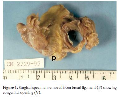
The anatomic pathology report on the broad ligament showed a gap of 1.5 cm x 1.0 cm surrounded by bleeding vessels (Figure 1).
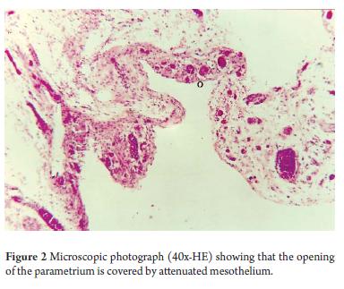
Microscopic study revealed a specimen composed of richly vascularized fibrous connective tissue with some enlarged vessels in the muscle walls. The fibrous connective tissue around the opening of the foramen was covered with mesothelial cells (Figure 2).
The patient was diagnosed as having a congenital opening in the broad ligament and a history of ileal protrusion into that opening.
CASE 2
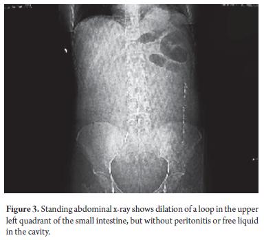
An otherwise healthy 51 year old woman was admitt ed to the hospital with severe lower abdominal pain, nausea. She had begun to vomit shortly before admission. Patient had no history of fever, vaginal trauma, or abdominal interventions, although she had vaginally born two children and had had an abortion. A physical examination showed that her vital signs were normal, but that she had a distended stomach with extreme periumbilical sensitivity and was suffering from some degree of hyperperistalsis. Blood and biochemical tests were normal. An abdominal x-ray taken while the patient was standing showed jejunal dilation.
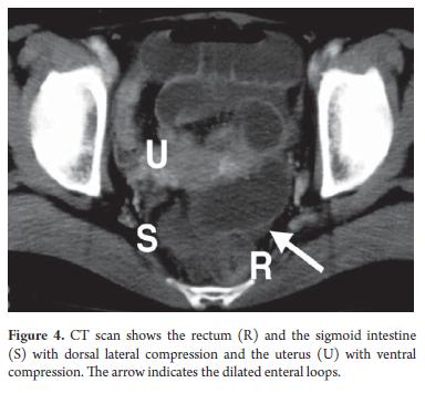
Initially the patient's condition was managed conservatively with parenteral liquids administered through a nasogastric tube. Patient's pain worsened and obstipation and tachycardia had developed by 24 hours after admission. A CT scan revealed an intestinal obstruction.
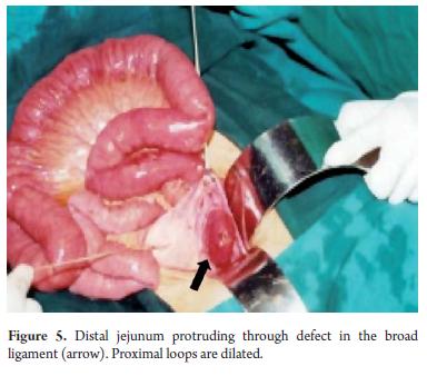
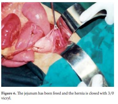
During the laparotomy it was found that a small jejunal loop had entered into small hernia in the broad ligament of the uterus. Congestion and contusion of the loop had caused the obstruction although there was no perforation. Once the loop had been released it recovered completely. The defect in the broad ligament was closed with one continuous vicryl suture. The patient recovered with problems (Figures 3 to 6).
EMBRYOLOGICAL AND ANATOMICAL CONSIDERATIONS
Embryology
The broad ligament is a fold in the peritoneum that connects the uterus, the fallopian tubes, and the ovaries to the pelvis. The broad ligament is formed as the result of the fusion of both paramesonephric ducts (Müllerian ducts). This fusion permits the union of two peritoneal folds to constitute the broad ligament at each side of the fused ducts. The fused paramesonephric ducts transform into the uterus, fallopian tubes and the cervix (17).
Anatomy and physiology
The broad ligament is composed of a double layer of mesothelial cells. It extends from both sides of the uterus to the lateral walls of the pelvis and to the pelvic floor. Its upper part extends from the anterior to the posterior round ligament of the uterus to the fallopian tubes and the ovarian ligament. In the middle of the ligament it surrounds the uterus and laterally surrounds the ovaries forming the suspensory ligament of the ovary (infundibulopelvic ligament). This ligament connects the ovaries to the lateral walls of the pelvis.
Extraperitoneal tissue called the parametrium is found between the two layers of the broad ligament. It consists of connective tissue, smooth muscle, nerves, and blood vessels. The mesovarium is a short peritoneal fold extending from the anterior ovary to the posterior face of the broad ligament. The mesoalpinx is the part of the broad ligament that lies between the ovarian ligament, the ovary and the fallopian tubes.
The uterus and the broad ligament together form a septum across the female pelvis which divides it into two compartments. The bladder is in the anterior compartment and the rectum is in the posterior compartment. It is believed that the broad ligament holds the uterus in its normal position and maintains its anatomical relation with the fallo-pian tubes and the ovaries. This is a very important role for reproduction. Nevertheless, its role in supporting the pelvis is minimal. This job is done primarily by the pelvic floor.
DISCUSSION
Anatomical defects of the broad ligament can be congenital or acquired. Different mechanisms capable of producing these defects have been described. They include the trauma of pregnancy and childbirth, infl ammatory diseases of the pelvis, and surgical damage. Congenital cysts in the broad ligament are remnants of the paramesonephric ducts. It is believed that when these cysts break they can leave a defect in the broad ligament which would explain defects in women who have not born children, undergone surgery or had inflammatory pelvic diseases (18).
Although unilateral defects are more common, bilateral defects also occur. Hunt has classified these into two types (19).
- Type 1: Fenestration in which the defect compromises the anterior and posterior layers of the broad ligament and closes an window between the two layers.
- Type 2: Bag type defects compromise only one of the two layers of the broad ligament.
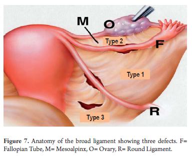
- Cilley has proposed another way of classifying these defects based on location (Figure 7) (20).
- Type 1: Defects which occur in the widest part of the broad are the most common type.
- Type 2: Defects which pass through the mesoalpinx and the mesovarium.
- Type 3: Defects which occur in the middle of the round ligament.
Internal herniation of the small intestine is a rare cause of intestinal obstructions. It represents only 1% to 4% of all cases of intestinal obstructions (3). The first description of a hernia passing through a defect in the broad ligament of the uterus was the result of an autopsy performed by Quain in 1861 and cited by Baron (21). Internal hernias passing through a defect in the broad ligament are even rarer and represent only 4% to 7% of all of these internal hernias (22, 23). Usually an ileal loop is herniated (24, 25), but herniated jejunal loops, such as the second case described above, have also been described (16, 26), as have herniated loops of the adnexa and colon (27-30.
Fenestrations account for the majority of the defects which occur (31). A study of 57 patients in Japan reported only three cases of obstruction with bag type defects (32). Herniation may occur anterior or posterior, or the herniated loop can displace the uterus in a contralateral direction (Figure 4).
Typically the patient presents with a case of peritonitis (acute abdomen), abdominal pain, nausea and vomiting. Early diagnosis is crucial for performing surgery to release the herniated loop and avoid ischemia, necrosis and perforation. A simple abdominal x-ray will show dilated intestinal loops and fluid levels. A diagnostic abdominal and pelvic CT scan can show a herniated loop terminating near the uterus which passes through the broad ligament (4, 33, 34) Emergency surgery includes reducing the size of the herniated loop and if necessary taking secondary measures to prevent a recurrence. These include repair measures such as in the first case presented above and include closure of the defect in the broad ligament as in the second case presented above. Laparoscopic procedures using absorbable clips or continuous sutures to close the defect in the ligament have been described in the literature (3, 29, 35-38).
Although physicians must be awake to the likelihood that intestinal obstructions in the pelvic area are due to adhesions or neoplasia as the primary suspects, internal herniation is a condition that must also be taken into account once the most frequent causes have been discarded. Having this in mind will allow for an early diagnosis and surgical intervention within a prudent period of time thus reducing the possibilities of morbidity and mortality for these patients.
REFERENCES
1. Castaño R, Oliveros R. Factores pronósticos en la obstrucción intestinal por cáncer. Revista del Instituto Nacional de Cancerología 2000; 4: 13-22.
2. Castaño R, Oliveros R. Obstrucción intestinal en el paciente con cáncer. Rev Col de Cirugía 2001; 16: 96-105.
3. Varela GG, Lopez-Loredo A, Garcia Leon JF. Broad ligament hernia-associated bowel obstruction. JSLS 2007; 11: 127-30.
4. Blachar A, Federle MP, Dodson SF. Internal hernia: clinical and imaging findings in 17 patients with emphasis on CT c riteria. Radiology 2001; 218: 68-74.
5. Langan RC, Holzman K, Coblentz M. Strangulated hernia through a defect in the broad ligament: a sheep in wolf 's cloth ing. Hernia 2010.
6. Lin CT, Hsu KF, Hong ZJ, et al. A paraduodenal hernia (Treitz's hernia) causing acute bowel obstruction. Rev Esp Enfe rm Dig 2010; 102: 220-1.
7. Khalaileh A, Schlager A, Bala M, et al. Left laparoscopic paraduodenal hernia repair. Surg Endosc 2010; 24: 1486-9.
8. Park CY, Kim JC, Choi SJ, Kim SK. A transmesenteric hernia in a child: gangrene of a long segment of small bowel thro ugh a large mesenteric defect. Korean J Gastroenterol 2009; 53: 320-3.
9. Capito C, Podevin J, Lascarrou JB, Lehur PA, Armstrong O. Large congenital transmesenteric hernia: a missed smallbowel atresia? Hernia 2009; 13: 209-11.
10. Cisse M, Konate I, Ka O, Dieng M, Dia A, Toure CT. Internal supravesical hernia as a rare cauase of intestinal obstruction: a case report. J Med Case Reports 2009; 3: 9333.
11. Ikeuchi K, Torii A, Kurita A, et al. [A case report: an ileus caused by cecal volvulus and intersigmoidal hernia due to mesenterium commune]. Nippon Shokakibyo Gakkai Zasshi 2006; 103: 415-9.
12. Osvaldt AB, Mossmann DF, Bersch VP, Rohde L. Intestinal obstruction caused by a foramen of Winslow hernia. Am J Surg 2008; 196: 242-4.
13. Le Moigne F, Lamboley JL, de Charry C, et al. An exceptional case of internal transomental hernia: correlation between CT and surgical findings. Gastroenterol Clin Biol 2010; 34: 562-4.
14. Choong AM, Carney L, Beaconsfield T, Shorvon PJ, Bhutiani RP. 'The ins and outs of abdominal pain': a case report of a transomental internal hernia. Ann R Coll Surg Engl 2010; 92: W35-6.
15. Aggarwal BK, Rajan S, Aggarwal A, Gothi R, Sharma R, Tandon V. CT diagnosis of Meckel diverticulum in a paracolic internal hernia. Abdom Imaging 2005; 30: 56-9.
16. Kanbur AS, Ahmed K, Bux B, Hande T. Jejunal obstruction and perforation resulting from herniation through broad ligam ent. J Postgrad Med 2000; 46: 189-90.
17. Miller A, Hong MK, Hutson JM. The broad ligament: a review of its anatomy and development in diff erent species and ho rmonal environments. Clin Anat 2004; 17: 244-51.
18. Guillem P, Cordonnier C, Bounoua F, Adams P, Duval G. Small bowel incarceration in a broad ligament defect. Surg Endo sc 2003; 17: 161-2.
19. Hunt AB. Fenestra and pouches in the broad ligament as an actual and potential cause of strangulated intraabdominal h ernia. Surg Gynecol Obstet 1934; 1934: 906-13.
20. Cilley R, Poterack K, Lemmer J, Dafoe D. Defects of the broad ligament of the uterus. Am J Gastroenterol 1986; 81: 38 9-91.
21. Baron A. Defect in the broad ligament and its association with intestinal strangulation. Br J Surg 1948; 36: 91-4.
22. Karaharju E, Hakkiluoto A. Strangulation of small intestine in an opening of the broad ligament. Int Surg 1975; 60: 4 30.
23. Mailleux P, Ramboux A. Small bowel obstruction due to an internal herniation through a defect of the broad ligament. JBR-BTR 2010; 93: 201-3.
24. Tanioka Y, Hirano A, Okita K, et al. A case report of internal hernia through an abnormal defect in the broad ligamen t of the uterus. Nippon Shokakibyo Gakkai Zasshi 2010; 107: 620-4.
25. Cisse M, Ka I, Konate I, et al. Incarcerated internal hernia through a breach of the broad ligament, a case report. Gynecol Obstet Fertil 2011; 39: e47-8.
26. Garcia-Fadrique A, Sospedra Ferrer R, Vazquez Tarragon A, Martinez Abad M. Intestinal obstruction due to a hernia across the broad ligament of the uterus. Cirugia espanola 2011.
27. Demir H, Scoccia B. Internal herniation of adnexa through a defect of the broad ligament: case report and literature review. J Minim Invasive Gynecol 2010; 17: 110-2.
28. Karcaaltincaba D, Avsar F, Iskender C, Korukluoglu B. Unusual mechanism of isolated torsion of fallopian tube followi ng minor trauma. Herniation through a broad ligament tear. Saudi Med J 2007; 28: 637-8.
29. Onida S, Lynes K, Ozdemir BA, Whitehouse PA. Unexpected findings at diagnostic laparoscopy: caecal incarceration with concurrent appendicitis in a patient with bilateral broad ligament defects. Ann R Coll Surg Engl 2010; 92: W19-20.
30. Vo TM, Gyaneshwar R, Mayer C. Concurrent sigmoid volvulus and herniation through broad ligament defect during pregnan cy: case report and literature review. J Obstet Gynaecol Res 2008; 34: 658-62.
31. Papas HN. Intestinal obstruction associated with pouches and fenestrae in the broad ligament: review of the literature and report of a case. Am J Obstet Gynecol 1960; 80: 172-5.
32. Terado M, Okazaki M, Shinozaki K. A case report of internal herniation through an abnormal defect in the broad liga-me nt. Shujutsu 2002; 56: 265-9.
33. Barbier Brion B, Daragon C, Idelcadi O, Mantion G, Kastler B, Delabrousse E. Small bowel obstruction due to broad ligament hernia: computed tomography findings. Hernia 2010.
34. Kosaka N, Uematsu H, Kimura H, Yamamori S, Hirano K, Itoh H. Utility of multi-detector CT for pre-operative diagnosis of internal hernia through a defect in the broad ligament (2007: 1b). Eur Radiol 2007; 17: 1130-3.
35. Garcia-Oria M, Inglada J, Domingo J, Biescas J, Ching C. Small bowel obstruction due to broad ligament hernia success fully treated by laparoscopy. J Laparoendosc Adv Surg Tech A 2007; 17: 666-8.
36. Leone V, Misuri D, Faggi U, Giovane A, Fazio C, Cardini S. Laparoscopic treatment of incarcerated hernia through righ t broad ligament in patients with bilateral parametrium defects. G Chir 2009; 30: 141-3.
37. Takayama S, Hirokawa T, Sakamoto M, et al. Laparoscopic management of small bowel incarceration caused by a broad ligament defect: report of a case. Surg Today 2007; 37: 437 -9.
38. Bangari R, Uchil D. Laparoscopic Management of Internal Hernia of Small Intestine through a Broad Ligament Defect. J Minim Invasive Gynecol 2012; 19: 122-4.
1. Castaño R, Oliveros R. Factores pronósticos en la obstrucción intestinal por cáncer. Revista del Instituto Nacional de Cancerología 2000; 4: 13-22. [ Links ]
2. Castaño R, Oliveros R. Obstrucción intestinal en el paciente con cáncer. Rev Col de Cirugía 2001; 16: 96-105. [ Links ]
3. Varela GG, Lopez-Loredo A, Garcia Leon JF. Broad ligament hernia-associated bowel obstruction. JSLS 2007; 11: 127-30. [ Links ]
4. Blachar A, Federle MP, Dodson SF. Internal hernia: clinical and imaging findings in 17 patients with emphasis on CT criteria. Radiology 2001; 218: 68-74. [ Links ]
5. Langan RC, Holzman K, Coblentz M. Strangulated hernia through a defect in the broad ligament: a sheep in wolf 's clothing. Hernia 2010. [ Links ]
6. Lin CT, Hsu KF, Hong ZJ, et al. A paraduodenal hernia (Treitz's hernia) causing acute bowel obstruction. Rev Esp Enfe rm Dig 2010; 102: 220-1. [ Links ]
7. Khalaileh A, Schlager A, Bala M, et al. Left laparoscopic paraduodenal hernia repair. Surg Endosc 2010; 24: 1486-9. [ Links ]
8. Park CY, Kim JC, Choi SJ, Kim SK. A transmesenteric hernia in a child: gangrene of a long segment of small bowel thro ugh a large mesenteric defect. Korean J Gastroenterol 2009; 53: 320-3. [ Links ]
9. Capito C, Podevin J, Lascarrou JB, Lehur PA, Armstrong O. Large congenital transmesenteric hernia: a missed smallbowel atresia? Hernia 2009; 13: 209-11. [ Links ]
10. Cisse M, Konate I, Ka O, Dieng M, Dia A, Toure CT. Internal supravesical hernia as a rare cauase of intestinal obstruction: a case report. J Med Case Reports 2009; 3: 9333. [ Links ]
11. Ikeuchi K, Torii A, Kurita A, et al. [A case report: an ileus caused by cecal volvulus and intersigmoidal hernia due to mesenterium commune]. Nippon Shokakibyo Gakkai Zasshi 2006; 103: 415-9. [ Links ]
12. Osvaldt AB, Mossmann DF, Bersch VP, Rohde L. Intestinal obstruction caused by a foramen of Winslow hernia. Am J Surg 2008; 196: 242-4. [ Links ]
13. Le Moigne F, Lamboley JL, de Charry C, et al. An exceptional case of internal transomental hernia: correlation between CT and surgical findings. Gastroenterol Clin Biol 2010; 34: 562-4. [ Links ]
14. Choong AM, Carney L, Beaconsfield T, Shorvon PJ, Bhutiani RP. 'The ins and outs of abdominal pain': a case report of a transomental internal hernia. Ann R Coll Surg Engl 2010; 92: W35-6. [ Links ]
15. Aggarwal BK, Rajan S, Aggarwal A, Gothi R, Sharma R, Tandon V. CT diagnosis of Meckel diverticulum in a paracolic internal hernia. Abdom Imaging 2005; 30: 56-9. [ Links ]
16. Kanbur AS, Ahmed K, Bux B, Hande T. Jejunal obstruction and perforation resulting from herniation through broad ligam ent. J Postgrad Med 2000; 46: 189-90. [ Links ]
17. Miller A, Hong MK, Hutson JM. The broad ligament: a review of its anatomy and development in diff erent species and ho rmonal environments. Clin Anat 2004; 17: 244-51. [ Links ]
18. Guillem P, Cordonnier C, Bounoua F, Adams P, Duval G. Small bowel incarceration in a broad ligament defect. Surg Endo sc 2003; 17: 161-2. [ Links ]
19. Hunt AB. Fenestra and pouches in the broad ligament as an actual and potential cause of strangulated intraabdominal hernia. Surg Gynecol Obstet 1934; 1934: 906-13. [ Links ]
20. Cilley R, Poterack K, Lemmer J, Dafoe D. Defects of the broad ligament of the uterus. Am J Gastroenterol 1986; 81: 38 9-91. [ Links ]
21. Baron A. Defect in the broad ligament and its association with intestinal strangulation. Br J Surg 1948; 36: 91-4. [ Links ]
22. Karaharju E, Hakkiluoto A. Strangulation of small intestine in an opening of the broad ligament. Int Surg 1975; 60: 4 30. [ Links ]
23. Mailleux P, Ramboux A. Small bowel obstruction due to an internal herniation through a defect of the broad ligament. JBR-BTR 2010; 93: 201-3. [ Links ]
24. Tanioka Y, Hirano A, Okita K, et al. A case report of internal hernia through an abnormal defect in the broad ligamen t of the uterus. Nippon Shokakibyo Gakkai Zasshi 2010; 107: 620-4. [ Links ]
25. Cisse M, Ka I, Konate I, et al. Incarcerated internal hernia through a breach of the broad ligament, a case report. Gynecol Obstet Fertil 2011; 39: e47-8. [ Links ]
26. Garcia-Fadrique A, Sospedra Ferrer R, Vazquez Tarragon A, Martinez Abad M. Intestinal obstruction due to a hernia across the broad ligament of the uterus. Cirugia espanola 2011. [ Links ]
27. Demir H, Scoccia B. Internal herniation of adnexa through a defect of the broad ligament: case report and literature review. J Minim Invasive Gynecol 2010; 17: 110-2. [ Links ]
28. Karcaaltincaba D, Avsar F, Iskender C, Korukluoglu B. Unusual mechanism of isolated torsion of fallopian tube followi ng minor trauma. Herniation through a broad ligament tear. Saudi Med J 2007; 28: 637-8. [ Links ]
29. Onida S, Lynes K, Ozdemir BA, Whitehouse PA. Unexpected findings at diagnostic laparoscopy: caecal incarceration with concurrent appendicitis in a patient with bilateral broad ligament defects. Ann R Coll Surg Engl 2010; 92: W19-20. [ Links ]
30. Vo TM, Gyaneshwar R, Mayer C. Concurrent sigmoid volvulus and herniation through broad ligament defect during pregnan cy: case report and literature review. J Obstet Gynaecol Res 2008; 34: 658-62. [ Links ]
31. Papas HN. Intestinal obstruction associated with pouches and fenestrae in the broad ligament: review of the literature and report of a case. Am J Obstet Gynecol 1960; 80: 172-5. [ Links ]
32. Terado M, Okazaki M, Shinozaki K. A case report of internal herniation through an abnormal defect in the broad liga-me nt. Shujutsu 2002; 56: 265-9. [ Links ]
33. Barbier Brion B, Daragon C, Idelcadi O, Mantion G, Kastler B, Delabrousse E. Small bowel obstruction due to broad ligament hernia: computed tomography findings. Hernia 2010. [ Links ]
34. Kosaka N, Uematsu H, Kimura H, Yamamori S, Hirano K, Itoh H. Utility of multi-detector CT for pre-operative diagnosis of internal hernia through a defect in the broad ligament (2007: 1b). Eur Radiol 2007; 17: 1130-3. [ Links ]
35. Garcia-Oria M, Inglada J, Domingo J, Biescas J, Ching C. Small bowel obstruction due to broad ligament hernia success fully treated by laparoscopy. J Laparoendosc Adv Surg Tech A 2007; 17: 666-8. [ Links ]
36. Leone V, Misuri D, Faggi U, Giovane A, Fazio C, Cardini S. Laparoscopic treatment of incarcerated hernia through righ t broad ligament in patients with bilateral parametrium defects. G Chir 2009; 30: 141-3. [ Links ]
37. Takayama S, Hirokawa T, Sakamoto M, et al. Laparoscopic management of small bowel incarceration caused by a broad ligament defect: report of a case. Surg Today 2007; 37: 437 -9. [ Links ]
38. Bangari R, Uchil D. Laparoscopic Management of Internal Hernia of Small Intestine through a Broad Ligament Defect. J Minim Invasive Gynecol 2012; 19: 122-4. [ Links ]

