


 text in
  English (pdf)
 Article in xml format
 Article references
 Send this article by e-mail
 Cited by SciELO
  Cited by Google
  Similars in
SciELO
Similars in
SciELO  Similars in Google
 Permalink
Introduction
Gastric cancer is the fifth most commonly diagnosed cancer and the third leading cause of death from cancer worldwide1. East Asia (China, Korea, Mongolia, and Japan)1, Eastern Europe, Central and South America2 have been described as high risk areas for developing this type of cancer.
In Colombia, according to the data available in population-based cancer registries, it has a high impact in the general population, and people living in the southwestern region of Colombia, especially men, are more affected by it. For example, an annual incidence rate of 26.7/100 000 inhabitants in men living in the city of Pasto has been reported3. In addition, other risk factors such as low education, poverty, high salt intake, low antioxidant intake, the presence of premalignant lesions such as intestinal atrophy and metaplasia, and Helicobacter pylori infection have been described4,5. However, its association with different risk factors depends on the tumor histotype.
According to the classification by Lauren and Jarvi, adenocarcinomas are classified as intestinal and diffuse6; this classification is very relevant because despite they are tumors of epithelial origin, there are some differences. In this sense, intestinal carcinomas occur more frequently in black men and in older people; also, their occurrence has been associated with environmental factors, poor public health conditions, premalignant lesions and H. pylori infection. This type of cancer also shows large geographical variations and is more frequently located in the antropyloric region7. On the other hand, diffuse type adenocarcinomas are not associated with environmental conditions, premalignant lesions or H. pylori infection; furthermore, a higher incidence in women and a common association with E-cadherin mutations have been reported for this type of tumor8.
Several studies exploring the sociodemographic and clinical characteristics of patients with gastric cancer or premalignant lesions have been conducted in Southwest Colombia9,10; However, based on a literature review conducted by us, so far there are no studies reporting the histopathological and sociodemographic characteristics of patients with gastric cancer in the department of Nariño, Colombia. Bearing this in mind, the present descriptive study was carried out in order to analyze the sociodemographic and microscopic characteristics of patients with gastric cancer and look into the alleged relationship between these variables and tumor histotype.
Materials and methods
Descriptive study. Sociodemographic and histopathological data were collected retrospectively. All patients diagnosed with gastric cancer and who underwent total and subtotal gastrectomy between 2014 and 2017 at the Clínica Oncológica Aurora (Aurora Cancer Clinic), located in Pasto, were included. Sociodemographic data were obtained from the patients’ medical records, while histopathological information was retrieved from pathology reports. Sociodemographic variables included age, sex, ethnicity, marital status, origin, health care plan enrollment in the Colombian health system and education level. Histopathological diagnosis was reached based on paraffin-embedded sections and subsequently stained with hematoxylin-eosin. Variables such as tumor location, presence of H. pylori infection, presence of associated premalignant lesions and tumor histotype were determined. Premalignant lesions were classified according to the Sydney system, while tumor histotypes were classified as intestinal or diffuse based on the Lauren’s classification system. When both components (intestinal and diffuse to an 50% extent each) were reported in the microscopic study, tumors were classified as mixed.
Means and standard deviations were calculated for the age variable. Categorical variables were described using absolute frequencies and proportions. A one-factor ANOVA was performed to determine the differences in the mean age between the different tumor histotypes. Also, a chi-square test (χ2) was performed to compare the proportions of the variables of interest between tumor histotypes. A significant level of p < 0.05 was considered. Statistical analysis was carried out using SPSS software, version 25.
Results
Data from 54 patients aged between 26 and 88 years were collected during the study period. Participants’ mean age was 64 years (standard deviation: 13.2 years). Most tumors occurred in male patients, with a male female ratio of 2.6:1. The other sociodemographic characteristics are shown in Table 1.
Table 1 Sociodemographic characteristics of the study population
| Variable | n | % | |
|---|---|---|---|
| Sex |
|
|
|
| Ethnicity |
|
|
|
| Marital status |
|
|
|
| Origin |
|
|
|
| Health care regime |
|
|
|
| Education level |
|
|
|
The youngest patient was a 26-year-old woman with diffuse adenocarcinoma, while the oldest patient was an 88-year-old man with intestinal adenocarcinoma.
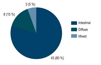
The microscopic classification of gastric tumors is shown in Figure 1.
The mean ages of patients with intestinal, diffuse and mixed tumors were 63, 64 and 73 years, respectively. No significant differences regarding mean age were found between the three tumor types in the one-factor ANOVA (p = 0.492).
Finally, the relationship of variables such as categorized age (< 50 years and ≥ 50 years), sex and histopathological variables with the different types of tumors was explored, but no statistically significant differences were found (Table 2). H. pylori was reported in 13 cases, that is 24.07 % of the patients.
Table 2 Relationship between age, sex, and anatomical and pathological characteristics, and tumor histotypes
| Variable | Intestinal | Diffuse | Mixed | p value* | |
|---|---|---|---|---|---|
| Age |
|
|
|
|
0.422 |
| Sex |
|
|
|
|
0.518 |
| Tumor location |
|
|
|
|
0.390 |
| H. pylori |
|
|
|
|
0.390 |
| Associated premalignant lesion |
|
|
|
|
0.970 |
*A p value < 0.05 was considered statistically significant.
Discussion
Gastric cancer is a multifactorial disease in which host-related and environmental aspects, as well as H. pylori infection11 have been described as factors associated with its occurrence. In the case of host-related factors, age > 50 years and being male have been reported as variables associated with the development of gastric cancer12, which is consistent with the results described in the present study. The prevalence of stomach cancer according to sex varies: in developed countries a 2.2:1 male-female ratio has been reported, while in developing countries this ratio is 1.8:113. In this regard, in the present study a male-female ratio of 2.6:1 was found, and male sex stood out as an important risk factor for gastric cancer. However, some authors have reported that diffuse adenocarcinoma occurs more frequently in young females14,15, yet such relationship was not observed here, since all patients with diffuse adenocarcinomas (n=8) were older than 50 years, of which 75 % (n=6) were men. Similarly, all patients with mixed histotype were men over 50 years of age. These findings highlight the importance of sex and age in gastric carcinogenesis.
The relationship between age and cancer development is associated with an increased genomic instability caused by factors such as cell aging, chronic inflammatory response in the gastric mucosa, tissue microenvironment modifications resulting from diet, and direct and indirect changes induced by H. pylori. This genomic instability would generate two molecular mechanisms: Microsatellite instability and intrachromosomal genomic instability; the first is related to the inability to repair damaged deoxyribonucleic acid (DNA), while the second, to alterations in oncogenes and tumor suppressor genes16,17. Some genetic alterations have been associated with tumor hystotype. In this sense, alterations in E-cadherin are more common in diffuse gastric carcinomas, while Erb gene mutations are more frequently associated with intestinal gastric adenocarcinomas7,8. These are very important findings, for they suggest two different pathways of tumor progression. In our study, the presence of diffuse adenocarcinoma in the youngest patient, a 26-year-old woman, stands out. The existence of this type of cancer in a young patient should encourage clinical suspicion because of the association between E-cadherin and cadherin-1 (CDH1) germline mutations with hereditary diffuse gastric cancer syndrome18. On the other hand, when tumors occur in people older than 50 years, these are probably the result of mutations and the cumulative genomic instability over the years in the somatic cell lineage.
The relative low prevalence of H. pylori infection (24 %), as reported in the histopathology reports, found in our study stands out. This finding differs from what was reported by Hooi et al. in a recent meta-analysis and in which the prevalence rates of H. pylori infection in Africa, Asia, and Latin America and the Caribbean were 79.1%, 54.7%, and 63.4%, respectively19. In Colombia, possibly the most relevant study in this topic is the one conducted by Bravo et al.20, where the general prevalence of H. pylori was 69.1 % and its prevalence in patients with cancer was 40.2 %21. According to Bravo et al., the following factors were considered as possible causes of the low prevalence of H. pylori found in their study: first, perioperative antimicrobial therapy; second, the presence of intestinal metaplasia in almost all patients, and third, the assessment of diffuse or mixed histotype tumors, which are not usually associated with bacterial infection. Unfortunately, we were not able to document the establishment of preoperative H. pylori infection treatments in the present study, since the clinic where it was conducted is a surgical oncology reference center and the complete medical records of patients treated in other institutions, where they presumably received pharmacological treatment, are not available. Similarly, it is well known that documenting H. pylori in gastric mucosa with intestinal metaplasia is difficult21, since it seems metaplastic intestinal epithelium is a hostile environment for the bacterium by limiting its ability to synthesize ammonium. In addition, it has been suggested that even if it were present in this facet of carcinogenesis, identifying it is very difficult due to chronic inflammation and the tissue damage caused by it. Finally, H. pylori infection has been associated with intestinal type tumors, but not with diffuse tumors22,23.
In our study, infection was reported in 28% of patients with intestinal gastric cancer and only in 12% of those with diffuse gastric cancer. In addition, contrary to what has been described in developed countries, where an increasing prevalence of carcinoma occurrence in the cardia, the body and the fundus has been reported24,25, tumor location in the antropyloric region was the most frequent in the present study, a finding consistent with what was reported by Adrada et al. in a study conducted in the department of Cauca and that has remained unchanged for many decades in southwestern Colombia9. In this regard, several authors have proposed two location-based types of carcinoma: on the one hand, cardia carcinoma, which is more frequent in developed countries, where public health conditions are better and it is associated with Western diet, alcohol consumption and obesity, and, on the other, non-cardia carcinomas, typical of developing countries and associated with poor public health conditions, premalignant lesions and H. pylori infection13,26.
One of the study limitations is its small sample size; however, the findings reported here are consistent with the sociodemographic and histopathological behavior described in other works conducted in other geographic areas and with larger sample sizes. Similarly, it was not possible to determine whether the patients who underwent gastrectomy were treated for H. pylori infection. Taking this into account, further research is needed to determine whether the microorganism was eradicated by prior treatment or not, which is why studies considering prior biopsies and breath tests to verify H. pylori infection would be relevant.
Conclusions
Our findings suggest that public health conditions, H. pylori infection control, and early screening of premalignant lesions must be improved, as they are determining factors in carcinogenesis in patients with non-cardia carcinomas and intestinal type tumors located in the antrum and body of the stomach. Age over 50 and male sex were factors associated with gastric cancer in the study population. Although no significant differences were found between tumor histotypes, intestinal adenocarcinoma located in the antropyloric region was the most frequent diagnosis.

