SciELO - Scientific Electronic Library Online

 
vol.36 issue1Sociodemographic and histopathological characteristics of patients with gastric cancer in a high-risk population in ColombiaClinical and epidemiological description of patients with chronic pancreatitis treated at a quaternary care hospital in Cali, 2011-2017 author indexsubject indexarticles search
Home Pagealphabetic serial listing  

Services on Demand

Journal

Article

Indicators

Related links

  • On index processCited by Google
  • Have no similar articlesSimilars in SciELO
  • On index processSimilars in Google

Share


Revista colombiana de Gastroenterología

Print version ISSN 0120-9957On-line version ISSN 2500-7440

Rev. colomb. Gastroenterol. vol.36 no.1 Bogotá Jan./Mar. 2021  Epub Oct 20, 2021

https://doi.org/10.22516/25007440.564 

Original articles

Validity of nodules detected at colonoscopy for the diagnosis of nodular lymphoid hyperplasia in children

Pablo Xavier Sempértegui-Cárdenas1  * 
http://orcid.org/0000-0001-7325-3082

José Francisco Cadena-León2 
http://orcid.org/0000-0002-3138-310X

Karen Rubí Ignorosa-Arellano2 
http://orcid.org/0000-0002-8364-6936

Erick Manuel Toro-Monjaraz2 
http://orcid.org/0000-0001-8701-4189

Flora Elva Zárate-Mondragón3 
http://orcid.org/0000-0001-5347-3889

Roberto Cervantes-Bustamante2 
http://orcid.org/0000-0003-4898-8815

Jaime Alfonso Ramírez-Mayans2 
http://orcid.org/0000-0003-4770-4809

Celso Tomás Corcuera-Delgado4 

Laura Becerril-Cholula4 
http://orcid.org/0000-0002-3944-9062

1MSc. Pediatrician, Specialist in Gastroenterology and Pediatric Nutrition. Department of Gastroenterology and Nutrition, Instituto Nacional de Pediatría, Mexico City, Mexico.

2Pediatrician, Specialist in Gastroenterology and Pediatric Nutrition. Department of Gastroenterology and Nutrition, Instituto Nacional de Pediatría, Mexico City, Mexico.

3PhD. Pediatrician, Specialist in Gastroenterology and Pediatric Nutrition. Department of Gastroenterology and Nutrition, Instituto Nacional de Pediatría, Mexico City, Mexico.

4MD, Specialist in pathological anatomy. Department of Pathological Anatomy, Instituto Nacional de Pediatría, Mexico City, Mexico.


Abstract

Introduction:

Nodular lymphoid hyperplasia of the colon is characterized by the presence of >10 lymphoid nodules visible in colonoscopy. There are no studies that confirm their validity when compared with histopathology.

Objective:

To determine the validity of nodules detected at colonoscopy for the diagnosis of nodular lymphoid hyperplasia in children.

Materials and methods:

Prospective study of diagnostic test accuracy. Colonoscopies performed consecutively from 2014 to 2018 using Olympus PCFQ150AI and GIFXP150N biopsy machines were included. The endoscopic criterion was the presence of >10 nodules from 2 to 10mm of diameter, while the histological criterion was presence of follicular lymphoid hyperplasia and lymphocyte mantles in lamina propia or submucosa. Data were analyzed in Epidat3.1. Sensitivity (SE), specificity (SP), positive predictive value (PPV), negative predictive value (NPV), positive likelihood ratio (LR+) and negative likelihood ratio (LR-) were obtained with their corresponding confidence intervals.

Results:

327 colonoscopies were included; the median age was 84 months. The main indication for colonoscopy was lower gastrointestinal bleeding (38.8 %). Nodules were found in 21 % of the patients, predominantly throughout the whole colon (46 %), whereas histopathology found nodular lymphoid hyperplasia in 38 %. SE for the finding of nodules was 32 % (95 % confidence interval [CI]: 24-140), SP was 84 % (95 % CI: 79-89), PPV was 56% (95 % CI: 44-68), NPV was 67 % (95 % CI: 61-72), LR+ was 2.04 (95 % CI: 1.4-3) and LR- was 0.8 (95 % CI: 0.8-0.9).

Conclusions:

The validity of the presence of nodules on colonoscopy for the diagnosis of nodular lymphoid hyperplasia is poor, so biopsy should always be performed.

Keywords: Children; Colonoscopy; Nodular lymphoid hyperplasia; Sensitivity; Specificity

Resumen

Introducción:

La hiperplasia nodular linfoide del colon se define como > 10 nódulos linfoides visibles en colonoscopia. No existen estudios de su validez al compararlo con la histopatología.

Objetivos:

Determinar la validez del hallazgo de nódulos en colonoscopia para el diagnóstico de hiperplasia nodular linfoide.

Material y métodos:

Estudio prospectivo de prueba diagnóstica. Se incluyeron colonoscopias realizadas consecutivamente de 2014 al 2018 con equipos Olympus PCFQ150AI y GIFXP150N con obtención de biopsias. El criterio endoscópico fue la presencia de > 10 nódulos de 2 a 10 mm y el criterio histológico fue hiperplasia de folículos linfoides y mantos de linfocitos en lámina propia o submucosa. Los datos se analizaron en Epidat3.1. Se obtuvo la sensibilidad (S), especificidad (E), valor predictivo positivo (VPP) y negativo (VPN), y coeficientes de probabilidad positivo (LR+) y negativo (LR-) con sus intervalos de confianza.

Resultados:

Se incluyeron 327 colonoscopias, la mediana de edad fue de 84 meses. La principal indicación para la colonoscopia fue sangrado digestivo bajo (38,8%). El hallazgo de nódulos se encontró en el 21% y el sitio de mayor frecuencia fue el colon total (46%), mientras que por histopatología se encontró hiperplasia nodular linfoide en el 38%. El hallazgo de nódulos obtuvo una S de 32% (intervalo de confianza [IC] del 95%: 24-140), E de 84% (IC 95%: 79-89), VPP de 56% (IC 95%: 44-68), VPN de 67% (IC 95%: 61-72), LR+ de 2,04 (IC 95%: 1,4-3) y LR- de 0,8 (IC 95%: 0,8-0,9).

Conclusiones:

La validez diagnóstica del hallazgo de nódulos en colonoscopia para hiperplasia nodular linfoide es pobre, por lo que la toma de biopsia debe recomendarse siempre.

Palabras clave: Niños; colonoscopia; hiperplasia nodular linfoide; sensibilidad; especificida

Introduction

Nodular lymphoid hyperplasia of the colon is defined as a group of > 10 lymphoid nodules observed during colonoscopy1, these have been reported most frequently in young children and without association with clinical symptoms, cases in which nodular lymphoid hyperplasia is considered to be physiological1-3; however, it has also been described in patients undergoing colonoscopy due to gastrointestinal bleeding, chronic abdominal pain4, refractory constipation, delayed growth, or anemia, in frequencies ranging from 12.8%5 to 49%1. In this regard, a study conducted in Cuba found that 63%, 33% and 4% of nodular lymphoid hyperplasia cases occurred in children under 6 years of age, children between 6 and 10 years old, and children older than 10 years, respectively6.

The clinical relevance and etiology of nodular lymphoid hyperplasia is unclear. For many years it was thought to be a casual finding during colonoscopy; somehow, it has been associated with infectious diseases such as Helicobacter pylori infection7, enterobiasis, amebiasis, Escherichia coli infection and Giardia lamblia infection8-11; Mediterranean family fever; immunodeficiency disorders such as common variable immunodeficiency12-14, IgA deficiency14,15, hypogammaglobulinemia12,13,16 and human immunodeficiency virus (HIV)17; food allergy; inflammatory bowel disease1,4,5,18; irritable bowel syndrome; celiac disease; and Behçet’s disease, among others5.

The diagnosis of nodular lymphoid hyperplasia is reached by means of an endoscopy, and histopathology studies are required for its confirmation19. Endoscopic features include nodules ranging from 2 to 10 mm and can occur in the stomach, the small intestine (most commonly in the terminal ileum), and the colon/rectum20. In the colon, they may look like red macules, circumferential target lesions, or raised papules21,22. When the large intestine is affected, the rectum is most frequently involved23,24. Their endoscopic appearance can be surprisingly similar to polyposis syndromes23, but histological findings can help reach a differential diagnosis. Histologically, it is defined by the presence of lymphoid follicles hyperplasia, germinal centers with mitotic activity, and well-defined mantle lymphocytes located in the lamina propria or the submucosa25; it may resemble a malignant lymphoma26, both clinically and histologically, but it can be differentiated by its polymorphic nature resulting from the infiltration, the absence of significant cytologic atypia, and the presence of reactive follicles within the lesion27.

Nodular lymphoid hyperplasa is considered a risk factor for the development of both intestinal28,29 and extraintestinal lymphomas30,31, adenomas, and carcinomas 32, and some authors recommend performing several barium swallows and capsule endoscopies for surveillance purposes24, although the duration and intervals of such surveillance studies have not been determined15. So far there are no studies evaluating the diagnostic validity of detecting nodules during colonoscopy compared to histological findings in the diagnosis of nodular lymphoid hyperplasia.

Objective

To determine the validity of detecting nodules during colonoscopy for the diagnosis of nodular lymphoid hyperplasia.

Materials and methods

A prospective diagnostic test accuracy study was conducted in a pediatric referral hospital. A total of 327 colonoscopies performed consecutively between January 1, 2014 and December 31, 2018 were included. The sample size was calculated in Epidat 3.2 based on an expected sensitivity of 40 %-60 %, an expected specificity of 70 %-90 %, a disease prevalence of 30.9 % (average of the prevalence rates of nodular lymphoid hyperplasia described in the literature)1,5, with a confidence interval (CI) of 95 % and a statistical power of 80 %. All colonoscopies were performed by a pediatric gastroenterologist trained in this type of procedure; an Olympus PCF-Q150AI colonoscope was used in children over 10 kg, while an Olympus GIF-XP150N neonatal endoscope was used in those under 10 kg; in all cases, biopsies were taken during colonoscopy using cold biopsy forceps and removing at least 2 samples from the cecum, 2 from the ascending colon, 2 from the transverse colon, 2 from the descending colon, and 2 from the sigmoid colon and rectum, which were analyzed by 2 pathologists from the histopathology department of the institution; it should be noted that the analysis was blind and was carried out within a maximum of 1 week. The pathologists were previously validated by interobserver agreement with a Cohen’s Kappa coefficient of 1.0 (p = 0.000). There were no adverse events during the performance of both colonoscopies or biopsies.

The colonoscopic criterion was defined according to what has been reported in the relevant literature, that is, detecting a group of >10 nodules with a size ranging from 2 to 10 mm at any site in the large bowel20. The histology study was carried out using hematoxylin-eosin staining with a light microscope at 5X, 10X and 40X; the histological criterion was defined based on what has been described in the literature, that is, the presence of lymphoid follicles hyperplasia and well-defined lymphocyte mantles in the lamina propria or submucosa25.

Data were recorded in a database in the SPSS software, version 22, where they were coded for tabulation. In the age variable, the Kolmogorov-Smirnov test was performed, and since data distribution was not normal, medians and interquartile ranges were used. On the other hand, qualitative variables such as age interval, sex, indication for colonoscopy, colonoscopic findings, site of colonoscopic finding, and histological findings are expressed by means of frequencies and percentages. Colonoscopic findings were dichotomized based on the presence or absence of visible nodules, while histological findings, based on the presence or absence of nodular lymphoid hyperplasia and were crossed in 2 x 2 tables. The proportion of nodular hyperplasia according to age and indication for colonoscopy was compared using chi-square (χ2). The Epidat 3.1 software was used to obtain the sensitivity, specificity, positive predictive value (PPV) and negative predictive value (NPV), and positive and negative likelihood ratios with their respective confidence intervals, as well as the overall value of the diagnostic test.

Results

Of the 327 colonoscopies, 188 were performed in boys (57.5 %) and the rest in girls. Participants’ median age was 84 months (7 years) with an interquartile range of 111 months, a minimum value of 1 month, and a maximum value of 216 months.

Lower gastrointestinal bleeding (with or without anemia) was the most frequent indication for colonoscopy (n=127, 38.8%), followed by suspicion or surveillance of inflammatory bowel disease (17 %), suspicion or monitoring of food allergy (14.4 %), chronic diarrhea (11.3 %), polyposis syndrome surveillance (6.1 %) and suspected rectal polyp (3.7 %); the remaining colonoscopies were performed due to several reasons including suspected graft versus host disease, chronic abdominal pain, protein-losing enteropathy, vascular malformation, foreign bodies, and suspected colon cancer, among others.

The prevalence of colonoscopic detection of nodules was 21.7 % (n = 71). The most frequent location of the nodules was the total colon, with 33 cases (46 %), followed by rectum limited involvement (17 %), left colon limited involvement (14 %) and sigmoid colon and rectum limited involvement (13 %). The proportion of nodules detection was similar in boys and girls (21.3 % vs 22.3%). Nodules occurred more frequently at a lower age (p = 0.016) (Table 1 and Figure 1).

Table 1 Distribution of patients undergoing colonoscopy according to the detection of nodules, sex, and age (2014-2018) 

Variable Nodules Total Pearson χ2 p value
Yes No
n % n % n
Sex
Male 40 21,3 148 78,7 188 0,049 0,892
Female 31 22,3 108 77,7 139
Age (years)
< 2 16 32,0 34 68,0 50 8,295 0,016
2-10 40 24,2 125 75,8 165
>10 15 13,4 97 86,6 112
Total 71 21,7 256 78,3 327

*There is a statistically significant difference. Source: Data collection forms. Own elaboration.

Figure 1 Flow diagram of the colonoscopies and biopsies performed in the study population. 

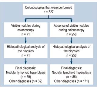
The prevalence of nodular lymphoid hyperplasia according to the histopathology report was 38 % (n = 124). The lowest frequency was found in patients who underwent colonoscopy due to recurrence of intussusception, unexplained weight loss, and suspected intestinal pseudo-obstruction (0%), followed by, in ascending order, those who underwent it due to polyposis syndromes surveillance (5%) and suspected graft versus host disease (11 %). On the other hand, the highest frequency of nodular lymphoid hyperplasia was observed in the cases in which colonoscopy was indicated due to colon cancer, suspected intestinal tuberculosis, and refractory constipation (100%), followed by, in descending order, suspected rectal fistula and suspected enteropathy (50%), suspected or monitoring of inflammatory bowel disease (49 %), suspected rectal polyp (42 %) and suspected or monitoring of food allergy (40 %).

Statistically significant differences regarding the frequency of nodular lymphoid hyperplasia were found according to the indication for colonoscopy (p=0.038). With regard to the final diagnosis, the condition in which nodular lymphoid hyperplasia was most frequently reported was food allergy (47 %, n = 81), followed by chronic nonspecific colitis (52 %, n = 105), inflammatory bowel disease (22 %, n = 27) and polyposis (22%, n = 59); suspected cases of colon cancer and intestinal tuberculosis were ruled out.

A nodular pattern was observed during colonoscopy in only 32% of patients with nodular lymphoid hyperplasia, while such pattern was not present in 84% of children without this disorder; 56% of patients with a nodular pattern had nodular lymphoid hyperplasia and 67% of those without such pattern did not have this disorder. In addition, children with nodular hyperplasia were 2 times more likely to have a nodular pattern on colonoscopy, while in those without nodular hyperplasia not having a nodular pattern was 1.25 more likely (Table 2). The overall validity of nodular pattern was 64 %.

Table 2 Distribution of patients undergoing colonoscopy according to the nodular pattern finding on colonoscopy and the histopathological finding of lymphoid nodular hyperplasia (2014-2018) 

Nodular pattern by colonoscopy Lymphoid nodular hyperplasia by histology S (95% CI) Sp (95% CI) PPV (95% CI) NPV (95% CI) LR+ (95% CI) LR- (95% CI)
Yes No Total
Yes 39 32 71 32,31 (23,8-40,7) 84,13 (78,9-89,3 56 (44,1-67,9) 66,5 (60,5-72,4) 2,04 (1,37-3,04) 0,8 (0,7-0,92)
No 85 171 256
Total 124 203 327

CI: Confidence intervals; LR: Likelihood ratio; NPV: Negative predictive value; PPV: Positive predictive value; S: Sensitivity; Sp: Specificity. Source: Data Collection forms. Own elaboration.

Discussion

In our study, lower gastrointestinal tract bleeding was the main indication for colonoscopy (38.8%), a percentage similar to the 32% described by Colon et al.4.

The prevalence of nodules detection on colonoscopy in our study is similar to what Zahmatkeshan et al. have reported33; however, in the present study, the prevalence of this finding confirmed by means of the histopathology study almost doubled the one reported on the endoscopic procedure, so it was probably underdiagnosed.

Total colonic involvement was observed in most of patients in whom nodules were detected, a finding that differs from what has been described in the literature, as it has been reported that the number of nodules in the colon is greater in the anorectal region20,23,24.

Regarding age, it was found that the younger the patient, the greater the presence of nodules, which is similar to what is described in the study conducted in Cuba6. There were significant differences regarding the presence of nodular hyperplasia according to the indication for colonoscopy, which could be related to a higher frequency of this disorder in specific conditions, including food allergy; however, the association between nodular hyperplasia and its specific causes exceeds the objectives of the present study, and therefore should be addressed in future works.

The majority of patients who were diagnosed with lymphoid nodular hyperplasia based on the histology report did not show a nodular pattern during colonoscopy, which points out the different endoscopic mucosal lesion patterns of nodular lymphoid hyperplasia21-23, an aspect that should be studied in future works. The PPV of the detection of nodules was low; when analyzing the positive likelihood ratio, it was found that the probability of having visible nodules during colonoscopy was only two times higher in patients with nodular hyperplasia, so the detection of nodules has a poor diagnostic value.

The specificity of absence of nodules during colonoscopy was high, which means that most patients without nodular hyperplasia do not have a nodular pattern; somehow, the NPV and the negative likelihood ratio were low.

The usefulness of endoscopic findings obtained through new endoscopic techniques such as Narrow Band Imaging (NBI® by Olympus Medical Systems Corporation), Fuji Intelligent Color Enhancement (FICE® by Fujinon Corporation) or iSCAN (iSCAN® by Pentax) for the diagnosis of nodular lymphoid hyperplasia was not assessed, so further research is needed to determine their diagnostic validity.

Regarding the poor overall validity of detecting nodules during colonoscopy for the diagnosis of nodular lymphoid hyperplasia, we consider it appropriate to stop using the current definition of nodular lymphoid hyperplasia as the detection of > 10 nodules during the endoscopic procedure1, and start using a definition based only on histopathological criteria, being biopsies indispensable during colonoscopy.

Ethical considerations

This study complied with both international ethical standards and those in force in our institution. No experiments involving human beings or animals were performed. Besides, since this is a descriptive study, informed consent was not required. The identity of participants has been kept strictly confidential.

REFERENCES

1. Mansueto P, Iacono G, Seidita A, D’Alcamo A, Sprini D, Carroccio A. Review article: intestinal lymphoid nodular hyperplasia in children--the relationship to food hypersensitivity. Aliment Pharmacol Ther. 2012;35(9):1000-9. https://doi.org/10.1111/j.1365-2036.2012.05062.xLinks ]

2. Colarian J, Calzada R, Jaszewski R. Nodular lymphoid hyperplasia of the colon in adults: is it common? Gastrointest Endosc. 1990;36(4):421-2. https://doi.org/10.1016/s0016-5107(90)71092-9Links ]

3. Bharadhwaj G, Triadafilopoulos G. Endoscopic appearances of colonic lymphoid nodules: new faces of an old histopathological entity. Am J Gastroenterol. 1995;90(6):946-50. [ Links ]

4. Colón AR, DiPalma JS, Leftridge CA. Intestinal lymphonodular hyperplasia of childhood: patterns of presentation. J Clin Gastroenterol. 1991;13(2):163-6. https://doi.org/10.1097/00004836-199104000-00009Links ]

5. Gurkan OE, Yilmaz G, Aksu AU, Demirtas Z, Akyol G, Dalgic B. Colonic lymphoid nodular hyperplasia in childhood: causes of familial Mediterranean fever need extra attention. J Pediatr Gastroenterol Nutr. 2013;57(6):817-21. https://doi.org/10.1097/MPG.0b013e3182a9083bLinks ]

6. Silverio C, García W, Andrade M. Diagnósticos colonoscópicos más frecuentes en pediatría. Rev Cubana Pediatr. 2001;73(1):28-33. [ Links ]

7. Khuroo MS, Khuroo NS, Khuroo MS. Diffuse duodenal nodular lymphoid hyperplasia: a large cohort of patients etiologically related to Helicobacter pylori infection. BMC Gastroenterol. 2011;11:36. [ Links ]

8. Rubio-Tapia A, Hernández-Calleros J, Trinidad-Hernández S, Uscanga L. Clinical characteristics of a group of adults with nodular lymphoid hyperplasia: a single center experience. World J Gastroenterol. 2006;12(12):1945-8. https://doi.org/10.3748/wjg.v12.i12.1945Links ]

9. Canto J, Arista J, Hernández J. Hiperplasia linfoide nodular intestinal. Características clínico-patológicas en 11 casos [Nodular lymphoid hyperplasia of the intestine. Clinico-pathologic characteristics in 11 cases]. Rev Invest Clin. 1990;42(3):198-203. [ Links ]

10. Olmez S, Aslan M, Yavuz A, Bulut G, Dulger AC. Diffuse nodular lymphoid hyperplasia of the small bowel associated with common variable immunodeficiency and giardiasis: a rare case report. Wien Klin Wochenschr. 2014 May;126(9-10):294-7. https://doi.org/10.1007/s00508-014-0525-5Links ]

11. Onbaşi K, Günşar F, Sin AZ, Ardeniz O, Kokuludağ A, Sebik F. Common variable immunodeficiency (CVID) presenting with malabsorption due to giardiasis. Turk J Gastroenterol. 2005;16(2):111-3. [ Links ]

12. Ajdukiewicz AB, Youngs GR, Bouchier IA. Nodular lymphoid hyperplasia with hypogammaglobulinaemia. Gut. 1972;13(8):589-95. https://doi.org/10.1136/gut.13.8.589Links ]

13. Hermans P, Huizenga K, Hoffman H, Brown A, Markowitz H. Dysgammaglobulinemia associated with nodular lymphoid hyperplasia of the small intestine. Am J Med. 1966;40(1):78-89. https://doi.org/10.1016/0002-9343(66)90189-6Links ]

14. Nazi N, Ladomenou F. Gastrointestinal manifestations of primary immune deficiencies in children. Int Rev Immunol. 2018;37(2):111-118. https://doi.org/10.1080/08830185.2017.1365147Links ]

15. Postgate A, Despott E, Talbot I, Phillips R, Aylwin A, Fraser C. An unusual cause of diarrhea: diffuse intestinal nodular lymphoid hyperplasia in association with selective immunoglobulin A deficiency (with video). Gastrointest Endosc. 2009;70(1):168-9; discussion 169. https://doi.org/10.1016/j.gie.2009.03.004Links ]

16. Webster AD, Kenwright S, Ballard J, Shiner M, Slavin G, Levi AJ, Loewi G, Asherson GL. Nodular lymphoid hyperplasia of the bowel in primary hypogammaglobulinaemia: study of in vivo and in vitro lymphocyte function. Gut. 1977;18(5):364-72. https://doi.org/10.1136/gut.18.5.364Links ]

17. Levendoglu H, Rosen Y. Nodular lymphoid hyperplasia of gut in HIV infection. Am J Gastroenterol. 1992;87(9):1200-2. [ Links ]

18. Iacono G, Ravelli A, Di Prima L, Scalici C, Bolognini S, Chiappa S, Pirrone G, Licastri G, Carroccio A. Colonic lymphoid nodular hyperplasia in children: relationship to food hypersensitivity. Clin Gastroenterol Hepatol. 2007;5(3):361-6. https://doi.org/10.1016/j.cgh.2006.12.010Links ]

19. Schwartz DC, Cole CE, Sun Y, Jacoby RF. Diffuse nodular lymphoid hyperplasia of the colon: polyposis syndrome or normal variant? Gastrointest Endosc. 2003;58(4):630-2. [ Links ]

20. Kuper CF. Histopathology of mucosa-associated lymphoid tissue. Toxicol Pathol. 2006;34(5):609-15. https://doi.org/10.1080/01926230600867735Links ]

21. Smith MB, Blackstone MO. Colonic lymphoid nodules: another cause of the red ring sign. Gastrointest Endosc. 1991;37(2):206-8. https://doi.org/10.1016/s0016-5107(91)70692-5Links ]

22. Straub RF, Wilcox CM, Schwartz DA. Variable endoscopic appearance of colonic lymphoid tissue. J Clin Gastroenterol. 1994;19(2):158-64; discussion 164-5. https://doi.org/10.1097/00004836-199409000-00018Links ]

23. Molaei M, Kaboli A, Fathi AM, Mashayekhi R, Pejhan S, Zali MR. Nodular lymphoid hyperplasia in common variable immunodeficiency syndrome mimicking familial adenomatous polyposis on endoscopy. Indian J Pathol Microbiol. 2009;52(4):530-3. https://doi.org/10.4103/0377-4929.56152Links ]

24. Bayraktar Y, Ersoy O, Sokmensuer C. The findings of capsule endoscopy in patients with common variable immunodeficiency syndrome. Hepatogastroenterology. 2007;54(76):1034-7. [ Links ]

25. Rambaud JC, De Saint-Louvent P, Marti R, Galian A, Mason DY, Wassef M, Licht H, Valleur P, Bernier JJ. Diffuse follicular lymphoid hyperplasia of the small intestine without primary immunoglobulin deficiency. Am J Med. 1982;73(1):125-32. https://doi.org/10.1016/0002-9343(82)90938-xLinks ]

26. Tomita S, Kojima M, Imura J, Ueda Y, Koitabashi A, Suzuki Y, Nakamura Y, Mitani K, Terano A, Fujimori T. Diffuse nodular lymphoid hyperplasia of the large bowel without hypogammaglobulinemia or malabsorption syndrome: a case report and literature review. Int J Surg Pathol. 2002;10(4):297-302. https://doi.org/10.1177/106689690201000411Links ]

27. Ranchod M, Lewin KJ, Dorfman RF. Lymphoid hyperplasia of the gastrointestinal tract. A study of 26 cases and review of the literature. Am J Surg Pathol. 1978;2(4):383-400. https://doi.org/10.1097/00000478-197812000-00005Links ]

28. Chiaramonte C, Glick SN. Nodular lymphoid hyperplasia of the small bowel complicated by jejunal lymphoma in a patient with common variable immune deficiency syndrome. AJR Am J Roentgenol. 1994;163(5):1118-9. https://doi.org/10.2214/ajr.163.5.7976886Links ]

29. Schaefer PS, Friedman AC. Nodular lymphoid hyperplasia of the small intestine with Burkitt’s lymphoma and dysgammaglobulinemia. Gastrointest Radiol. 1981;6(4):325-8. https://doi.org/10.1007/BF01890278Links ]

30. Monsanto P, Lérias C, Almeida N, Lopes S, Cabral JE, Figueiredo P, Silva M, Julião M, Gouveia H, Sofia C. Intestinal nodular lymphoid hyperplasia and extraintestinal lymphoma--a rare association. Acta Gastroenterol Belg. 2012;75(2):260-2. [ Links ]

31. Jonsson OT, Birgisson S, Reykdal S. Resolution of nodular lymphoid hyperplasia of the gastrointestinal tract following chemotherapy for extraintestinal lymphoma. Dig Dis Sci. 2002;47(11):2463-5. https://doi.org/10.1023/a:1020547723325Links ]

32. Rubio CA. Nonprotruding colorectal neoplasms: epidemiologic viewpoint. World J Surg. 2000;24(9):1098-103. https://doi.org/10.1007/s002680010147Links ]

33. Zahmatkeshan M, Fallahzadeh E, Najib K, Geramizadeh B, Haghighat M, Imanieh MH. Etiology of lower gastrointestinal bleeding in children:a single center experience from southern iran. Middle East J Dig Dis. 2012;4(4):216-23. [ Links ]

Citation: Sempértegui-Cárdenas PX, Cadena-León JF, Ignorosa-Arellano KR, Toro-Monjaraz EM, Zárate-Mondragón FE, Cervantes-Bustamante R, Ramírez-Mayans JA, Corcuera-Delgado CT, Becerril-Cholula L. Validity of nodules detected at colonoscopy for the diagnosis of nodular lymphoid hyperplasia in children. Rev Colomb Gastroenterol. 2021;36(1):24-29. https://doi.org/10.22516/25007440.564

Authorship

Pablo Xavier Sempertegui-Cárdenas and José Francisco Cadena-León declare authorship regarding the design and concept of the study, data recording, statistical analysis, data interpretation and writing of the paper.

Co-authors Karen Rubí Ignorosa-Arellano, Flora Elva Zárate-Mondragón, Roberto Cervantes-Bustamante, Jaime Alfonso Ramírez-Mayans, Celso Tomás Corcuera-Delgado and Laura Becerril-Cholula declare authorship in relation to the critical review of the paper by contributing important intellectual content to it.

Funding sources

None declared by the authors.

Received: May 11, 2020; Accepted: July 21, 2020

*Correspondence: Pablo Xavier Sempértegui-Cárdenas. troverospa@gmail.com

This paper was presented only as a poster at the Semana Nacional de Gastroenterología (National Gastroenterology Week) in Mexico. Cancún, held from November 16 to November 20, 2019.

Conflicts of interest

None declared by the authors.

Creative Commons License This is an open-access article distributed under the terms of the Creative Commons Attribution License