SciELO - Scientific Electronic Library Online

 
vol.36 issue1Malacoplakia in a teenager: A case reportGastric perforation caused by fish bone. Case report and literature review author indexsubject indexarticles search
Home Pagealphabetic serial listing  

Services on Demand

Journal

Article

Indicators

Related links

  • On index processCited by Google
  • Have no similar articlesSimilars in SciELO
  • On index processSimilars in Google

Share


Revista colombiana de Gastroenterología

Print version ISSN 0120-9957On-line version ISSN 2500-7440

Rev. colomb. Gastroenterol. vol.36 no.1 Bogotá Jan./Mar. 2021  Epub Oct 21, 2021

https://doi.org/10.22516/25007440.477 

Report of cases

Esophageal tuberculosis associated with HIV immunosuppression: Case report

Edwar Jassir Rozo-Ortiz1  * 

Mónica Paola Quemba-Mesa2 
http://orcid.org/0000-0001-5646-6123

Juan David Bustos-Acosta3 

Iván Yesid Castellanos-Pedroza3 

Jamir Muñoz-Torres3 
http://orcid.org/0000-0002-1950-4618

Ledmar Jovanny Vargas-Rodríguez4 
http://orcid.org/0000-0001-6001-5720

1 MD, Internist, Hospital San Rafael de Tunja and Clínica Los Andes. Tunja, Colombia.

2 Nurse, Clinical Epidemiologist, MSc in chronic care. Associate professor, Universidad de Boyacá. Tunja, Colombia.

3 Medical student, Universidad de Boyacá. Tunja, Colombia.

4 MD, Universidad de Boyacá. Tunja, Colombia.


Abstract

Introduction:

Tuberculosis is an infectious disease that usually affects the lungs and is caused by Mycobacterium tuberculosis. It is transmitted from one person to another through droplets generated in the respiratory system of patients with active lung disease. Esophageal tuberculosis is a rare condition (accounting for only 2.8 % of all cases of gastrointestinal tuberculosis). It usually occurs as a result of the direct spread from the mediastinal nodes, but rarely from the lungs or bloodstream. Its definitive diagnosis requires the isolation of tuberculous bacilli, which is rarely achieved in clinical practice. However, clinically speaking, patients present with symptoms of dysphagia, odynophagia, and weight loss.

Case study:

This is the case of a 40-year-old patient with symptoms of 6 months of evolution with progressive dysphagia exacerbated to dysphagia for liquids, asthenia, adynamia, hyporexia, pleuritic pain, epigastric pain, and cough with whitish sputum. An endoscopy of the upper digestive tract was performed, showing severely inflamed mucosa with irregular ulcerations up to 2 cm deep, which suggested esophageal TB. The diagnosis was confirmed by histopathology, which reported chronic granulomatous esophagitis. Tetraconjugated treatment was initiated, including rifampicin, isoniazid, pyrazinamide, and ethambutol.

Conclusions:

Gastrointestinal tuberculosis is a rare entity. It should be noted that the severity of the symptoms is usually associated with HIV co-infection, and their association is a frequent occurrence.

Keywords: Esophagus; Tuberculosis; Immunosuppression; HIV; Endoscopy; Dysphagia

Resumen

Introducción:

La tuberculosis es una enfermedad infecciosa que suele afectar a los pulmones y es causada por Mycobacterium tuberculosis. Se transmite de una persona a otra a través de gotículas generadas en el sistema respiratorio de pacientes con enfermedad pulmonar activa. La tuberculosis esofágica es una afección rara (representa solo el 2,8% de todos los casos de tuberculosis gastrointestinal). Por lo general, ocurre como resultado de la diseminación directa desde los ganglios mediastínicos (rara vez desde los pulmones o el torrente sanguíneo). Su diagnóstico definitivo requiere el aislamiento de los bacilos tuberculosos, lo que rara vez se logra en la práctica clínica; aun así, clínicamente los pacientes presentan síntomas de disfagia, odinofagia y adelgazamiento.

Caso clínico:

Se presenta un paciente de 40 años de edad con cuadro clínico de 6 meses de evolución con disfagia progresiva exacerbada a disfagia para líquidos, astenia, adinamia, hiporexia, dolor pleurítico, epigastralgia y tos con expectoración blanquecina; se realizó una endoscopia de vías digestivas altas que mostró una mucosa gravemente inflamada con ulceraciones irregulares de hasta 2 cm de profundidad, lesiones que sugirieron tuberculosis esofágica, confirmada por el estudio histopatológico: esofagitis crónica granulomatosa; por tanto, se inició el tratamiento tetraconjugado: rifampicina, isoniazida, pirazinamida y etambutol.

Conclusiones:

La tuberculosis con afección del tubo digestivo es una entidad de baja frecuencia; adicionalmente, es importante recordar que la gravedad del cuadro clínico usualmente está relacionada con la coinfección por el virus de la inmunodeficiencia humana (VIH), y que la asociación de ambas patologías es frecuente.

Palabras clave: Esófago; tuberculosis; inmunosupresión; virus de la inmunodeficiencia humana (VIH); endoscopia; disfagia

Introducction

According to the World Health Organization (WHO), tuberculosis continues to be a public health problem. It is estimated that, by 2015, there were 10.4 million new cases of tuberculosis (regardless of the type) worldwide, while only 6.1 million cases were reported. In the case of Colombia, 13 626 tuberculosis cases were reported in the Colombian Public Health Surveillance System (Sivigila for its acronym in Spanish) in 2016, of which 2147 were tuberculosis and human immunodeficiency virus (HIV) coinfection cases and the reported incidence in the country was 27.9 cases per 100 000 inhabitants1. By the end of 2015, there were already 36.7 million people with HIV, 2.1 million had been infected with HIV, and 1.1 had died from acquired immunodeficiency syndrome (AIDS)-related diseases2,3.

Extrapulmonary tuberculosis diagnosis is confirmed when the infection occurs in organs other than the lungs (pleura, lymph nodes, abdomen, genitourinary tract, skin, joints, bones, esophagus, or meninges)4,5. Esophageal tuberculosis is a rare condition (accounting for only 2.8 % of all gastrointestinal tuberculosis cases). It usually occurs as a result of the direct spread of the infection from mediastinal lymph nodes (rarely from the lungs or the bloodstream)6. Primary esophageal tuberculosis rarely occurs due to protective mechanisms such as multilayered stratified squamous epithelium, mucus, saliva, peristalsis, and upright posture, which cause Mycobacterium tuberculosis bacteria to remain only temporarily in the esophagus7. Esophageal tuberculosis has no clinical, laboratory, radiological or endoscopic characteristics. Its definitive diagnosis requires isolating the tubercle bacilli, which is rarely achieved in clinical practice8; even so, clinically speaking, patients present with dysphagia, odynophagia and weight loss. These clinical findings, together with endoscopic findings, may mimic an esophageal carcinoma diagnosis9. In this paper, the case of a patient with esophageal tuberculosis associated with immunosuppression caused by HIV infection is presented.

Case presentation

This is the case of a 40-year-old male with a 30-year history of chronic guarapo (a fermented alcoholic drink produced from sugarcane) consumption and who was diagnosed with HIV infection one week prior to being admitted to the emergency department due to having experienced the following symptoms for six months: progressive weight loss (approximately 40 kg) associated with progressive dysphagia and worsened by dysphagia to liquids, asthenia, adynamia, hyporexia, pleuritic pain, blood in stools and epigastric pain, the latter being present within the last 10 days before his admission to the emergency service. In addition, he reported having coughed up whitish mucus on several occasions.

The following vital signs were reported on his physical examination on admission to the emergency department: blood pressure (BP): 80/60 mm Hg; mean arterial pressure (MAP): 80 mm Hg; heart rate (HR): 90 beats per minute (bpm); respiratory rate (RR): 22 breaths per minute (rpm); body temperature (°C): 36.5; oxygen saturation (O2Sat): 93%. The patient was alert, had a distant and hostile attitude, with evident signs of attention deficit and expressing thoughts of helplessness and hopelessness. In addition, he was hypotymic with signs of irritability, as well as introspective, showing impaired judgment and suicidal tendencies and thoughts. Physically, he was cachectic and oral mucosa with whitish plaques was found in the palate and the cheeks. Enophthalmia, abdominal distension, and tenderness to palpation in the left hypochondrium, without signs of peritoneal irritation, were also reported. Finally, marked muscle atrophy in the limbs, protein-calorie malnutrition, shiny and scaly skin were also described.

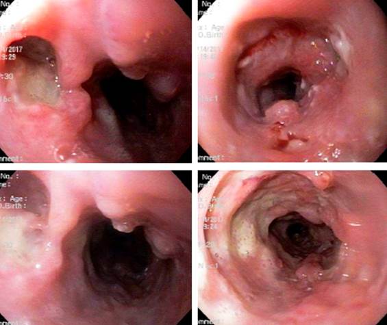
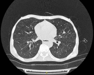
An Acid-Fast Bacillus (AFB) smear and an upper gastrointestinal endoscopy were performed as diagnostic support tests. The AFB smear was positive and the following findings were observed in the upper endoscopy: severely inflamed mucosa with irregular ulcerations up to 2 cm deep, dirty and covered with fibrin, up to 1 cm size irregular elevated lesions, suggestive of gastrointestinal tuberculosis. This was later confirmed by the histopathology report: chronic granulomatous esophagitis suggestive of tuberculosis infection (Figure 1), and a chest CT scan showing centrilobular nodules with tree-in-bud opacity pattern and characteristics suggestive of atypical or typical mycobacteria, and an infiltrate-like space-occupying lesion in the right upper lobe (Figure 2). Additionally, the following findings were reported in the complete blood count tests: leukocytes: 5040/mm3, neutrophils: 70 %, lymphocytes: 19 %, hemoglobin: 9.6 %, platelets: 271 000/mm3, prothrombin time: 18 seconds, international normalized ratio (INR): 1.45, blood urea nitrogen (BUN): 12 mg/dL, creatinine: 0.6 mg/dL, aspartate aminotransferase (AST): 123 IU/L, alanine aminotransferase (ALT): 44 IU/L, lactate dehydrogenase (LDH): 129 IU/L, CRP: 2.07. Complementary tests: HIV 191 (positive), HCV (non-reactive), HBV 0.4 (negative), treponema (negative), toxoplasma immunoglobulin M (IgM) 0.2 (negative).

Figure 1 Findings suggestive of gastrointestinal tuberculosis. 

Figure 2 Centrilobular nodules with a tree-in-bud opacity pattern. 

Based on these findings, treatment consisting in the administration of rifampicin (150 mg) + isoniazid (75 mg) + pyrazinamide (400 mg) + ethambutol (275 mg) every 24 hours was started. Somehow, on the sixth day of treatment the patient experienced systemic deterioration and died.

Discussion

Regarding clinical manifestations, 10 to 20% of all tuberculosis cases are extrapulmonary; somehow, this percentage increases considerably (45%-75%) in patients with HIV infection10. The high frequency of extrapulmonary tuberculosis in HIV-infected patients is caused by their compromised immune system and the absence of immune response to Mycobacterium tuberculosis, which allows hematogenous spread of the bacteria and the subsequent bacterial invasion of one or multiple extrapulmonary tissues.

The clinical presentation of esophageal tuberculosis has not yet been determined, in fact it can be described as a spectrum of diffuse clinical features; however, a large proportion of esophageal tuberculosis cases reported in the scientific literature coincide in describing the presence of dysphagia and odynophagia, together with decreased appetite and involuntary weight loss, as shown in the cases reported by Garg R & Carter S in 201711 and Martins M et al. in 20169. Even so, the occurrence of such manifestations is not always well defined, resulting in misdiagnosis, as the cases in which the presentation occurs along retrosternal pain, epigastric pain and vomiting, according to Kumar et al., 201612. Due to its gastrointestinal location, the clinical manifestations of esophageal tuberculosis are frequently related to this system, such as purulent rectal discharge and hematemesis, as reported by Zuleta et al., 201213 and Manrique et al., 201414, respectively, although they are non-specific for its detection and diagnosis.

With regard to paraclinical tests, in general terms, reaching an extrapulmonary tuberculosis diagnosis is difficult due to the fact that this is a rare disease and there is little familiarity with its clinical presentation. In addition, inaccessibility to the infection sites and the low bacterial load also make even more difficult its diagnosis10. Upper endoscopy and the collection of biopsy samples during the procedure is the most useful diagnostic tool, even though there are no characteristic findings and pathological anatomy is often inconclusive9.

Usually, a solitary crater-like ulcer with raised edges located in the middle third of the esophagus is observed in upper GI endoscopy9,15; however, as shown above, there is no classic or typical lesion of esophageal tuberculosis. According to the scientific literature, esophageal endoscopic findings vary from superficial ulcers to raised lesions suggestive of neoplastic lesions, as well as strictures and fistulas(16, 17). In contrast, Hun Seo et al. reported that endoscopic findings of esophageal tuberculosis include ulcerative, granular or hypertrophied lesions, being linear ulceration with irregular margins and a grayish base with an irregular infiltrated edge in the mid-esophagus the most common finding7.

Furthermore, endosonography, also known as endoscopic ultrasound, is currently a test with a high impact as it allows observing characteristic findings of this disease, such as the subcarinal or periesophageal presence of heterogeneous matted lymph nodes adjacent to the esophageal pathology8,25 with a predominantly hypoechogenic26 or anechoic echotexture, particularly in the center and some hyperechogenic threads and foci (spots)7,8,25. Other typical findings include the thickening of the esophageal wall and the formation of masses disrupting the adventitia7,27.

Regarding treatment, the standard pharmacological regimen recommended for treating esophageal tuberculosis is the same than the preferred regimen for treating tuberculosis, that is, an intensive phase of four anti-tuberculosis drugs (rifampicin, isoniazid, pyrazinamide and ethambutol)10,13,14,22, plus a 4-month continuation phase of isoniazid and rifampicin14; however, streptomycin can be used instead of ethambutol23. Some authors affirm the use of a treatment regimen based on three drugs21: isoniazid, rifampicin and pyrazinamide17, and some others decide to add pyridoxine9. Generally, these patients have a good prognosis if an early diagnosis is made21,23 with treatment success rates of 90%14; somehow, immunocompromised patients, particularly those with AIDS17,21,23, fail to respond adequately to antituberculosis treatment regimens and may die due to the severity of the infection21,23. This is why it has been described that the efficacy of treatment depends on the status of the patient’s immune system13. In the case reported here, despite the patient received timely pharmacological treatment (rifampicin 150 mg + isoniazid 75 mg + pyrazinamide 400 mg + ethambutol 275 mg), his health condition worsened progressively and systemically resulting in his death.

Conclusion

Esophageal tuberculosis is a rare type of extrapulmonary tuberculosis. Despite its wide spectrum of clinical manifestations, dysphagia is one of the most representative. Coinfection with HIV has been frequent in recent years. Timely use of upper gastrointestinal endoscopy and esophageal ultrasound is very useful to suspect its diagnosis, which is confirmed through the histopathological analysis of the lesions.

REFERENCES

1. Fadul-Pérez S. Protocolo de Vigilancia en Salud Pública, tuberculosis, código: 813. INS; 2020 [consultado el 30 de enero de 2021]. Disponible en: Disponible en: https://www.ins.gov.co/buscador-eventos/Lineamientos/PRO_Tuberculosis.pdfLinks ]

2. Onusida. Hoja informativa - Últimas estadísticas sobre el estado de la epidemia de sida. [consultado el 30 de mayo de 2018]. Disponible en: Disponible en: http://www.unaids.org/es/resources/fact-sheetLinks ]

3. OMS. VIH/sida. [consultado el 30 de mayo de 2018]. Disponible en: Disponible en: https://www.who.int/es/news-room/fact-sheets/detail/hiv-aidsLinks ]

4. Tang N, Frank A, Pahalawatta V, Lampinen J, Coblenz-Korte A, Dunn C, Li C, Cloherty G, Abravaya K, Leckie G. Analytical and clinical performance of Abbott RealTime MTB, an assay for detection of Mycobacterium tuberculosis in pulmonary specimens. Tuberculosis (Edinb). 2015;95(5):613-9. https://doi.org/10.1016/j.tube.2015.05.010Links ]

5. Procedimientos para la prevención y control de la tuberculosis. Manual 2017. Quito: Ministerio de Salud Pública de Ecuador; 2017 [consultado el 30 de mayo de 2018]. Disponible en: Disponible en: https://www.salud.gob.ec/wp-content/uploads/2017/07/MANUAL-DE-PROCEDIMIENTOS-DE-TB-FINAL.pdfLinks ]

6. Khanna V, Kumar A, Alexander N, Surendran P. A case report on esophageal tuberculosis - A rare entity. Int J Surg Case Rep. 2017;35:41-43. https://doi.org/10.1016/j.ijscr.2017.03.042Links ]

7. Seo JH, Kim GH, Jhi JH, Park YJ, Jang YS, Lee BE, Song GA. Endosonographic features of esophageal tuberculosis presenting as a subepithelial lesion. J Dig Dis. 2017;18(3):185-188. https://doi.org/10.1111/1751-2980.12454Links ]

8. Tang Y, Shi W, Sun X, Xi W. Endoscopic ultrasound in diagnosis of esophageal tuberculosis: 10-year experience at a tertiary care center. Dis Esophagus. 2017;30(8):1-6. https://doi.org/10.1093/dote/dox031Links ]

9. Martins M, Juliana O, Carvalho A, Santos A, Santos-Costa C, Monteiro J, Abreu A, Esteves J, Garcia J, Pinto-Correia J. Tuberculose esofágica primária: desafio para o cirurgião. Rev Port Cir. 2016;(38):31-35. [ Links ]

10. Bresky G, Silva N, Madariaga J. Esofagitis tuberculosa en un paciente HIV positivo. Rev Méd Chile. 2007;135(10):1323-1326. http://dx.doi.org/10.4067/S0034-98872007001000014Links ]

11. Garg R, Carter S, Barawi M. Dysphagia from esophageal tuberculosis in a patient with undiagnosed HIV infection. Endoscopy. 2017;49(8):E195-E196. https://doi.org/10.1055/s-0043-110667Links ]

12. Kumar S, Minz M, Sinha SK, Vaiphei K, Sharma A, Singh S, Kenwar DB. Esophageal tuberculosis with coexisting opportunistic infections in a renal allograft transplant recipient. Transpl Infect Dis. 2017;19(1). https://doi.org/10.1111/tid.12640Links ]

13. Zuleta MA, Viveros D, Cañón DP. Tuberculosis intestinal: reporte de caso y revisión de la literatura. Infectio. 2012;16(3):178-182. https://doi.org/10.1016/S0123-9392(12)70009-7Links ]

14. Manrique CA, Salinas DC, Martínez JD, Garzón OM, Hernández CG, Hormaza N, Lizarazo J, Marulanda J, Molano JC, Rey M. Tuberculosis esofágica: informe de un caso. Rev Col Gastroenterol. 2014;29(4):446-448. [ Links ]

15. Sala A, Leibovich N, Souto G, Sabatini C, Brodersen C, Segal E. Tuberculosis esofágica: presentación de un caso y revisión de la literatura. Acta Gastroenterol Latinoam. 2011;41(1):47-51. [ Links ]

16. Salinas DC, Moreno DC, Dennis RJ. Tuberculosis pulmonar, esofágica y laríngea. Acta Médica Colombiana. 2014;39(1):85-9. [ Links ]

17. Baños R, Serrano A, Alberca F, Alajarín M, Albaladejo A, Vargas A, Molina J. Tuberculosis esofágica. Rev Gastroenterol Perú. 2006;26:200-202. [ Links ]

18. Laverdure N, Lepilliez V, Mion F. Esophageal tuberculosis: A rather specific endoscopic lesion? Clin Res Hepatol Gastroenterol. 2016;40(6):e69-e70. https://doi.org/10.1016/j.clinre.2016.05.009Links ]

19. Lagos AC, Marques I, Reis J, Esteves P, Costeiro J, Afonso A, Costa B. Tuberculose primária do esófago. GE J Port Gastrenterol. 2012;19(3):143-145. https://doi.org/10.1016/j.jpg.2010.08.001Links ]

20. Momin RN, Chong VH. Oesophageal tuberculosis: rare but not to be forgotten. Singapore Med J. 2012;53(9):e192-4. [ Links ]

21. Pioner SR, Rech D; Menegotto L, Pioner F, Santos C. Tuberculose esofágica primária - relato de caso. Rev Col Bras Cir. 2007;34(5):357-358. [ Links ]

22. Mohan P, Myilsamy S, Leelakrishnan V. Esophageal tuberculosis. Clin Res Hepatol Gastroenterol. 2012;36(4):312-3. https://doi.org/10.1016/j.clinre.2011.12.017Links ]

23. Perdomo JA, Naomoto Y, Haisa M, Yamatsuji T, Kamikawa Y, Tanaka N. Tuberculosis of the esophagus. Dis Esophagus. 1998;11(1):72-4. [ Links ]

24. Montoya M, Chumbiraico R, Ricalde M, Cazorla E, Hernandez G. Blastomicosis Oral, Papilomatosis Laríngea y Tuberculosis Esofágica. Rev Peru Med Exp Salud Pública. 2012;29(2):250-254. [ Links ]

25. Puri R, Khaliq A, Kumar M, Sud R, Vasdev N. Esophageal tuberculosis: role of endoscopic ultrasound in diagnosis. Dis Esophagus. 2012;25(2):102-6. https://doi.org/10.1111/j.1442-2050.2011.01223.xLinks ]

26. Li YX, Nian WD, Wang HH. A case of esophageal tuberculosis with unusual endoscopic feature. Clin Res Hepatol Gastroenterol. 2018;42(1):e5-e6. https://doi.org/10.1016/j.clinre.2017.04.010Links ]

27. Mou Y, Zeng H, Wang QM, Yi H, Liu W, Wen D, Hu B. Esophageal tuberculosis initially misdiagnosed by endoscopy as a submucosal tumor. Endoscopy. 2015;47 Suppl 1 UCTN:E30-1. https://doi.org/10.1055/s-0034-1391133Links ]

Citation: Rozo-Ortiz EJ, Quemba-Mesa MP, Bustos-Acosta JD, Castellanos-Pedroza IY, Muñoz-Torres J, Vargas-Rodríguez LJ. Esophageal tuberculosis associated with HIV immunosuppression: Case report. Rev Colomb Gastroenterol. 2021;36(1):109-114. https://doi.org/10.22516/25007440.477

Funding

Universidad de Boyacá.

Authors’ contributions Counsel and case construction from the clinical perspective: Edwar Jassir Rozo-Ortiz. Counsel and case construction from a methodological point of view: Mónica Paola Quemba-Mesa. Field work data collection and writing of the case report: the other authors.

Received: October 30, 2019; Accepted: March 02, 2020

*Correspondence: Edwar Jassir Rozo Ortiz. eduarrozo@gmail.com

Conflicts of interest

None stated by the authors.

Creative Commons License This is an open-access article distributed under the terms of the Creative Commons Attribution License