


 texto en
  Inglés (pdf)
 Articulo en XML
 Referencias del artículo
 Enviar articulo por email
 Citado por SciELO
  Citado por Google
  Similares en
SciELO
Similares en
SciELO  Similares en Google
 Permalink
Clinical case
The patient was a 68-year-old woman with a history of diabetes mellitus and arterial hypertension, who was admitted due to abdominal pain in the right hypochondrium associated with jaundice. An ultrasound of the abdomen showed cholelithiasis and dilatation of the bile duct. It was considered that she was at high risk of choledocholithiasis, so an endoscopic retrograde cholangiopancreatography (ERCP) was scheduled, which revealed a large radiolucent image in the common bile duct, compatible with Mirizzi syndrome grade IV. Endoscopic extraction of the stone was not achieved, so a biliary stent was inserted, and purulent drainage was obtained (Figure 1).

Figure 1 ERCP - Radiolucent image in the common bile duct, compatible with Mirizzi syndrome grade IV.
Subsequently, the patient was scheduled for open cholecystectomy with biliary tract exploration. The procedure revealed a gallbladder with thickened walls, with Mirizzi syndrome grade IV; a large gallstone was extracted from the biliary tract using Randall forceps, an exhaustive lavage of the biliary tract was performed, and it was decided to leave a T-tube. The patient had a torpid evolution during the early postoperative period, so on the third day, it was decided to perform a computerized axial tomography (CAT) of the abdomen, finding an anterior perihepatic collection, which was successfully drained percutaneously; biliary material was extracted. After 3 days of drainage, the abdomen was acute, requiring a re-operation, in which bilioperitoneum and dislodged T-tube were observed; the peritoneal fluid was evacuated and a choledochorraphy with omental patch was performed. She was transferred to the intensive care unit (ICU) with mechanical ventilation and vasopressor support; Klebsiella pneumoniae and Enterobacter cloacae appeared in the peritoneal fluid and were treated with targeted antibiotic. In the following days, she presented hematemesis and fall of hemoglobin levels, so she was taken to endoscopy, finding a bleeding through the papilla; therefore, management with sclerotherapy was carried out, without achieving bleeding control. An angiotomography of the abdomen was then performed in which a pseudoaneurysm of the right hepatic artery with communication biliary tract was found (Figure 2).
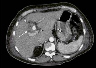
Figure 2 Angiotomography of the abdomen Pseudoaneurysm of the right hepatic artery. Hypodense area surrounding the vessel in contact with the bile duct (arrow).
An interventional radiology assessment was requested, in which Onyx embolization of small branches of the right hepatic artery was performed. The patient continued bleeding and also presented with hypotension and vasopressor support requirement; therefore, she was taken to a new emergency CT angiography of the celiac axis, and superselective embolization of the hepatic right hepatic artery pseudoaneurysm was attempted, which was unsuccessful. Due to this result, percutaneous embolization of the pseudoaneurysm was performed, with subsequent fluoroscopic evidence of its correction, without complications. The patient had an adequate course without bleeding or anemization and without requiring further interventions.
Discussion
Hemobilia is defined as bleeding from the biliary tract and is a rare cause of upper gastrointestinal bleeding, accounting for less than 5 % of all cases1. The first case was described in 1654 by Francis Glisson, a British anatomist, who reported following a blunt trauma to the right upper quadrant of the abdomen. The term hemobilia has been used since 1948, when Philip Sandbrom used it in patients presenting with such bleeding. Series of cases of hemobilia following trauma, endoscopic or percutaneous procedures have been reported over the years, and their diagnosis is easier when patients have a history of any of these events, given that clinical suspicion is low due to their low incidence2,3.
Its etiology is variable and occurs when there is a fistula between a blood vessel and the biliary tract. This may be caused by an obstruction or a lesion of the pancreaticobiliary tract organs. Normally, high-pressure arterial vessels are involved, which leads to abundant bleeding, although cases in which portal hypertension occurs have been described, and the direct causes are venous vessels4. The anatomical proximity of the bile ducts and the vasculature of the biliary tract make them more prone to iatrogenic lesions, so hemobilia will be related to percutaneous, endoscopic, or surgical invasive procedures in approximately 50 % of the cases. The literature reports that hemobilia in 38% of patients are associated with percutaneous biliary tract drainage or percutaneous liver biopsies, while 19% are occur after open and laparoscopic surgical procedures, and 10% after endoscopic procedures. During surgical procedures, biliovascular lesions may be generated by the passage of sutures, diathermy, by clips, or by instruments that injure the walls of the vessels and the biliary tract. Other less frequent etiologies have been described such as trauma, mainly blunt trauma, neoplasms, biliary lithiasis, inflammatory diseases, infections by parasites, or anatomical malformations5,6.
Regarding pathophysiology, it should be noted that the mixture between bile and blood creates a dispersion between particles that prevents the proper formation of clots, so they are weak and with little adhesion. In addition, bile is considered to contain fibrinolytic factors that limit fibrin production. It is believed that, when bleeding is abundant, it passes into the duodenum and manifests with melena or hematemesis; on the other hand, in minor bleeding, weak clots may form, obstructing the biliary tract, generating jaundice, and predisposing to the subsequent formation of stones7.
Their clinical manifestations depend on the amount and frequency of bleeding. Quincke’s triad (upper abdominal pain, upper gastrointestinal hemorrhage, and jaundice) is only observed in one-third of cases.8 Bleeding manifests as melena in 90 % of the cases and hematemesis in 60 %, whereas jaundice and pain in the upper hemiabdomen are present in 60 % and 70 % of cases, respectively. In some cases, hemobilia has been associated with pancreatitis or cholangitis. Chronic anemia is reported in few patients, taking into account blood losses secondary to hemobilia9. When hemobilia is secondary to trauma, it may develop immediately after the trauma or weeks later, and generally the origin is 50 % intrahepatic and 50 % extrahepatic, with the presence of abdominal pain in most cases, but with variable onset of jaundice6.
The diagnosis of hemobilia is complex and requires a high diagnostic suspicion given its low incidence It should always be suspected in the patient with upper GI bleeding, with a history of instrumentation or manipulation of the biliary tract, or with a previous diagnosis of biliopancreatic disease. Initial laboratory tests include a complete blood count and liver profile (transaminases, bilirubin, and its differentials), because a significant percentage of patients present with anemia and hyperbilirubinemia; it is also important to request clotting times10,11. The initial approach to gastrointestinal bleeding includes an endoscopic study, with evidence of bleeding or release of clots through the ampulla of Vater in up to 90% of patients. ERCP is useful when hemobilia is associated with obstructive jaundice because it shows bleeding from the papilla and allows the evacuation of clots causing the obstruction12,13.
When hemobilia is suspected, angiography is often performed in search of vascular abnormalities such as pseudoaneurysms or fistulas that may explain the bleeding, allowing the correction of these abnormalities in the same procedure. Currently, CT angiography can be performed as the initial study, with sensitivities and specificities similar to the gold standard, which allows observing the vascular alteration with subsequent therapeutic planning14.
Treatment is based on interventional radiology by angiography and embolization, with autologous blood, coils, balloon, or cyanoacrylate, which require the use of vascular stents in selected cases. This procedure has a low rate of morbidity and mortality, is well tolerated and successful in most patients. In very few cases, and as a last resort, surgery is used to treat hemobilia, during which direct examination of the liver, ligation of arterial vessels, resection of the pseudoaneurysm and cholecystectomy are performed, and resolution of the obstruction of the bile ducts is achieved15.
Conclusion
Hemobilia should be suspected in patients with evidence of gastrointestinal bleeding in the postoperative period following any endoscopic, percutaneous, or surgical procedure of the liver, biliary tract, and pancreas. The initial approach to these patients is CT angiography, which confirms the diagnosis to plan further angiographic treatment, while surgery is reserved as the last therapeutic alternative.

