SciELO - Scientific Electronic Library Online

 
vol.36 número2Gastrostomía descompresiva en obstrucción gastrointestinal maligna: reporte de caso y revisión de la literaturaÚlcera rectal solitaria como simulador de cáncer colorrectal y enfermedad de Crohn: reporte de caso índice de autoresíndice de materiabúsqueda de artículos
Home Pagelista alfabética de revistas  

Servicios Personalizados

Revista

Articulo

Indicadores

Links relacionados

  • En proceso de indezaciónCitado por Google
  • No hay articulos similaresSimilares en SciELO
  • En proceso de indezaciónSimilares en Google

Compartir


Revista colombiana de Gastroenterología

versión impresa ISSN 0120-9957versión On-line ISSN 2500-7440

Rev. colomb. Gastroenterol. vol.36 no.2 Bogotá abr./jun. 2021  Epub 25-Nov-2021

https://doi.org/10.22516/25007440.638 

Report of cases

Appendiceal intussusception: a rare finding during colonoscopy. Case report

Luis Jorge Lombana-Amaya1  * 
http://orcid.org/0000-0002-7165-2124

Saúl Rugeles2 
http://orcid.org/0000-0003-0401-0743

Jorge Alejandro González-González3 
http://orcid.org/0000-0002-7604-0493

1 MD, Professor of Surgery, Universidad Javeriana. Chief of the Colon and Rectum Unit, Department of Surgery, San Hospital Universitario San Ignacio. Bogotá, Colombia.

2 MD, Professor of Surgery, Universidad Javeriana. Gastrointestinal Surgeon, Department of Surgery, Hospital Universitario San Ignacio. Bogotá, Colombia.

3 MD, General Surgery Resident, Department of Surgery, Hospital Universitario San Ignacio, Faculty of Medicine, Pontificia Universidad Javeriana. Bogotá, Colombia.


Abstract

This is the case of a young patient with an incidental endoscopic finding of a lesion in the cecum during follow-up colonoscopy with benign histology. The diagnostic process and surgical treatment are described. The existing literature was reviewed and the incidence, symptoms and indications of surgical treatment of this rare condition are discussed.

Keywords: Intussusception; Appendiceal neoplasm; Colonoscopy; Endometriosis

Resumen

Se presenta el caso de una mujer joven con hallazgo endoscópico incidental en una colonoscopia de seguimiento, que consistía en una lesión en el ciego con histología benigna. Se describe el proceso diagnóstico y el tratamiento quirúrgico. Se revisa la literatura existente y se discuten la incidencia, el cuadro clínico y las indicaciones de tratamiento quirúrgico de una patología rara de baja aparición.

Palabras clave: Intususcepción; neoplasia apendicular; colonoscopia; endometriosis

Introduction

Appendiceal intussusception is defined as the partial or complete invagination of the appendix and its corresponding mesentery within the adjoining intestinal lumen. In 1958, McKidd reported the first case in the literature, involving a 7-year-old patient.1 It is a rare condition with few cases reported in the literature, with an incidence of 0.01 % according to a cohort study conducted by Collins, which included 71 000 patients who underwent appendectomy over a 40-year period.2,3 It is most common in women in the fourth or fifth decades of life. Diagnosing this condition is difficult due to its non-specific symptoms and low incidence. The purpose of this publication is to present a case treated at our institution.

Case report

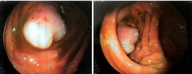
This is the case of a 43-year-old female patient with a history of a low-grade neuroendocrine tumor in the upper rectum, resected endoscopically 3 years earlier, who was undergoing clinical and endoscopic follow-up. She was completely asymptomatic but, during her the last total colonoscopy, a lesion of 2 cm in size was found at the level of the meatus of the appendix (Figure 1). A biopsy of the lesion was performed, revealing normal-looking mucosa with no signs suggesting malignancy or other pathological changes. CT scan of the abdomen and pelvis, as well as 5-hydroxyindoleacetic acid levels were normal. Her clinical examination was normal.

Figure 1 Invagination of cecal appendix, colonoscopy view of the cecal pole. 

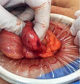
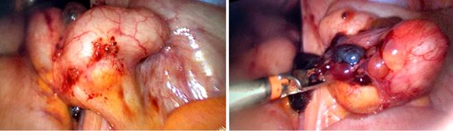
Although the histopathological study was inconclusive, it was decided to perform minimally invasive surgery. Surgery was started with a laparoscopy in which no cecal appendix was observed; a Rockey Davis incision was then made using an Alexis wound retractor to expose the cecum. Appendicular invagination was confirmed through palpation and visualization (Figure 2).

Figure 2 Cecum without appendix due to invagination. Surgical finding. 

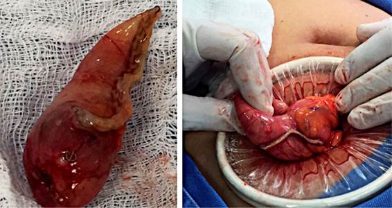
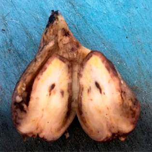
A cecectomy was performed using a mechanical linear cutter and the specimen was sent to pathology (Figure 3). Postoperative evolution was adequate and without complications. Pathology confirmed invagination secondary to foci of endometriosis (Figure 4).

Figure 3 Cecectomy with mechanical suture. 

Figure 4 Pathology specimen showing the complete invaginated appendix and its lumen. 

During postoperative follow-up, it was possible to obtain the report of the colonoscopy performed 3 years earlier when the neuroendocrine tumor in the rectum was treated, with a report of cecum and meatus of the appendix within normal limits, although without photographic documentation.

Discussion

The pathophysiology of this condition is unknown; however, it is associated with the presence of appendix alterations, most notably a mass at that level. Altered peristalsis is thought to occur and is associated with inflammation, foreign body, Crohn’s disease, parasites, polyps, lymphoid hyperplasia, ecchymoses, mucosa-associated lymphoid tissue lymphoma (MALT), adenoma, papilloma, adenocarcinoma, endometriosis, and mucocele4-7; the latter 2 are the main causes of invagination in adults, while inflammatory processes are the most common in children8-10.

Given its low incidence and the multiple and unspecific symptoms, the diagnosis of this condition poses a challenge for physicians. Three types of symptoms have been described in these patients:

  • symptoms of typical acute appendicitis;

  • nonspecific symptoms of intussusception (severe abdominal pain, vomiting, diarrhea, and melena); and

  • sensation of a mass, pain in the lower right quadrant, and melena for several months.

Most patients are asymptomatic with incidental findings during colonoscopy, which can also be found intraoperatively (57 %) or by pathology (11 %)1,7,8,11.

Regarding its etiology, the most common cause of appendiceal intussusception in adults is endometriosis, a benign entity first described in 1860 by Von Rokitansky12. It is defined as the presence of endometrial glands and stroma outside the uterine cavity. Between 10 % and 15 % of women suffer from endometriosis.11,13-15. The rectum and sigmoid colon are the main sites of intestinal implantation, followed by the ascending colon, small intestine, and appendix, which is observed in 0.08 % to 8 % of cases (Figure 5)8,10,12,16.

Figure 5 Foci of endometriosis involving a cecal appendix. 

Histologically, it is characterized by the presence of endometrial glands and stroma, which may be associated with macrophages. Atypical benign changes include necrotic pseudoxanthomatous nodules; vascular, perineural and nodal involvement; and metaplastic changes in both glands and stroma. In addition, it may contain premalignant changes such as polypoid endometriosis, stromal endometriosis, mesothelial hyperplasia associated with endometriosis, and atypical endometriosis with a 1% risk of conversion to cancer15.

The diagnostic methods used include enhanced abdominal tomography and colon enema, followed by ultrasound, which is very useful in the pediatric population. In colonoscopies performed in adults for various reasons, it is described as a pseudopedunculated polyp17-19 or mass in the meatus of the appendix, as in this case. No more than 200 cases have been reported in the literature; however, it is impossible to know the incidence of this finding. If it is approached as a polypoid lesion, performing a polypectomy may lead to the risk of perforation and peritonitis20. It is important to bear this entity in mind when evaluating lesions at the meatus of the appendix. To this end, a report by McSwain proposed a classification of intussusception depending on the anatomy found, simplified years later by Langsman17,21:

  • Type I: invagination of the tip of the appendix into the proximal appendix,

  • Type II: invagination begins at any point of the appendix other than the tip and base of the appendix,

  • Type III: invagination begins at the appendicular base into the most distal part of the cecum,

  • Type IV: retrograde invagination of a proximal segment within a distal segment of the same appendix,

  • Type V: complete invagination of the appendix into the cecum (as in this case).

Surgical treatment is the standard management, considering that there is always the probability of a malignant disease causing the intussusception. In most cases, patients are taken to cecectomy, but some cases may undergo a right hemicolectomy22. Endometriosis is the most common cause described in the reports of the last 10 years.

This case illustrates this disease very well. However, two more cecectomies with appendicular invagination secondary to endometriosis were performed in the following two years on the service, implying that the frequency is not as low as reported in the literature. For this reason, appendicular intussusception should be listed as one of the differential diagnoses of lesions in the meatus of the appendix.

Referencias

1. McKidd J. Case of invagination of caecum and appendix. Edinburgh Med J. 1858;4:793-7. [ Links ]

2. Chaar CI, Wexelman B, Zuckerman K, Longo W. Intussusception of the appendix: comprehensive review of the literature. Am J Surg. 2009;198(1):122-8. https://doi.org/10.1016/j.amjsurg.2008.08.023Links ]

3. Collins DC. 71,000 human appendix specimens. A final report, summarizing forty years’ study. Am J Proctol. 1963;14:265-81. [ Links ]

4. Atkinson GO, Gay BB Jr, Naffis D. Intussusception of the appendix in children. AJR Am J Roentgenol. 1976;126(6):1164-8. https://doi.org/10.2214/ajr.126.6.1164Links ]

5. Gilpin D. Intussusception of the appendix. Ulster Med J. 1989;58(2):193-5. [ Links ]

6. Komine N, Yasunaga C, Nakamoto M, Shima I, Iso Y, Takeda Y, et al. Intussusception of the appendix that reduced spontaneously during follow-up in a patient on hemodialysis therapy. Intern Med. 2004;43(6):479-83. https://doi.org/10.2169/internalmedicine.43.479Links ]

7. Tăban S, Dema A, Lazăr D, Sporea I, Lazăr E, Cornianu M. An unusual «tumor» of the cecum: the inverted appendiceal stump. Rom J Morphol Embryol. 2006;47(2):193-6. [ Links ]

8. Lee DJ, Kim HC, Yang DM, Kim SW, Ryu JK, Won KY, et al. A case of intussusception of the appendix secondary to endometriosis: US and CT findings. J Clin Ultrasound. 2015;43(7):443-6. https://doi.org/10.1002/jcu.22188Links ]

9. Duncan JE, DeNobile JW, Sweeney WB. Colonoscopic diagnosis of appendiceal intussusception: case report and review of the literature. JSLS. 2005;9(4):488-90. [ Links ]

10. Liang HH, Huang MT, Wei PL, Weu W, Lin YH, Tiang C, et al. Endometriosis-induced appendiceal intussusception. Am J Surg. 2009;197(6):e66-8. https://doi.org/10.1016/j.amjsurg.2008.04.024Links ]

11. Sánchez Santamaría M, Sánchez Gualberto A, Alcóucer Díaz MR, Creagh Cerquera R, Pujol de la Llave E. Endometriosis como causa de invaginación intestinal. Rev Clin Esp. 2007;207(10):536-7. Spanish. https://doi.org/10.1157/13111561Links ]

12. Costa M, Bento A, Batista H, Oliveira F. Endometriosis-induced intussusception of the caecal appendix. BMJ Case Rep. 2014;2014:bcr2013200098. https://doi.org/10.1136/bcr-2013-200098Links ]

13. Pérez Cabanas I, Zornoza G, Arconada FA, Oroz F, Voltas F. Endometriósis del apéndice vermiforme. Rev Med Univ Navarra. 1974;18(1-2):69-76. [ Links ]

14. Gustofson RL, Kim N, Liu S, Stratton P. Endometriosis and the appendix: a case series and comprehensive review of the literature. Fertil Steril. 2006;86(2):298-303. https://doi.org/10.1016/j.fertnstert.2005.12.076Links ]

15. McCluggage WG. Endometriosis-related pathology: a discussion of selected uncommon benign, premalignant and malignant lesions. Histopathology. 2020;76(1):76-92. https://doi.org/10.1111/his.13970Links ]

16. Iqbal CW, Kamath AS, Zietlow SP. Appendiceal intussusception masquerading as an ileocolic intussusception. J Gastrointest Surg. 2012;16(5):1076-7. https://doi.org/10.1007/s11605-012-1848-zLinks ]

17. Salehzadeh A, Scala A, Simson JN. Appendiceal intussusception mistaken for a polyp at colonoscopy: case report and review of literature. Ann R Coll Surg Engl. 2010;92(6):W46-8. https://doi.org/10.1308/147870810X12699662981591Links ]

18. Birriel TJ, Smith E, Eyvazzadeh D. Appendiceal intussusception from endometriosis: endoscopic and laparoscopic approach. Ann R Coll Surg Engl. 2017;99(1):e1-e2. https://doi.org/10.1308/rcsann.2016.0244Links ]

19. Sriram PV, Seitz U, Soehendra N, Schroeder S. Endoscopic appendectomy in a case of appendicular intussusception due to endometriosis, mimicking a cecal polyp. Am J Gastroenterol. 2000;95(6):1594-6. https://doi.org/10.1111/j.1572-0241.2000.02108.xLinks ]

20. Tavakkoli H, Sadrkabir SM, Mahzouni P. Colonoscopic diagnosis of appendiceal intussusception in a patient with intermittent abdominal pain: a case report. World J Gastroenterol. 2007;13(31):4274-7. https://doi.org/10.3748/wjg.v13.i31.4274Links ]

21. Betancourth-Alvarenga JE, Vázquez-Rueda F, Murcia-Pascual FJ, Ayala-Montoro J. Abdomen agudo secundario a invaginación apendicular. An Pediatr (Barc). 2015;82(1):e56-9. https://doi.org/10.1016/j.anpedi.2014.01.016Links ]

22. Suazo C, Burdiles P, Larach A. Invaginación apendicular. Revista Chilena de Cirugía. 2012;64(1):76-8. https://doi.org/10.4067/S0718-40262012000100013Links ]

Citation: Lombana-Amaya LJ, Rugeles S, González-González JA. Appendiceal intussusception: a rare finding during colonoscopy. Case report. Rev Colomb Gastroenterol. 2021;36(2):275-279. https://doi.org/10.22516/25007440.638

Received: August 13, 2020; Accepted: September 02, 2020

*Correspondence: Luis Jorge Lombana-Amaya. ljlombana@hotmail.com

Creative Commons License This is an open-access article distributed under the terms of the Creative Commons Attribution License