SciELO - Scientific Electronic Library Online

 
vol.36 suppl.1Cystic intestinal pneumatosis in a young adult with no clinical history: Case reportSubcapsular hepatic hematoma after ERCP. Case report and literature review author indexsubject indexarticles search
Home Pagealphabetic serial listing  

Services on Demand

Journal

Article

Indicators

Related links

  • On index processCited by Google
  • Have no similar articlesSimilars in SciELO
  • On index processSimilars in Google

Share


Revista colombiana de Gastroenterología

Print version ISSN 0120-9957On-line version ISSN 2500-7440

Rev. colomb. Gastroenterol. vol.36  supl.1 Bogotá Apr. 2021  Epub Feb 21, 2022

https://doi.org/10.22516/25007440.550 

Clinical case

Sphincter of Oddi dysfunction: Case report

Arecio Peñaloza-Ramírez1  * 
http://orcid.org/0000-0001-9052-530X

Esteban Coral-Argoty2 
http://orcid.org/0000-0002-6154-5149

María Castro-Rodríguez3 
http://orcid.org/0000-0003-3356-3224

Johann Álvarez-Gil4 
http://orcid.org/0000-0003-3808-9429

Pedro Aponte-Ordóñez5 
http://orcid.org/0000-0001-5148-7232

1 Associate researcher, Colciencias. Head, Gastroenterology and digestive endoscopy specialization program, Fundación Universitaria Ciencias de la Salud, Sociedad de Cirugía de Bogotá. Head, Gastroenterology and digestive endoscopy service, Hospital de San José. Bogotá, Colombia.

2 General surgeon. Gastroenterology and digestive endoscopy resident, Fundación Universitaria de Ciencias de la Salud, Sociedad de Cirugía de Bogotá. Bogotá, Colombia.

3 Internal medicine specialist. Gastroenterology and digestive endoscopy resident, Fundación Universitaria de Ciencias de la Salud, Sociedad de Cirugía de Bogotá. Bogotá, Colombia.

4 Internal medicine specialist. Gastroenterology and digestive endoscopy resident, Fundación Universitaria de Ciencias de la Salud, Sociedad de Cirugía de Bogotá. Bogotá, Colombia.

5 General surgeon, Gastroenterology and digestive endoscopy specialist. Associate professor, Gastroenterology and digestive endoscopy specialization program, Fundación Universitaria Ciencias de la Salud, Sociedad de Cirugía de Bogotá. Bogotá, Colombia.


Abstract

Sphincter of Oddi dysfunction is a clinical syndrome caused by functional (dyskinesia) or structural (stenosis) disease. The estimated prevalence of this condition in the general population is 1%, reaching 20% in patients with persistent pain after cholecystectomy and 70% in patients with idiopathic recurrent acute pancreatitis. It is clinically characterized by the presence of abdominal pain, similar to biliary colic or pancreatic pain in the absence of organic biliary disease. It is also observed in patients with idiopathic recurrent pancreatitis, associated with elevated pancreatic or hepatic enzymes, and bile duct and/or pancreatic duct dilatation. Treatment for sphincter of Oddi dysfunction type I is based on endoscopic sphincterotomy, but there is controversy regarding the management of sphincter of Oddi dysfunction types II and III. This article presents the clinical case of a 67-year-old female patient with a history of cholecystectomy by laparotomy. After the surgical procedure, she reported abdominal pain predominantly in the right hypochondrium, colicky, associated with emesis of biliary characteristics. Cholangioresonance report revealed mild intrahepatic bile duct dilatation, and scintigraphy with HIDA scan showed sphincter of Oddi dysfunction. Endoscopic sphincterotomy was performed. The patient was asymptomatic and the sphincter of Oddi dysfunction had resolved at two-year follow-up.

Keywords: Sphincter of Oddi dyskinesia; Endoscopic sphincterotomy; Scintigraphy; Endoscopic retrograde cholangiopancreatography

Resumen

La disfunción del esfínter de Oddi es un síndrome clínico causado por una enfermedad funcional (discinesia) o estructural (estenosis). La prevalencia estimada de disfunción del esfínter de Oddi en la población en general es del 1 %; aumentando a 20 % para pacientes con dolor persistente posterior a colecistectomía y a 70 % en pacientes con pancreatitis aguda recurrente idiopática. Se caracteriza clínicamente por la presencia de dolor abdominal, similar al cólico biliar o dolor tipo pancreático en ausencia de patología biliar orgánica; así como en pacientes con pancreatitis recurrente idiopática asociada con elevación de enzimas pancreáticas o hepáticas, y dilatación del conducto biliar o pancreático. El tratamiento para la disfunción del esfínter de Oddi tipo I se basa en la realización de esfinterotomía endoscópica, pero existe controversia en el manejo de la disfunción del esfínter de Oddi tipo II y III. En este artículo se presenta el caso clínico de una paciente de 67 años con antecedente de colecistectomía por laparotomía. Después del procedimiento quirúrgico refirió un dolor abdominal de predominio en el hipocondrio derecho tipo cólico asociado con emesis de características biliares. En el reporte de colangiorresonancia se encontró una ligera dilatación de la vía biliar intrahepática y gammagrafía con ácido iminodiacético hepatobiliar (HIDA) diagnóstica de disfunción del esfínter de Oddi. Se realizó una esfinterotomía endoscópica. En el seguimiento, dos años después, la paciente se encontraba asintomática con la disfunción del esfínter de Oddi resuelta.

Palabras clave: Discinesia del esfínter de Oddi; esfinterotomía endoscópica; gammagrafía; colangiopancreatografía retrógrada endoscópica

Introduction

The sphincter of Oddi, first described by Ruggero Oddi in 18871, is a system of sphincters composed of connective tissue, circular and longitudinal smooth muscle fibers mostly found within the duodenal wall. The system is composed of three sphincters: the sphincter of the common bile duct, the pancreatic sphincter and a common sphincter where the muscle fibers of the other two sphincters intertwine in an 8 shape way2.

The sphincter of Oddi is responsible for regulating the flow of bile and the exocrine secretion of the pancreas; it prevents the reflux of enteric contents from the duodenum into the pancreatic and biliary system, and it diverts bile into the gallbladder between meals, which facilitates its physiological concentration. During fasting, the sphincter of Oddi maintains pressures ranging from 3 mm Hg to 35 mm Hg (a pressure > 40 mm Hg is considered to be abnormal), with contractions that promote the flow into the duodenum, with an average frequency of 4 contractions per minute with an average duration of 6 seconds. Although there is a relationship with the migrating motor complex of the duodenum, most of the bile flow seems to be passive between the contractions of the sphincter of Oddi and increase due to gallbladder contraction, antral distention, the effect of cholecystokinin, and a decreased amplitude of the contractions of the sphincter in the postprandial state3.

Sphincter of Oddi dysfunction is a clinical syndrome caused by a functional (dyskinesia) or a structural (stenosis) disease4. It is clinically characterized by the presence of abdominal pain, similar to biliary colic or pancreatitis-like pain in the absence of an organic biliopancreatic disease. It should also be suspected in patients with recurrent idiopathic acute pancreatitis; pain and pancreatitis must be associated with elevated levels of pancreatic or hepatic enzymes, or with dilatation of the common bile duct or the pancreatic duct5.

This syndrome occurs more frequently in women between 20 and 50-years old who have undergone a cholecystectomy. The estimated prevalence of sphincter of Oddi dysfunction in the general population is 1%, but in patients with persistent postcholecystectomy pain and in those with recurrent idiopathic acute pancreatitis it increases to 20 % and 70%, respectively. However, its true prevalence cannot be easily determined due to the lack of definitive biomarkers or diagnostic criteria6-10.

Risk factors of sphincter of Oddi dysfunction include the presence of choledocholithiasis, cholesterosis, pancreatitis, ascaridiasis and malignancy, and the performance of endoscopic retrograde cholangiopancreatography (ERCP) or the surgical manipulation of the bile duct5.

The Milwaukee classification for sphincter of Oddi dysfunction, which divides patients into 3 types based on its clinical presentation and altered laboratory and imaging findings, is used for its diagnosis (Table 1)11-13. Somehow, the Rome IV classification, which describes 2 types of biliary sphincter of Oddi dysfunction and one third type corresponding to episodic functional abdominal pain, was published in 2016 (Table 2)14.

Table 1 Milwaukee classification for sphincter of Oddi dysfunction15

Biliary sphincter of Oddi dysfunction Pancreatic sphincter of Oddi dysfunction

  • Type I:

  • Biliary colic-like pain

  • Elevated liver enzymes (AST, ALT, FA) 2 times above the normal limits and documented at least 2 times during the pain episode

  • Dilatation of the common bile duct (> 12 mm) or delayed biliary drainage (> 45 min)

  • Type I:

  • Recurrent pancreatitis or pain possibly associated with pancreatitis.

  • Elevated pancreatic enzymes (amylase, lipase) 2 times above normal limits and documented at least 2 times during the pain episode

  • Dilatation of the pancreatic duct (head > 6 mm and body > 5 mm) or delayed pancreatic fluid drainage (> 9 min).

  • Type II:

  • Biliary colic-like pain and at least 1 of the other 2 criteria described above

  • Type II:

  • Pain possibly associated with pancreatitis and at least 1 of the other 2 criteria described above

  • Type III:

  • Biliary colic-like pain without any other alteration

  • Type III:

  • Pain of possible pancreatic origin without any other alteration

ALT: alanine aminotransferase; AST: aspartate aminotransferase; ALP: alkaline phosphatase. Taken from: Madura JA 2nd et al. Surg Clin North Am. 2007;87(6):1417-29, ix.

Table 2 Criteria for the diagnosis of both biliary and pancreatic sphincter of Oddi dysfunction (ROMA IV classification)14

Biliary sphincter of Oddi dysfunction Pancreatic sphincter of Oddi dysfunction

  • Diagnosis

  • The following criteria must be met:

  • Biliary colic-like pain

  • Elevated hepatic enzymes or dilatation of the common bile duct

  • Absence of choledocholithiasis or other structural abnormalities.

  • Supporting criteria:

  • Normal amylase and lipase levels

  • Abnormal sphincter of Oddi manometry

  • Hepatobiliary scintigraphy

  • Diagnosis

  • The following criteria must be met:

  • Documented recurrent episodes of pancreatitis (typical pain with amylase or lipase levels > 3 times the upper normal limit or imaging findings of acute pancreatitis).

  • Exclusion of other etiologies of pancreatitis.

  • Negative EUS

  • Abnormal sphincter of Oddi manometry

  • Suspected biliary sphincter of Oddi dysfunction:

  • Biliary colic-like pain and at least 1 associated target finding.

  • Episodic functional abdominal pain:

  • Biliary-colic pain without any other alteration.

To the best of our knowledge, to date, in Colombia there are no case reports describing a sphincter of Oddi dysfunction case; moreover, there is controversy regarding the diagnosis and treatment of this condition.

Clinical case

This is the case of a 67-year-old woman who received treatment for cholelithiasis in another health institution and who underwent a laparoscopic cholecystectomy in 2013; besides, she had a history of dyslipidemia which was being treated with atorvastatin. After the surgical procedure, she repeatedly visited the emergency department due to abdominal colicky pain predominantly located in the right hypochondrium, together with bile vomiting; however, normal liver function tests values were found in all these episodes. In December 2016, a magnetic resonance cholangiopancreatography (MRCP) showed the surgical absence of the gallbladder, a dilatation of the intrahepatic bile duct in the left lobe with an 8 mm common bile duct, and absence of choledocholithiasis (Figure 1). Normal values were reported in both the complete blood count test and the liver function tests (Table 3).

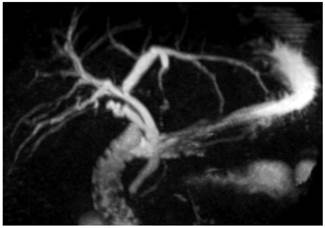
Figure 1 MRCP. Postcholecystectomy status, dilatation of the intrahepatic bile duct in the left lobe, common bile duct of 8 mm. 

Table 3 Postcholecystectomy laboratory tests results 

Laboratory test Results
Complete blood count

  • Leukocytes: 10 100 x 103/µL

  • Neutrophils: 7600 x 103/µL

  • Hemoglobin: 15.5 g/dL

  • Hematocrit: 45.7%.

  • Platelets: 307 000 x 103/µL

Liver function tests

  • AST: 19 U/L

  • ALT: 36 U/L

  • Alkaline phosphatase: 84 U/L

  • Total bilirubin: 0.7 mg/dL

  • Direct bilirubin: 0.3 mg/dL

  • Indirect bilirubin: 0.4 mg/dL

In October 2017, she was evaluated in a medical consultation by our service, where, due to her clinical signs and her surgical history, sphincter of Oddi dysfunction was suspected. A hepatobiliary iminodiacetic acid (HIDA) scan was then requested. The HIDA scan was performed in November 2017 and the following findings were reported: “elimination to the intrahepatic duct before 15 minutes with abundant retention in the major hepatic ducts and the common bile duct. A score of 9 points was obtained in the Sostre scale for sphincter of Oddi dysfunction (scores > 5 points are consistent)” (Table 4,Figure 2).

Table 4 Sostre scale score. 

Oddi’s dysfunction. Sostre Score Points Patient

  • Common bile duct peak time

  • < 10 minutes

  • > 10 minutes

  • 0

  • 1

1

  • Biliary visualization time

  • < 15 min

  • > 15 min

  • 0

  • 1

1

  • Biliary tree prominence

  • Not prominent

  • Prominence of major intrahepatic ducts

  • Prominence of minor intrahepatic ducts

  • 0

  • 1

  • 2

1

  • Intestinal visualization

  • < 15 minutes

  • 15-30 minutes

  • > 30 minutes

  • 0

  • 1

  • 2

2

  • Evacuation of the common bile duct

  • > 50 %

  • < 50 %

  • Absence of changes

  • Increases

  • 0

  • 1

  • 2

  • 3

1

  • Common bile duct/liver ratio

  • Common bile duct 60 min < liver 60 min

  • Common bile duct 60 min > liver 60 < liver 15 min

  • Common bile duct 60 min > liver 60 e = liver 15 min

  • Common bile duct 60 min > liver 60 y > liver 15 min

  • 0

  • 1

  • 2

  • 3

3
Total 9

A score of 9 points was obtained in the Sostre scale for sphincter of Oddi dysfunction (scores > 5 points are consistent).

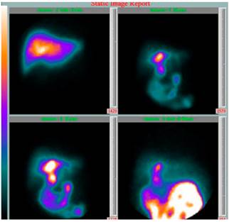
Figure 2 HIDA scan, elimination to the intrahepatic duct before 15 minutes with abundant retention in the major hepatic ducts and the common bile duct. 

Based on these findings, a type II sphincter of Oddi dysfunction diagnosis was considered and an ERCP plus a biliary endoscopic sphincterotomy were performed in February 2018, in which a normal major papilla and dilated intra- and extrahepatic bile ducts were documented (1.3 cm). The biliary endoscopic sphincterotomy was carried out and the bile duct was explored with a basket up to the hepatic duct, obtaining a clear biliary fluid outflow.

After performing the ERCP, the patient’s condition improved satisfactorily and there were no complications. In February 2020 (2 years after the ERCP), date in which the patient attended her last follow-up visit, she was asymptomatic and normal values were reported in her complete blood count test. In addition, the patient did not visit the emergency department anymore after the ERCP was performed.

Discussion

We present the case of a patient in which a postcholescytectomy sphincter of Oddi dysfunction diagnosis was suspected. Since diagnosis was supported by the HIDA scan findings, an ERCP was performed achieving a complete resolution of the patient’s symptoms and clinical signs.

Clinically, differentiating sphincter of Oddi dysfunction from other biliopancreatic diseases, as well as from other functional digestive tract disorders is not an easy task. However, the medical record and physical examination, laboratory and imaging findings must be carefully evaluated in order to suspect its diagnosis.

The referral of patients with this condition to health centers with expertise in the treatment of biliopancreatic diseases is fundamental for their outcome, where laboratory tests (hepatic and pancreatic enzymes) and imaging studies, including abdominal ultrasound, abdominal tomography, MRCP, biliopancreatic endosonography (EUS) or isotopic techniques such as hepatobiliary scintigraphy, must be requested prior to the assessment of the patient’s symptoms10.

Regarding the analysis of the sphincter of Oddi dysfunction diagnosis, it can be noted that in patients with bile duct dilatation, diagnosis is suspected based on the symptomatology and complete blood count results; when there is a high probability of biliary disease and other causes have been ruled out based on previous imaging findings, performing an ERCP is suggested16. On the contrary, in the case of bile duct dilatation, but low probability of biliary disease (symptomatology and complete blood count results), performing an EUS or a MRCP is suggested, since the diagnostic performance of these procedures is similar to that of ERCP, but they don’t have the morbidity and mortality associated with the latter16. Specifically, in the context of bile duct assessment, EUS has a diagnostic performance of only 53 % in patients with bile duct dilatation and abnormal complete blood count results16, which decreases to 6% in patients with bile duct dilatation and normal complete blood count results16. This 6% is associated with factors such as age over 65 years and having undergone a cholecystectomy16.

The use of ERCP plus sphincter of Oddi manometry have been recommended for studying sphincter of Oddi dysfunction. In patients with clinical suspicion of the condition and any objective alteration associated with a biliary disease (type II sphincter of Oddi dysfunction), an elevated sphincter of Oddi pressure (> 40 mm Hg) while in resting position is a diagnostic criterion for sphincter of Oddi dysfunction, as well as a good predictor of symptom resolution after undergoing a biliary endoscopic sphincterotomy. However, the probability of developing post-ERCP pancreatitis and sphincter of Oddi manometry pancreatitis can be up to 30 %11,17-19. In our opinion, this level of risk associated with the performance of a diagnostic study is unacceptable, not to mention the fact that the possibility of false negatives can be up to 65%20,21.

Research has been conducted on diagnostic and therapeutic methods including Nardi test, secretin-enhanced MRCP, cholecystokinin scintigraphy, functional luminal imaging probe, calcium channel blockers, tricyclic antidepressants, nitroglycerin, somatostatin and botulinum toxin. To date, none of the above alternatives has been shown to have relevant clinical usefulness22-29.

Biliary endoscopic sphincterotomy is indicated in patients with type I sphincter of Oddi dysfunction; no other additional studies are required in these patients20,30. On the contrary, in those with type II and III sphincter of Oddi dysfunction, diagnosis and management are controversial. In our opinion, a hepatobiliary scintigraphy should be performed before ERCP in all patients with suspected type II and III sphincter of Oddi dysfunction, provided that this nuclear medicine procedure has, at least, a specificity of 90%14,31-34. In addition, in the evaluation of postcholecystectomy biliary colic-like pain, scintigraphy is included in the diagnostic algorithm14.

Treatment in patients with type II sphincter of Oddi dysfunction consists of biliary endoscopic sphincterotomy, because clinical response, despite being variable, is generally favorable when the procedure is performed by an expert, with some reports describing that this treatment is successful in up to 90% of cases. We do not suggest performing a sphincter of Oddi manometry prior to the biliary endoscopic sphincterotomy because of the aforementioned risks of complications and the debatable relationship between sphincter of Oddi manometry findings and sphincter of Oddi dysfunction12,35-37.

Patients with type III sphincter of Oddi dysfunction are the most difficult to diagnose due to the absence of abnormal laboratory and imaging finings; in addition, there is controversy regarding their treatment. These patient do not benefit from the performance of ERCP and biliary endoscopic sphincterotomy (the severity of symptoms decrease only in approximately 20%)38-41; on the contrary, they are exposed to the risks inherent to these procedures42. Therefore, treatment in these patients must be symptomatic and based on medications used to treat functional gastrointestinal disorders43.

In conclusion, we suggest suspecting sphincter of Oddi dysfunction in patients who present with symptomatology compatible with biliary colic-like pain or pain of pancreatic origin after having undergone a cholecystectomy; likewise, the diagnostic suspicion should be based on tools other than sphincter of Oddi manometry (scintigraphy) that, based on their specificity, allow making a decision regarding the performance of an ERCP plus a biliary endoscopic sphincterotomy to achieve the resolution of symptoms.

Finally, this case serves as a starting point for conducting prospective studies in other population groups.

REFERENCIAS

1. Avisse C, Flament JB, Delattre JF. Ampulla of Vater. Anatomic, embryologic, and surgical aspects. Surg Clin North Am. 2000;80(1):201-12. https://doi.org/10.1016/s0039-6109(05)70402-3Links ]

2. HAND BH. An anatomical study of the choledochoduodenal area. Br J Surg. 1963;50:486-94. https://doi.org/10.1002/bjs.18005022303Links ]

3. Guelrud M, Mendoza S, Rossiter G, Villegas MI. Sphincter of Oddi manometry in healthy volunteers. Dig Dis Sci. 1990;35(1):38-46. https://doi.org/10.1007/BF01537220Links ]

4. Behar J, Corazziari E, Guelrud M, Hogan W, Sherman S, Toouli J. Functional gallbladder and sphincter of oddi disorders. Gastroenterology. 2006;130(5):1498-509. https://doi.org/10.1053/j.gastro.2005.11.063Links ]

5. Moody F, Poots J. Dysfunction of the ampulla of Vater. En: Braasch J, Tompkins R (editores). Surgical Diseases of the Biliary Tract and Pancreas, multidisciplinary management. St. Louis: Mosby; 1994. p. 334-48. [ Links ]

6. George J, Baillie J. Biliary and gallbladder dyskinesia. Curr Treat Options Gastroenterol. 2007;10(4):322-7. https://doi.org/10.1007/s11938-007-0075-2Links ]

7. Toouli J. What is sphincter of Oddi dysfunction? Gut. 1989;30(6):753-61. https://doi.org/10.1136/gut.30.6.753Links ]

8. Drossman DA, Li Z, Andruzzi E, Temple RD, Talley NJ, Thompson WG, Whitehead WE, Janssens J, Funch-Jensen P, Corazziari E, et al. U.S. householder survey of functional gastrointestinal disorders. Prevalence, sociodemography, and health impact. Dig Dis Sci. 1993;38(9):1569-80. https://doi.org/10.1007/BF01303162Links ]

9. Eversman D, Fogel EL, Rusche M, Sherman S, Lehman GA. Frequency of abnormal pancreatic and biliary sphincter manometry compared with clinical suspicion of sphincter of Oddi dysfunction. Gastrointest Endosc. 1999;50(5):637-41. https://doi.org/10.1016/s0016-5107(99)80011-xLinks ]

10. Kaw M, Brodmerkel GJ Jr. ERCP, biliary crystal analysis, and sphincter of Oddi manometry in idiopathic recurrent pancreatitis. Gastrointest Endosc. 2002;55(2):157-62. https://doi.org/10.1067/mge.2002.118944Links ]

11. Hogan WJ, Geenen JE. Biliary dyskinesia. Endoscopy. 1988;20 Suppl 1:179-83. https://doi.org/10.1055/s-2007-1018172Links ]

12. Sherman S, Troiano FP, Hawes RH, O’Connor KW, Lehman GA. Frequency of abnormal sphincter of Oddi manometry compared with the clinical suspicion of sphincter of Oddi dysfunction. Am J Gastroenterol. 1991;86(5):586-90. [ Links ]

13. Solano J. Disfunción del esfínter de Oddi. En: Aponte D, Cañadas R (editores). Técnicas de endoscopia digestiva. 3.a edición. Bogotá: Asociación Colombiana de Endoscopia Digestiva; 2018. p. 515-9. [ Links ]

14. Elta G, Cotton P, Carter C, Corraziari E, Pasricha P. Gallbladder and sphincter of Oddi Disorders. En: Drossman D (ed). Rome IV Functional gastrointestinal disorders. 4.a edición. Raleigh: The Rome Foundation; 2016. p. 1117-78. [ Links ]

15. Madura JA 2nd, Madura JA. Diagnosis and management of sphincter of Oddi dysfunction and pancreas divisum. Surg Clin North Am. 2007;87(6):1417-29, ix. https://doi.org/10.1016/j.suc.2007.08.011Links ]

16. Malik S, Kaushik N, Khalid A, Bauer K, Brody D, Slivka A, McGrath K. EUS yield in evaluating biliary dilatation in patients with normal serum liver enzymes. Dig Dis Sci. 2007;52(2):508-12. https://doi.org/10.1007/s10620-006-9582-6Links ]

17. Fogel EL, Eversman D, Jamidar P, Sherman S, Lehman GA. Sphincter of Oddi dysfunction: pancreaticobiliary sphincterotomy with pancreatic stent placement has a lower rate of pancreatitis than biliary sphincterotomy alone. Endoscopy. 2002;34(4):280-5. https://doi.org/10.1055/s-2002-23629Links ]

18. Steinberg WM. Controversies in clinical pancreatology: should the sphincter of Oddi be measured in patients with idiopathic recurrent acute pancreatitis, and should sphincterotomy be performed if the pressure is high? Pancreas. 2003;27(2):118-21. https://doi.org/10.1097/00006676-200308000-00002Links ]

19. Calzadilla J, Sanhueza N, Farías S, González F. Pancreatitis recurrente secundaria a disfunción del esfínter de Oddi: presentación de un caso [Recurrent pancreatitis secondary to sphincter of Oddi dysfunction: case report]. Medwave. 2016;16(9):e6585. https://doi.org/10.5867/medwave.2016.09.6585Links ]

20. Madura JA, Madura JA 2nd, Sherman S, Lehman GA. Surgical sphincteroplasty in 446 patients. Arch Surg. 2005;140(5):504-11; discussion 511-3. https://doi.org/10.1001/archsurg.140.5.504Links ]

21. Cicala M, Habib FI, Vavassori P, Pallotta N, Schillaci O, Costamagna G, Guarino MP, Scopinaro F, Fiocca F, Torsoli A, Corazziari E. Outcome of endoscopic sphincterotomy in post cholecystectomy patients with sphincter of Oddi dysfunction as predicted by manometry and quantitative choledochoscintigraphy. Gut. 2002;50(5):665-8. https://doi.org/10.1136/gut.50.5.665Links ]

22. Nardi GL, Acosta JM. Papillitis as a cause of pancreatitis and abdominal pain: role of evocative test, operative pancreatography and histologic evaluation. Ann Surg. 1966;164(4):611-21. https://doi.org/10.1097/00000658-196610000-00008Links ]

23. Piccinni G, Angrisano A, Testini M, Bonomo GM. Diagnosing and treating Sphincter of Oddi dysfunction: a critical literature review and reevaluation. J Clin Gastroenterol. 2004;38(4):350-9. https://doi.org/10.1097/00004836-200404000-00010Links ]

24. Pereira SP, Gillams A, Sgouros SN, Webster GJ, Hatfield AR. Prospective comparison of secretin-stimulated magnetic resonance cholangiopancreatography with manometry in the diagnosis of sphincter of Oddi dysfunction types II and III. Gut. 2007;56(6):809-13. https://doi.org/10.1136/gut.2006.099267Links ]

25. Sharma BC, Agarwal DK, Dhiman RK, Baijal SS, Choudhuri G, Saraswat VA. Bile lithogenicity and gallbladder emptying in patients with microlithiasis: effect of bile acid therapy. Gastroenterology. 1998;115(1):124-8. https://doi.org/10.1016/s0016-5085(98)70373-7Links ]

26. Kunwald P, Drewes AM, Kjaer D, Gravesen FH, McMahon BP, Madácsy L, Funch-Jensen P, Gregersen H. A new distensibility technique to measure sphincter of Oddi function. Neurogastroenterol Motil. 2010;22(9):978-83, e253. https://doi.org/10.1111/j.1365-2982.2010.01531.xLinks ]

27. Khuroo MS, Zargar SA, Yattoo GN. Efficacy of nifedipine therapy in patients with sphincter of Oddi dysfunction: a prospective, double-blind, randomized, placebo-controlled, cross over trial. Br J Clin Pharmacol. 1992;33(5):477-85. https://doi.org/10.1111/j.1365-2125.1992.tb04074.xLinks ]

28. Sand J, Nordback I, Koskinen M, Matikainen M, Lindholm TS. Nifedipine for suspected type II sphincter of Oddi dyskinesia. Am J Gastroenterol. 1993;88(4):530-5. [ Links ]

29. Nakeeb A. Sphincter of Oddi dysfunction: how is it diagnosed? How is it classified? How do we treat it medically, endoscopically, and surgically? J Gastrointest Surg. 2013;17(9):1557-8. https://doi.org/10.1007/s11605-013-2280-8Links ]

30. Small AJ, Kozarek RA. Sphincter of Oddi Dysfunction. Gastrointest Endosc Clin N Am. 2015;25(4):749-63. https://doi.org/10.1016/j.giec.2015.06.009Links ]

31. Corazziari E, Cicala M, Scopinaro F, Schillaci O, Habib IF, Pallotta N. Scintigraphic assessment of SO dysfunction. Gut. 2003;52(11):1655-6. https://doi.org/10.1136/gut.52.11.1655Links ]

32. Craig AG, Peter D, Saccone GT, Ziesing P, Wycherley A, Toouli J. Scintigraphy versus manometry in patients with suspected biliary sphincter of Oddi dysfunction. Gut. 2003;52(3):352-7. https://doi.org/10.1136/gut.52.3.352Links ]

33. Sostre S, Kalloo AN, Spiegler EJ, Camargo EE, Wagner HN Jr. A noninvasive test of sphincter of Oddi dysfunction in postcholecystectomy patients: the scintigraphic score. J Nucl Med. 1992;33(6):1216-22. [ Links ]

34. Corazziari E, Cicala M, Habib FI, Scopinaro F, Fiocca F, Pallotta N, Viscardi A, Vignoni A, Torsoli A. Hepatoduodenal bile transit in cholecystectomized subjects. Relationship with sphincter of Oddi function and diagnostic value. Dig Dis Sci. 1994;39(9):1985-93. https://doi.org/10.1007/BF02088136Links ]

35. Sherman S. What is the role of ERCP in the setting of abdominal pain of pancreatic or biliary origin (suspected sphincter of Oddi dysfunction)? Gastrointest Endosc. 2002;56(6 Suppl):S258-66. https://doi.org/10.1067/mge.2002.129016Links ]

36. Geenen JE, Nash JA. The role of sphincter of Oddi manometry and biliary microscopy in evaluating idiopathic recurrent pancreatitis. Endoscopy. 1998;30(9):A237-41. https://doi.org/10.1055/s-2007-1001447Links ]

37. Petersen BT. Sphincter of Oddi dysfunction, part 2: Evidence-based review of the presentations, with “objective” pancreatic findings (types I and II) and of presumptive type III. Gastrointest Endosc. 2004;59(6):670-87. https://doi.org/10.1016/s0016-5107(04)00297-4Links ]

38. Baillie J. Sphincter of Oddi dysfunction. Curr Gastroenterol Rep. 2010;12(2):130-4. https://doi.org/10.1007/s11894-010-0096-1Links ]

39. Hyun JJ, Kozarek RA. Sphincter of Oddi dysfunction: sphincter of Oddi dysfunction or discordance? What is the state of the art in 2018? Curr Opin Gastroenterol. 2018;34(5):282-287. https://doi.org/10.1097/MOG.0000000000000455Links ]

40. Shah T, Zfass A, Schubert ML. Management of Sphincter of Oddi Dysfunction: Teaching an Old SOD New Tricks? Dig Dis Sci. 2016;61(9):2459-61. https://doi.org/10.1007/s10620-016-4252-9Links ]

41. Wilcox CM. Sphincter of Oddi dysfunction Type III: New studies suggest new approaches are needed. World J Gastroenterol. 2015;21(19):5755-61. https://doi.org/10.3748/wjg.v21.i19.5755Links ]

42. Peñaloza-Ramírez A, Leal-Buitrago C, Rodríguez-Hernández A. Adverse events of ERCP at San José Hospital of Bogotá (Colombia). Rev Esp Enferm Dig. 2009;101(12):837-49. https://doi.org/10.4321/s1130-01082009001200003Links ]

43. Pérez LV. Disfunción del esfínter de Oddi y trastornos biliares funcionales. Revista andaluza de patología digestiva. 2017;40(5):233-42. [ Links ]

Citation: Peñaloza-Ramírez A, Coral-Argoty E, Castro-Rodríguez M, Álvarez-Gil J, Aponte-Ordóñez P. Sphincter of Oddi dysfunction: Case report. Rev Colomb Gastroenterol. 2021;36(Supl.1):52-58. https://doi.org/10.22516/25007440.550

Funding None declared by the authors.

Received: April 24, 2020; Accepted: October 26, 2020

*Correspondence: Arecio Peñaloza-Ramírez. apenaloza@fucsalud.edu.co

Conflicts of interest

None declared by the authors.

Creative Commons License This is an open-access article distributed under the terms of the Creative Commons Attribution License