SciELO - Scientific Electronic Library Online

 
vol.37 issue1Usefulness of the Liver Fibrosis Index (LFI) Measured during Endoscopic Ultrasonography in Evaluating the Pancreatic ParenchymaExperience in the Use of Video Capsule Endoscopy in Patients with Unexplained Iron Deficiency Anemia author indexsubject indexarticles search
Home Pagealphabetic serial listing  

Services on Demand

Journal

Article

Indicators

Related links

  • On index processCited by Google
  • Have no similar articlesSimilars in SciELO
  • On index processSimilars in Google

Share


Revista colombiana de Gastroenterología

Print version ISSN 0120-9957On-line version ISSN 2500-7440

Rev. colomb. Gastroenterol. vol.37 no.1 Bogotá Jan./Mar. 2022  Epub May 16, 2022

https://doi.org/10.22516/25007440.703 

Original article

Survival in Patients with Cirrhosis According to Etiology. Retrospective Cohort

Jhon Edison Prieto-Ortiz1  * 
http://orcid.org/0000-0002-8009-5999

Nathaly Garzón-Orjuela2 
http://orcid.org/0000-0001-6181-8154

Santiago Sánchez-Pardo3 
http://orcid.org/0000-0003-3869-6907

Robin German Prieto-Ortiz4 
http://orcid.org/0000-0001-7605-760X

Javier Eslava-Schmalbach5 
http://orcid.org/0000-0003-1502-2918

1Surgeon, specialist in Internal Medicine, Gastroenterology and Gastrointestinal Endoscopy, Hepatology, and liver transplant. Center for Liver and Digestive Diseases (CEHYD). Bogotá, Colombia.

2Master’s in Clinical Epidemiology, Universidad Nacional, and member of the Equity and Health research group. Universidad Nacional de Colombia. Bogotá, Colombia.

3Surgeon, specialist in Internal Medicine, Universidad Industrial de Santander. Bucaramanga, Santander.

4Gastroenterologist, specialist in General Surgery, Gastroenterology and Gastrointestinal Endoscopy, Center for Liver and Digestive Diseases (CEHYD), Hospital Central de la Policía (HOCEN). Bogotá, Colombia.

5Physician, Professor of the Surgery Department, Universidad Nacional de Colombia. Bogotá, Colombia.


Abstract

Introduction:

Cirrhosis is the final stage of chronically progressive liver diseases of various etiologies. It is a common disease, with a variable prevalence in each country. Its peak incidence occurs between 40 and 50 years of age, predominantly in men.

Aims:

To compare a cohort of patients diagnosed with cirrhosis, evaluate their complications and survival according to etiology, describe clinical and laboratory aspects, and determine the role of a fatty liver.

Materials and methods:

A retrospective cohort study was carried out with patients who held a specialized hepatology consultation in the center of liver and digestive diseases (CEHYD) in Bogotá, Colombia, between January 2010 and June 2019.

Results:

We reviewed a total of 1,200 medical records (56.8 % women). There were no statistically significant differences in median survival between groups by etiology, sex, presence or absence of complications, or Child. We noted that the older the age at the diagnosis of cirrhosis, the higher the risk of death; HR 1.04 (95 % CI 1.02-1.075). For each month that follow-up increases, the risk of death decreases by 90 %; HR 0.1 (95 % CI 0.03-0.29). For each month that the follow-up of complications increases, the risk of death is reduced by 2 %; HR 0.98 (95 % CI 0.97-0.99).

Conclusions:

Survival by etiology was similar in the different groups. Nonalcoholic steatohepatitis (NASH) was the leading cause of cirrhosis in this cohort. Efforts should focus on its diagnosis and management in the early stages.

Keywords: Liver cirrhosis; survival; fatty liver; nonalcoholic fatty liver disease

Resumen

Introducción:

la cirrosis es el estadio final de enfermedades hepáticas crónicamente progresivas de diferentes etiologías. Es una enfermedad frecuente, con una prevalencia variable en cada país. Su pico de incidencia se presenta entre los 40 y 50 años, predominantemente en hombres.

Objetivos:

comparar una cohorte de pacientes con diagnóstico de cirrosis, evaluar sus complicaciones y sobrevida de acuerdo con su etiología, describir los aspectos clínicos y de laboratorio, y determinar el papel del hígado graso.

Materiales y métodos:

se realizó un estudio de cohorte retrospectiva, en donde se incluyeron pacientes que asistieron a consulta especializada de hepatología en el centro de enfermedades hepáticas y digestivas (CEHYD), en la ciudad de Bogotá, durante enero de 2010 y junio de 2019.

Resultados:

se revisaron un total de 1200 historias clínicas (56,8 % mujeres). No se evidenció diferencias estadísticamente significativas en las medianas de sobrevida entre los grupos por etiologías, sexo, presencia o no de complicaciones, o Child. Se evidenció que entre mayor edad en el diagnóstico de cirrosis, el riesgo de muerte es mayor; HR 1,04 (IC 95 % 1,02-1,075). Por cada mes que aumenta el seguimiento se reduce el riesgo de muerte en 90 %; HR 0,1 (IC 95 % 0,03-0,29). Por cada mes que aumenta el seguimiento de las complicaciones se reduce el riesgo de muerte en 2 %; HR 0,98 (IC 95 % 0,97-0,99).

Conclusiones:

La sobrevida por etiología fue similar en los diferentes grupos. La esteatohepatitis no alcohólica (NASH) fue la principal causa de cirrosis en esta cohorte. Se deben orientar esfuerzos a su diagnóstico y manejo en fases tempranas.

Palabras clave: Cirrosis hepática; sobrevida; hígado graso; enfermedad del hígado graso no alcohólico

Introduction

Cirrhosis is the final phase of progressive and long-term liver disease1,2. The prevalence varies in each country, with a maximum incidence between 40 and 50 years, affecting more men2. In Western countries, 90% of cases are due to alcohol abuse, non-alcoholic fatty liver disease (NAFLD), and chronic viral hepatitis, including hepatitis B (HBV) and hepatitis C (HCV). About 10% of the etiology of this entity is unknown1-3.

Cirrhosis is the consequence of the continuous death of hepatocytes, with loss of parenchyma, inflammation, fibrogenesis, changes in cell regeneration, and alterations in macro- and microcirculation4-6. It is a dynamic and reversible process at some point7-9, which has motivated studies in the search for adequate management by identifying and monitoring asymptomatic patients and preventing the entity’s complications. Four of them are classic decompensations: Ascites, variceal bleeding, encephalopathy, and jaundice10-12.

The manifestation of any of them markedly decreases the survival of cirrhotic patients, being worse with hepatocarcinoma (HCC)12-15, which has an approximate annual incidence of 7%1,12,15 and is associated with the same causes of cirrhosis, HCV, alcohol, and NAFLD1,16-20. Fatty liver is a cause of cirrhosis on the rise21,22, directly correlated with elements of the metabolic syndrome. For example, type 2 diabetes mellitus, obesity, and high blood pressure, all with increased cardiovascular risk23. Therefore, this study aims to compare a cohort of patients diagnosed with cirrhosis, evaluate their complications and survival according to their etiology, describe clinical and laboratory aspects, and determine the role of fatty liver.

Materials and methods

We carried out a retrospective cohort study. It included patients who attended a specialized Hepatology consultation at the Center for Liver and Digestive Diseases (CEHYD) in Bogotá between January 2010 and June 2019. Confirmed diagnosis of cirrhosis by the clinic, radiology or liver biopsy, and available medical records were the only inclusion criteria.

Based on the Child-Pugh (CP)24 cirrhosis severity scale, we defined:

  • Compensated cirrhosis: A Child A stage, without any additional decompensation

  • Decompensated cirrhosis: A Child B or C stage, or a Child A cirrhosis with ascites, variceal bleeding, encephalopathy, or jaundice

Additionally, ascites, variceal bleeding, encephalopathy, jaundice, HCC, hepatorenal syndrome, or coagulopathy were defined as complications of cirrhosis.

The information collected was summarized using means and standard deviations for a normal distribution (Shapiro-Wilks test); otherwise, it was summarized with medians and interquartile ranges. We performed the analysis with non-parametric statistics and summarized categorical variables as proportions. Survival was assessed based on medians and interquartile ranges. We used the Kaplan Meier estimator of the survival function and the log-rank test. A Cox hazard model was made for the analysis adjusted by the effect of confounding covariates. STATA v15.1 and R were employed for statistical analysis.

Ethical considerations

As a retrospective study, no intentional intervention or modification of the individuals’ biological, physiological, psychological, or social variables was carried out. It was conducted under the principles declared at the 18th WMA General Assembly (Helsinki, 1964) as a world reference for research on human beings. As set forth in Article 11(a), Title II (about research on human beings), Chapter I (about the ethical aspects of research on human beings) of Resolution 008430 dated October 4, 1993, issued by the Ministry of Health of the Republic of Colombia, this study constitutes research without risk. It uses retrospective documentary research techniques and methods, including medical records, interviews, questionnaires, and others, in which sensitive data on illnesses are not identified or processed.

Since it has no risk and only uses documentary research techniques and methods, this study is understood to follow the fundamental principles of ethics: Beneficence, autonomy, justice, and non-maleficence.

Results

We reviewed a total of 1,200 medical records, of which 681 (56.8%) were women. The mean age at diagnosis of cirrhosis was 63 years (interquartile range [IQR] 56-71). The death occurred in 51 patients, with a median age of 75 years (IQR 65-80). The patients were monitored over time, measured in months, with a median of 17.4 (IQR 5.5-45.3). The first complication of cirrhosis occurred in 545 patients (45.4%) 0.7 months after diagnosis (IQR 0-18.1). Complications were, in order of importance, ascites (33.8%), variceal bleeding (22.2%), HCC (17.4%), jaundice (14.3%), encephalopathy (7.3%), ascites plus encephalopathy (2.6%), coagulopathy (2%), and hepatorenal syndrome (0.4%). The clinical and demographic characteristics are shown in Table 1.

Table 1 Clinical and laboratory variables in patients with cirrhosis 

Variable Number Median (IQR)*
BMI kg/m2 1139 27 (24 to 30)
White cells (cell/mL) 1154 5500 (4400 to 6890)
Hemoglobin (g/dL) 1153 14 (13 to 16)
Hematocrit % 1153 43 (38 to 47)
Platelets (cell/mm3) 1155 158 000 (111 650 to 224 500)
Glycemia mg/dL 1087 98 (88 to 115)
AST IU/dL 1158 54 (34 to 90)
ALT IU/dL 1161 50 (31 to 87)
GGT IU/dL 1049 120 (56 to 259)
Alkaline phosphatase IU/dL 1125 132 (95 to 211)
Total bilirubin (mg/dL) 1143 1 (0,6 to 1,7)
Direct bilirubin (mg/dL) 1139 0,4 (0,2 to 0,8)
Indirect bilirubin (mg/dL) 1137 0,5 (0,3 to 0,9)
Total protein (g/dL) 1044 7,3 (6,9 to 7,8)
Albumin (g/dL) 1100 4 (3,4 to 4,3)
INR 1117 1,1 (1 to 1,2)

BMI: Body mass index; INR: International normalized ratio; IQR: Interquartile range tested for normality (Shapiro-Wilk test).

According to the cirrhosis etiology, the patients were classified into five groups (Table 2). The lowest median age at diagnosis of cirrhosis was for the cholestatic group. Furthermore, the alcohol group had a lower median age at death, 64 years. The etiological group with the highest percentage of complications was again alcohol (63.5%), and the lowest was the non-alcoholic steatohepatitis (NASH) group, with 31.6%.

Table 2 Differences between groups by etiology 

Etiology of cirrhosis NASH n = 399 (33.2%) Cholestatic n = 245 (20.4%) Alcohol n = 230 (19.2%) Other n = 191 (15.9%) HCV n = 135 (11.2%) Total n = 1,200 (100%)
Sex*

  • - Woman

  • - Man

  • 257 (64.4)

  • 142 (35.6)

  • 208 (84.9)

  • 37 (15.1)

  • 27 (11.7)

  • 203 (88.3)

  • 91 (47.9)

  • 100 (52.1)

  • 98 (72.6)

  • 37 (27.4)

  • 681 (56.8)

  • 519 (43.2)

  • Age at first consultation (years)

  • Median (IQR)**

  • 64

  • (57.8 to 71)

  • 59

  • (48.7 to 69)

  • 64

  • (56 to 70)

  • 62

  • (55 to 70.8)

  • 64

  • (58.5 to 71)

  • 63

  • (55 to 70)

  • n = 1200

  • Age at diagnosis of cirrhosis (years)

  • Median (IQR)**

  • 65

  • (59 to 72)

  • 60

  • (48 to 69)

  • 63

  • (55 to 71)

  • 63

  • (54.3 to 70.8)

  • 64

  • (60 to 70)

  • 63

  • (56 to 71)

  • n = 1200

  • Age at death (years)

  • Median (IQR)

  • 70

  • (67.5 to 78.5)

  • 77.5

  • (72.3 to 80.8)

  • 64

  • (60 to 78)

  • 74.5

  • (64.3 to 79.8)

  • 78

  • (71 to 81.3)

  • 75

  • (65 to 80)

  • n = 51

Complicatión*

  • - Yes

  • - No

  • 126 (31,6)

  • 273 (68,4)

  • 121 (49,4)

  • 124 (50,6)

  • 146 (63,5)

  • 84 (36,5)

  • 94 (49,2)

  • 97 (50,8)

  • 58 (43)

  • 77 (57)

  • 545 (45,4)

  • 655 (54,6)

  • Follow-up of cirrhosis from its diagnosis in months

  • Median (IQR)**

  • (6.3 to 31.3)

  • 28.4

  • (9.7 to 59.2)

  • 17.3

  • (3.7 to 47.1)

  • 12.9

  • (4 to 35)

  • (4.5 to 48.3)

  • 17.4

  • (5.5 to 45.3)

  • n = 1186

  • Follow-up of complications from the diagnosis of cirrhosis in months

  • Median (IQR)**

  • 0

  • (0 to 12)

  • 3.8

  • (0 to 30.6)

  • 0

  • (0 to 12)

  • 0.02

  • (0 to 33.13)

  • 5.3

  • (0 to 33.13)

  • 0.7

  • (0 to 18.1)

  • n = 545

Child*

  • - A

  • - B

  • - C

  • 326 (84,2)

  • 50 (12,9)

  • 11 (2,9)

  • 170 (70,8)

  • 64 (26,7)

  • 6 (2,5)

  • 125 (58,4)

  • 69 (32,2)

  • 20 (9,3)

  • 116 (69,1)

  • 47 (28)

  • 5 (2,9)

  • 98 (76)

  • 26 (20,2)

  • 5 (3,8)

  • 835 (69,6)

  • 256 (21,3)

  • 47 (3,9)

Other: HBV, HCV, and B coinfection, medications, or toxic substances, three or more mixed, HBV plus alcohol

*Significant differences (p < 0.005) between etiological groups of cirrhosis, Fisher’s exact test.

**Significant differences (p < 0.005) between etiological groups of cirrhosis, Kruskal-Wallis test.

IQR: Interquartile range.

Regarding the severity of cirrhosis at admission, 69.6% had Child A (higher in the NASH group), 21.3% Child B, and 3.9% Child C (higher in the alcohol group).

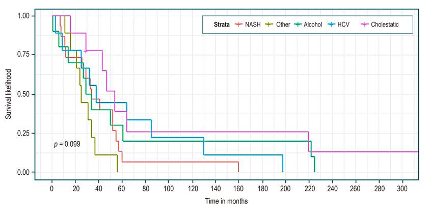
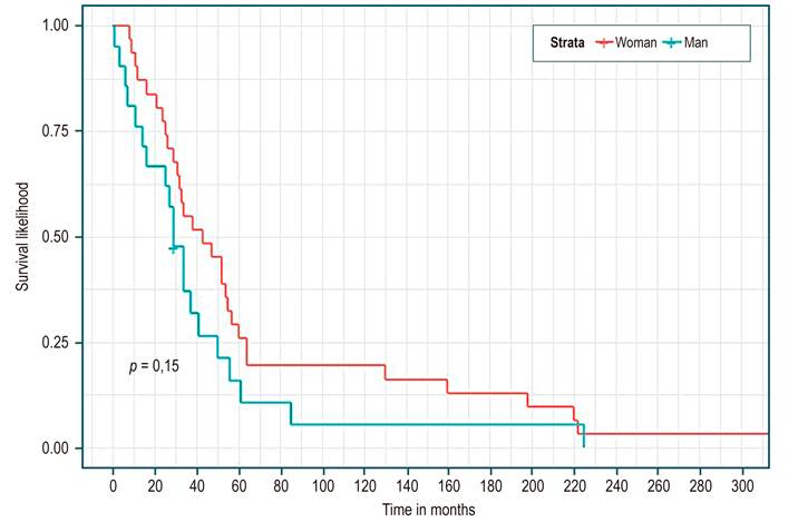
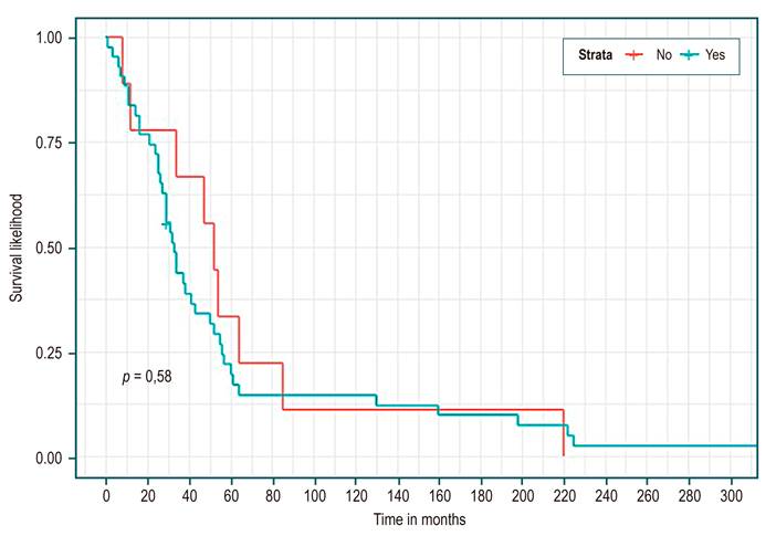
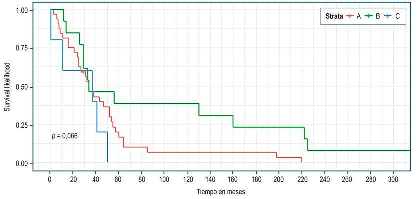
Table 3 shows, in all patients, a median survival of 34 months (95% CI 29-52). There were no statistically significant differences in median survival between the groups of etiology, sex, presence or absence of complications, or Child. However, higher survival was identified in the cholestatic group (Figure 1), being a woman (Figure 2), not having a complication (Figure 3), and Child C (Figure 4), the median survival being 54, 43, 52, and 37 months, respectively.

Table 3 Survival by group and difference comparison 

Number of events Median survival (95% CI) (months) Log-rank test
Total 51 34 (29 to 52)
Etiology of cirrhosis (Figure 1)

  • HCV

  • Alcohol

  • Cholestatic

  • NASH

  • Other

  • 9

  • 10

  • 8

  • 15

  • 9

  • 38 (25 to NC)

  • 31.5 (14 to NC)

  • 54 (43 to NC)

  • 34 (26 to 57)

  • 25 (21 to NC)

p = 0.099
Gender (Figure 2)

  • Woman

  • Man

  • 31

  • 20

  • 43 (31 to 60)

  • 29 (16 to 50)

p = 0.15
Complications (Figure 3)

  • - Yes

  • - No

  • 42

  • 9

  • 33 (27 to 50)

  • 52 (34 to NC)

p = 0.58
Child* (Figure 4)

  • - A

  • - B

  • - C

  • 31

  • 13

  • 5

  • 34 (25 to 54)

  • 34 (29 to NC)

  • 37 (11 to NC)

p = 0.066

NC: Not calculated.

*In two patients, it was not calculated due to a lack of data

Figure 1 Survival curves by cirrhosis etiology - Kaplan Meier. 

Figure 2 Survival curves by sex - Kaplan-Meier. 

Figure 3 Survival curves by presence or absence of complications - Kaplan Meier. 

Figure 4 Survival curves by Child - Kaplan Meier. 

Considering the variables and the follow-up time illustrated in Table 4, the univariate analysis found that as the age at diagnosis of cirrhosis increases, the risk of death is higher; HR 1.04 (95% CI 1.02-1.075; p = 0.000633). For each month that the follow-up of cirrhotic patients increases, the risk of death is reduced by 90%; HR 0.1 (95% CI 0.03-0.29; p < 0.00). Similarly, a longer follow-up of complications reduces the risk of death by 2%; HR 0.98 (95% CI 0.97-0.99; p = 0.000695). Finally, the group of other causes of cirrhosis presents a risk of death 3.87 times compared to the cause of cirrhosis due to cholestatic disease (p = 0.0386), statistically significant estimates.

Table 4 Differences between cirrhosis groups and follow-up 

Follow-up variables HR (95% CI) (univariate)
Age at first consultation in years 1.02 (0.99 to 1.04) p = 0.096
Age of diagnosis of cirrhosis in years 1.04 (1.02 to 1.075) p = 0.000633
Follow-up of cirrhosis from its diagnosis in months 0.1 (0.03 to 0.29) p <0.00
Follow-up of complications after the diagnosis of cirrhosis in months 0.98 (0.97 to 0.99) p = 0.000695
Etiology of cirrhosis

  • Cholestatic

  • Alcohol

  • HCV

  • NASH

  • Other

  • 1.56 (0.59 to 4.12)

  • 1.62 (0.58 to 4.44)

  • 2.36 (0.93 to 5.99)

  • 3.87 (1.36 to 11)

  • p = 0.3681

  • p = 0.3498

  • p = 0.0695

  • p = 0.0110

Sex p = 0,156

  • Woman

  • Man

  • 1

  • 1.51 (0.85 to 2.68)

Complications p = 0,591

  • - No

  • - Yes

  • 1.22 (0.58 to 2.53)

Child

  • - A

  • - B

  • - C

  • 1

  • 0,48 (0,23 a 1,01)

  • 1,59 (0,60 a 4,18)

  • p = 0,0556

  • p = 0,3462

Discussion

Studies in the United States showed that in 2010, chronic liver diseases and cirrhosis were the cause of 31,903 deaths, with age-adjusted mortality of 9.4 per 100,000 individuals, and decompensated cirrhosis accounted for more than 150,000 hospitalizations, with a cost close to 4 billion US dollars25,26. These data highlight the importance of monitoring the cirrhotic patient.

When reviewing this cohort, the average age of cirrhosis was 63 years, as reported in the Swedish population study by Nilsson et al26, with a follow-up of more than 1,000 patients. The highest incidence of the disease occurred in the group of 60-64 years, with a predominance in the female sex, as in our series. In other international series, the male gender predominates, probably due to etiological differences27,28.

D’Amico emphasizes a different prognosis in terms of mortality according to the presence or absence of decompensations12,28,29. In our cohort, the analysis focused on the presence or absence of complications. Classic ascites, variceal bleeding, jaundice, and encephalopathy12 were added to HCC, hepatorenal syndrome, and coagulopathy. In this series, complications occurred in 45.9% of all patients shortly after the diagnosis of cirrhosis (0.7 months) (IQR 0-18.1) and with a total follow-up of 17.4 months (IQR 5.5-45.3). European series show follow-up periods of up to 10 years or more27,28. These two pieces of data alert us to the lateness of our diagnoses since, in most cases, the complication leads to the diagnosis of cirrhosis, and advanced disease does not allow for further follow-up.

In different studies, the survival of patients with compensated cirrhosis is relatively good, with a median of 12 +/- 2 years on average30-32. Our data in patients without complications show a median survival of four years and four months (52 months), well below international data. Although it is higher than the group with complications, it does not present statistically significant differences and could be explained by the number of events analyzed. Moreover, the mortality rate (HR) in patients with complications was 1.22 (CI 0.58-2.53; p not significant), similar to Nilson’s study after the first year (HR 1 .44, CI 1.23-1.68)26. Again, these results suggest that our population probably requires more medical follow-up for their cirrhosis.

According to Child (Table 3), the results are similar when analyzing survival between 34 and 37 months for the three groups. Although it seemed to be longer for Child C, it favored Child B in the survival curve (Figure 4). No statistically significant differences were found in any case. These findings are explained by the low number of events (n = 51); however, it is expected that this cohort of patients will be monitored to clarify these results.

Although the survival data by cirrhosis etiology (Table 3 and Figure 1) did not present statistically significant differences, they seemed to favor cholestatic disease (HR reference pattern in Table 4). Apparently, it is an etiological cause of cirrhosis with a better prognosis, probably due to its onset in childhood or younger populations, with earlier diagnosis, better follow-up, and, in theory, less liver damage31. In a study with 9,261 patients, the cholestatic disease had a mortality rate per 100 patients/year of 6.3 (3.1-12.5) below alcohol, virus, and NASH, this being the highest at 15.2 (12.9-17.8)32. These trends are similar to our study, with HR for alcohol, HCV, and NASH of 1.56, 1.62, and 2.36, respectively (Table 4).

Having other causes of cirrhosis increases the risk of death 3.87 times (Table 4). This group comprises etiological combinations that result in more progressive liver damage. European HCV studies21-23 mention that having multiple risk factors, such as viral factors, fatty liver, and alcohol, increases the likelihood of death, as suggested in other studies12,15,27.

Our data show NASH as the primary etiology of cirrhosis (33.2%). The importance of fatty liver as a cause of liver disease and cirrhosis is corroborated by local and international series, where it is shown to displace other etiologies11,19,33-37). It must be remembered that worldwide, between 20% and 40% of the population suffers from it. A meta-analysis involving 8,515,431 estimated this global prevalence at 25%, with prevalence rates in South America of 31%38. Therefore, NASH as a cause of cirrhosis and its complications could even be underdiagnosed.

Despite being a single-center study, this is a Colombian cohort of patients under follow-up. It provides research options to learn about our reality and evidence of the fatty liver pandemic and its association with cirrhosis.

Conclusions

In the present cohort of cirrhotic patients, survival by etiology, gender, or presence/absence of complications did not show statistically significant differences; however, these complications manifest very quickly and alert us to late diagnoses. NASH was the leading cause of cirrhosis, and efforts should be directed towards its diagnosis and management in the early stages.

References

1. Tsochatzis EA, Bosch J, Burroughs AK. Liver cirrhosis. Lancet. 2014;383(9930):1749-61. https://doi.org/10.1016/S0140-6736(14)60121-5Links ]

2. Lefton HB, Rosa A, Cohen M. Diagnosis and epidemiology of cirrhosis. Med Clin North Am. 2009;93(4):787-99. https://doi.org/10.1016/j.mcna.2009.03.002Links ]

3. Hatzakis A, Wait S, Bruix J, Buti M, Carballo M, Cavaleri M, et al. The state of hepatitis B and C in Europe: Report from the hepatitis B and C summit conference. J Viral Hepat. 2011:18(Suppl 1):1-16. https://doi.org/10.1111/j.1365-2893.2011.01499.xLinks ]

4. Anthony PP, Ishak KG, Nayak NC, Poulsen HE, Scheuer PJ, Sobin LH. The morphology of cirrhosis. Recommendations on definition, nomenclature, and classification by a working group sponsored by the World Health Organization. J Clin Pathol. 1978;31(5):395-414. https://doi.org/10.1136/jcp.31.5.395Links ]

5. Lee UE, Friedman SL. Mechanisms of hepatic fibrogenesis. Best Pract Res Clin Gastroenterol. 2011;25(2):195-206. https://doi.org/10.1016/j.bpg.2011.02.005Links ]

6. Friedman SL. Mechanisms of hepatic fibrogenesis. Gastroenterology. 2008;134(6):1655-69. https://doi.org/10.1053/j.gastro.2008.03.003Links ]

7. García-Tsao G, Friedman S, Iredale J, Pinzani M. Now there are many (stages) where before there was one: In search of a pathophysiological classification of cirrhosis. Hepatology. 2010;51(4):1445-9. https://doi.org/10.1002/hep.23478Links ]

8. Desmet VS, Roskams T. Cirrhosis reversal: A duel between dogma and myth. J Hepatol. 2004;40(5):860-7. https://doi.org/10.1016/j.jhep.2004.03.007Links ]

9. Friedman SL, Bansal MB. Reversal of hepatic fibrosis-fact or fantasy? Hepatology. 2006;43(2 Suppl 1):S82-8. https://doi.org/10.1002/hep.20974Links ]

10. Benvegnù L, Gios M, Boccato S, Alberti A. Natural history of compensated viral cirrhosis: A prospective study on the incidence and hierarchy of major complications. Gut. 2004;53(5):744-9. https://doi.org/10.1136/gut.2003.020263Links ]

11. Asrani SK, Kamath PS. Natural history of cirrhosis. Curr Gastroenterol Rep. 2013;15(2):308. https://doi.org/10.1007/s11894-012-0308-yLinks ]

12. D’Amico G, García-Tsao G, Pagliaro L. Natural history and prognostic indicators of survival in cirrhosis: A systematic review of 118 studies. J Hepatol. 2006;44(1):217-31. https://doi.org/10.1016/j.jhep.2005.10.013Links ]

13. Schuppan D, Afdhal NH. Liver cirrhosis. Lancet. 2008;371(9615):838-51. https://doi.org/10.1016/S0140-6736(08)60383-9Links ]

14. D’Amico G, Pagliaro L, Bosch J. Pharmacological treatment of portal hypertension: An evidence-based approach. Semin Liver Dis. 1999;19(4):475-505. https://doi.org/10.1055/s-2007-1007133Links ]

15. Nusrat S, Khan MS, Fazili J, Madhoun MF. Cirrhosis and its complications: Evidence based treatment. World J Gastroenterol. 2014;20(18):5442-60. https://doi.org/10.3748/wjg.v20.i18.5442Links ]

16. Starr SP, Raines D. Cirrhosis: Diagnosis, management, and prevention. Am Fam Physician. 2011;84(12):1353-9. [ Links ]

17. Parkin DM, Bray F, Ferlay J, Pisani P. Estimating the world cancer burden: Globocan 2000. Int J Cancer. 2001;94(2):153-6. https://doi.org/10.1002/ijc.1440Links ]

18. Bruix J, Sherman M. Management of hepatocellular carcinoma. Hepatology. 2005;42(5):1208-36. https://doi.org/10.1002/hep.20933Links ]

19. White DL, Kanwal F, El-Serag HB. Association between nonalcoholic fatty liver disease and risk for hepatocellular cancer, based on systematic review. Clin Gastroenterol Hepatol. 2012;10(12):1342-1359.e2. https://doi.org/10.1016/j.cgh.2012.10.001Links ]

20. Bruix J. Hepatocellular carcinoma: Paving the road for further developments. Semin Liver Dis. 2014;34(4):361. https://doi.org/10.1055/s-0034-1395181Links ]

21. Bose SK, Ray R. Hepatitis C virus infection and insulin resistance. World J Diabetes. 2014;5(1):52-8. https://doi.org/10.4239/wjd.v5.i1.52Links ]

22. Adinolfi LE, Gambardella M, Andreana A, Tripodi MF, Utili R, Ruggiero G. Steatosis accelerates the progression of liver damage of chronic hepatitis C patients and correlates with specific HCV genotype and visceral obesity. Hepatology. 2001;33(6):1358-64. https://doi.org/10.1053/jhep.2001.24432Links ]

23. Haga Y, Kanda T, Sasaki R, Nakamura M, Nakamoto S, Yokosuka O. Nonalcoholic fatty liver disease and hepatic cirrhosis: Comparison with viral hepatitis-associated steatosis. World J Gastroenterol. 2015;21(46):12989-95. https://doi.org/10.3748/wjg.v21.i46.12989Links ]

24. Kamath PS, Wiesner RH, Malinchoc M, Kremers W, Therneau TM, Kosberg CL, et al. A model to predict survival in patients with end-stage liver disease. Hepatology. 2001;33(2):464-70. https://doi.org/10.1053/jhep.2001.22172Links ]

25. Asrani SK, Larson JJ, Yawn B, Therneau TM, Kim WR. Underestimation of liver-related mortality in the United States. Gastroenterology. 2013;145(2):375-82.e.2. https://doi.org/10.1053/j.gastro.2013.04.005Links ]

26. Nilsson E, Anderson H, Sargenti K, Lindgren S, Prytz H. Incidence, clinical presentation and mortality of liver cirrhosis in Southern Sweden: a 10-year population-based study, Aliment Pharmacol Ther. 2016;43(12):1330-9. https://doi.org/10.1111/apt.13635Links ]

27. Dam Fialla A, Schaffalitzky de Muckadell OB, Touborg Lassen A. Incidence, etiology and mortality of cirrhosis: A population-based cohort study. Scand J Gastroenterol. 2012;47(6):702-9. https://doi.org/10.3109/00365521.2012.661759Links ]

28. D’Amico G, Morabito A, Pagliaro L, Marubini E. Survival and prognostic indicators in compensated and decompensated cirrhosis. Dig Dis Sci. 1986;31(5):468-75. https://doi.org/10.1007/BF01320309Links ]

29. D’Amico G. Natural history of compensated cirrhosis and varices. En: Boyer TD, Groszmann RJ (editores). Complications of cirrhosis: pathogenesis, consequences and therapy. VA: American Association for the Study of Liver Diseases; 2001. p. 118-23. [ Links ]

30. Rodríguez-Osorio I, Mena A, Meijide H, Morano L, Delgado M, Cid P, et al. Liver related events and mortality among elderly patients with advanced chronic hepatitis C treated with direct-acting antivirals. PLoS One. 2019;14(6):e0217052. https://doi.org/10.1371/journal.pone.0217052Links ]

31. Wang X, Lin SX, Tao J, Wei XQ, Liu YT, Chen YM, et al. Study of liver cirrhosis over ten consecutive years in Southern China. World J Gastroenterol. 2014;20(37):13546-55. https://doi.org/10.3748/wjg.v20.i37.13546Links ]

32. Orman ES, Roberts A, Ghabril M, Nephew L, Desai A, Patidar K, et al. Trends in characteristics, mortality, and other outcomes of patients with newly diagnosed cirrhosis. JAMA Netw Open. 2019;5;2(6):e196412. https://doi.org/10.1001/jamanetworkopen.2019.6412Links ]

33. Giraldo AM, Barraza M, Villa H, Martínez JW, García G. Caracterización epidemiológica de pacientes con cirrosis en una consulta de gastroenterología en Pereira, Colombia, 2009-2012. Rev Med Risaralda. 2014;20(2):86-94. [ Links ]

34. Prieto JE, Sánchez S, Prieto RG, Rojas E, González L, Mendivelso F. Características clínicas y descompensación en pacientes con cirrosis hepática atendidos en dos centros de hepatología en la ciudad de Bogotá D.C., 2010-2014. Rev Col Gastroenterol. 2016;31(1):1-8. https://doi.org/10.22516/25007440.66. [ Links ]

35. Escorcia Charris E, Marrugo Balceiro W. Caracterización epidemiológica y clínica de la cirrosis hepática en un centro regional del caribe colombiano: clínica general del norte. Enero 2012 a marzo 2017. Bioc. 2018;13(1):17-30. https://doi.org/10.18041/2390-0512/bioc..1.2242Links ]

36. Debes JD, Chan J, Balderramo D, Kikuchi L, Gonzalez E, Prieto JE, et al. Hepatocellular carcinoma in South America: Evaluation of risk factors, demographics and therapy. Liver International. 2018;38(1):136-43. https://doi.org/10.1111/liv.13502Links ]

37. Zhou F, Zhou J, Wang W, Zhang XJ, Ji YX, Zhang P, et al. Unexpected rapid increase in the burden of NAFLD in China from 2008 to 2018: A systematic review and meta-analysis. Hepatology. 2019;70(4):1119-33. https://doi.org/10.1002/hep.30702Links ]

38. Younossi ZM, Golabi P, de Avila L, Paik JM, Srishord M, Fukui N, et al. The global epidemiology of NAFLD and NASH in patients with type 2 diabetes: A systematic review and meta-analysis. J Hepatol. 2019;71(4):793-801. https://doi.org/10.1016/j.jhep.2019.06.021Links ]

Citation: Prieto-Ortiz JE, Garzón-Orjuela N, Sánchez-Pardo S, Prieto-Ortiz RG, Eslava-Schmalbach J. Survival in Patients with Cirrhosis According to Etiology. Retrospective Cohort. Rev Colomb Gastroenterol. 2022;37(1):24-32. https://doi.org/10.22516/25007440.703

Funding sources The present study was carried out with its own resources only, with no other source of financing

Received: December 11, 2020; Accepted: December 20, 2021

*Correspondence: Jhon Edison Prieto-Ortiz. prieto.jhon@gmail.com

Conflict of interests

The authors declare no conflict of interest in conducting this study

Creative Commons License This is an open-access article distributed under the terms of the Creative Commons Attribution License