


 text in
  English (pdf)
 Article in xml format
 Article references
 Send this article by e-mail
 Cited by SciELO
  Cited by Google
  Similars in
SciELO
Similars in
SciELO  Similars in Google
 Permalink
Introduction
Intestinal volvuli are considered the cause of intestinal obstruction in 5%-15% of cases1, although their incidence is believed to increase in the Middle East, Asia, and Africa2 in the geographical area considered the “volvulus belt”1. This twisting is not frequent in our environment. Synchronous or simultaneous volvuli cases are considered rare3. Few cases are reported in the literature. This case report presents a patient with a cecum and sigmoid colon synchronous volvulus. This is currently one of the few cases reported in world literature.
Case report
A 48-year-old male patient arrived at the emergency department with his sister. The patient has a cognitive deficit, but his guardian reports 7 days of abdominal distension worsening, associated with hyporexia, having a stool of normal consistency the previous day. There was no fever or apparent pain on admission. Fecal incontinence history without previous studies was reported.
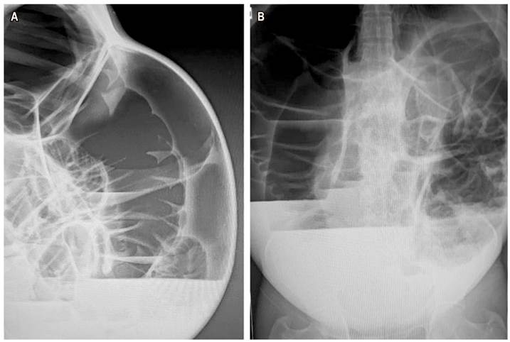
Upon admission, the patient was afebrile, hydrated, tachycardic with a frankly distended, tympanitic abdomen, and peristaltic noises with decreased tone and frequency. He presents no pain. Digital rectal examination performed with empty rectal ampulla and hypotonic anal sphincter. Abdominal X-rays are performed (Figure 1 ). Clear loop distension is considered (which conditions a critical colon), so urgent surgical intervention is indicated.
Despite decompression with a nasogastric tube, the patient has severe abdominal distension in the operating room (Figure 2). A xypho-pubic laparotomy is performed. Upon entering the cavity, two volvuli are found: one of the cecum and the other of the sigmoid colon, devolvulated with vascular pedicle control (Figure 3A-C). Distension that conditions a critical colon is found, so a subtotal colectomy plus ileostomy is performed (Figure 3D).
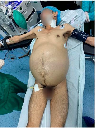
Figure 2 Photograph of the patient’s condition before the procedure started: Clear abdominal distension, despite decompression with a nasogastric tube and urinary catheter.
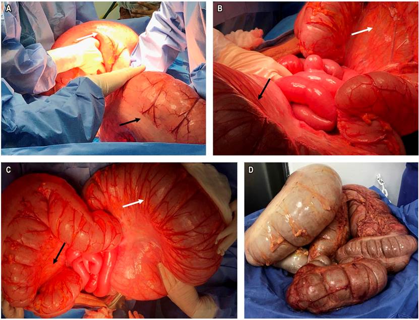
Figure 3 Intraoperative images. A. Synchronous volvulus of the cecum (white arrow) and sigmoid (black arrow). B and C. Results of the devolvulation: sigmoid (white arrow) and cecum (black arrow). D. Product of subtotal colectomy, surgical specimen.
The patient is extubated and transferred to the floor with expected clinical evolution. During the immediate postoperative period, 2 packaged red blood cells are transfused. On the second day, the patient has an adequate intestinal transit. The nasogastric tube is removed, and a liquid diet is started. On postoperative day 4, a nursery call is received since the patient has emetic episodes of feculent content with abundant bleeding from the ileostomy. The patient is tachycardic with great abdominal distension and generalized pain on palpation, so he is urgently taken to the operating room.
During the second surgical procedure, hemoperitoneum is found in the patient without apparent active bleeding or its cause, also a large dilation of thin loops with no transition zone. A non-ischemic permeable ileostomy is left with packaging. After 2 days, a new surgical procedure is carried out having turbid peritoneal fluid with fibrinopurulent membranes in thin intestinal loops. The wall is closed, but the patient requires vasopressor and invasive ventilatory support because extubation is not possible. The patient is transferred to the intensive care unit.
The patient evolves in very bad general conditions with vasopressor support in high doses and oligoanuric. He presents multiorgan dysfunction associated with septic diseases, which does not respond to medical or antibiotic management, and dies.
Discussion
The term volvulus is derived from Latin volvere “rotate on its own axis”1. It was first described in the Ebers papyrus in 1550 BC with a description of the natural course of the volvulus with spontaneous torsion and intestinal reduction or rotting1. The presentation of each volvulus is considered an independent entity3. Volvuli are more common in the sigmoid colon (75%) and the cecum (15%-22%)4. However, there are reports of volvuli in the transverse colon (3%) and the splenic flexure (2%)4.
The double colonic volvulus has been considered a rare entity3. The literature records this presentation as synchronous volvuli, compound volvuli, simultaneous volvuli, or double volvulus. Singh et al documented the first case in 19855. There are also reports by Theuer et al (1991)6, Moore et al (1992)3, Kellil et al (2017)7), and Islam et al (2016)4.
Volvuli occur mainly in the Middle East, Asia, and Africa2,3. This zone is known as the “volvulus belt”1) in the literature, which is associated with a fiber-rich diet. Additionally, other risk factors are considered: a long and mobile mesentery, redundant sigmoid, and chronic intestinal distension2. Likewise, there are individualized risk factors for each of its presentations. The sigmoid volvulus is related to a fiber-rich diet, previous abdominal surgery, psychiatric disorders, Chagas disease, Parkinson’s disease, ischemic colitis, and megacolon3. Additionally, it frequently affects the elderly population in nursing homes and patients with neurological pathologies3.
Symptoms include abdominal pain (100%), abdominal distension (94%-100%), nausea and vomiting (87% -100%), and rebound tenderness (69%)2. However, it is not an easily pre-surgically diagnosed condition. Only 20% of patients are diagnosed2.
Timely management is essential in these patients. Compromised blood circulation leads to a rapid progression of necrosis of the volvulated segments (75%-79%), which can be life-threatening3. These patients must receive aggressive fluid resuscitation, broad-spectrum antibiotics, and definitive surgical management4,6. According to the intraoperative findings and the patient’s conditions, it is decided whether the patient benefits from resection and anastomosis or whether a terminal ostomy and delayed anastomosis should be performed2,3.
Complications in these patients are associated with peritonitis, sepsis, and dehydration because of the third spaces generated2. Although described at 15%, mortality can be as high as 73% of cases. It is associated with septic shock, caused by bacterial translocation and intraoperative colon manipulation2.
Conclusion
Synchronous presentation of intestinal volvuli is an uncommon occurrence in patients with intestinal obstruction. It is an intraoperative diagnosis performed in a patient undergoing a surgical abdomen procedure in most cases. These patients’ management should be based on their conditions. However, the septic commitment and broad spectrum of antibiotic coverage that must be received in the postoperative period associated with strict clinical and paraclinical monitoring should be considered.

