


 text in
  English (pdf)
 Article in xml format
 Article references
 Send this article by e-mail
 Cited by SciELO
  Cited by Google
  Similars in
SciELO
Similars in
SciELO  Similars in Google
 Permalink
Introduction
According to data from GLOBOCAN 20181, colorectal cancer ranks third in morbidity and fourth in cancer-associated mortality after lung and prostate cancer. Identifying advanced diseases (synchronous or metachronous) is becoming increasingly common with new, better-performing diagnostic methods. Intra-abdominal organs represent one of the greatest challenges in imaging suggestive of metastases from a primary colon lesion1.
Enterobius vermicularis, also known as Oxyurid vermicularis, is a frequent helminthic infection in the United States and Eastern Europe. It predominantly affects the pediatric population2. The parasite lives and reproduces in the gastrointestinal tract. It usually lays eggs in the anal region with approximately 10,000-15,000 eggs overnight, causing itching and discomfort3. This parasite has different ectopic locations in the human body, including the omentum, lungs, and liver, the latter, with very few cases reported in the literature3.
The following is a patient’s case with adenocarcinoma of the sigmoid colon with images suggesting liver metastases, with subsequent histological confirmation of E. vermicularis infection.
Case presentation
A 39-year-old Hispanic female patient from Villavicencio (Meta, Colombia) was admitted to the emergency room for a 1-year history of colicky abdominal pain on the left hemiabdomen, associated with a 20 kg weight loss in 4 months, self-limiting emesis, and rectal bleeding. She underwent a colonoscopy, in which an infiltrating mass with stenosis of 80% of the lumen of the sigmoid colon located 30 cm from the anal ridge was reported. Biopsies of the lesion reported moderately differentiated adenocarcinoma originating in tubulovillous adenoma.
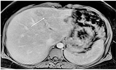
Extension and staging studies were conducted, including a carcinoembryonic antigen quantified at 1.35 ng/mL, a computed axial tomography (CT) of the abdomen with contrast showing a polypoid mass dependent on the descending colon of roughly 45 mm in diameter, irregular, ulcerated, associated with stranding of the perilesional fat and adenopathies at the mesenteric edge. In addition, the liver showed a 98 mm focal lesion in segment VII and an approximately 10 mm focal lesion in segment IV (Figure 1). A magnetic resonance imaging (MRI) of the abdomen showed a 7 mm focal lesion in the IV liver segment and a 7.4 mm focal lesion in the right lobe, indicating liver metastases (Figure 2).
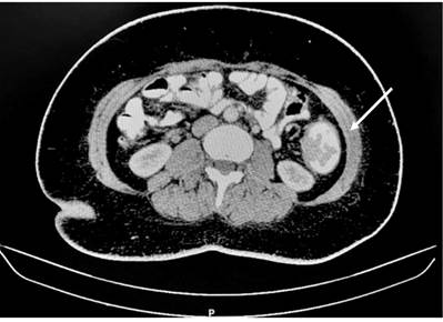
Figure 1 Abdomen CT scan using contrast, performed during admission, showing a sigmoid colon-dependent tumor with a large involvement of intestinal lumen.
Based on these findings, anterior resection of the rectum and sigmoid was performed with high anastomosis and liver biopsy, as well as high ligation of the vascular pedicle. After a one-week hospital stay, the patient showed a good clinical evolution and was discharged for an outpatient check-up. Subsequently, after receiving the surgical pathology report, it showed a distinguishable adenocarcinoma of the sigmoid colon infiltrating up to the muscle layer, without lymphovascular or perineural invasion, as well as tumor-free resection edges and 0/16 positive nodes for a T2N0. As for the liver biopsy, a “necrosis and acute inflammation with E. vermicularis eggs, without evidence of malignancy in the material examined” report was obtained. Thus, the disease was classified as T2N0M0 for stage IB due to the absence of metastatic lung lesions. During outpatient follow-up, the patient received anthelmintic treatment; a check-up abdomen MRI was performed, showing a residual scar lesion in segment VII with scarring characteristics and another subcapsular lesion in segment IV.
Discussion
There are few cases described of liver infection caused by E. vermicularis. In developed countries, this nematode, also known as Oxyurid vermicularis, produces a frequent helminthic infection and predominantly affects the pediatric population2. The parasite lives and reproduces in the gastrointestinal tract. It usually lays eggs in the anal region overnight, causing itching and discomfort3. The entrance route is through the ingestion of pinworm eggs through the fecal-oral route. They enter the stomach and small intestine, and the larvae migrate to the ileum, cecum, and appendix. The adult female settles in the ileum, where she copulates. Around the fifth week, she begins laying eggs and migrates toward the colon and anus. Then, she releases thousands of eggs onto the perianal skin. The eggs survive in a humid environment and a low temperature4. It is unclear how the nematode can reach the liver. However, it is believed that it cannot penetrate intact tissues, so the presence of non-intact or injured intestinal mucosa (as in colorectal cancer) can facilitate its migration5.
Liver infection and abscess formation caused by E. vermicularis are extremely rare. Fewer than 10 cases have been reported in the literature, from which approximately 5 cases have been reported in patients with colorectal cancer and liver infection simulating liver metastases in imaging studies2-4,6,7.
Colorectal cancer ranks third in cancer morbidity; 1.4 million patients with this disease are diagnosed each year globally. Patients with metastatic disease or stage IV have an overall 5-year survival1,8. Nonetheless, depending on the stage, up to 50% of patients will at some point develop liver metastases8,9.
Abdominal ultrasound has a limited role in the complete characterization of liver lesions. Contrasting abdominal CT allows adequate identification and even volumetric calculations of liver lesions. Generally speaking, images suggesting metastasis describe a peripheral reinforcement in the arterial phase, and 11% may present calcifications10. Liver metastases are hypovascular in the arterial phase with hypervascular peripheral enhancement and heterogeneous hypoattenuation in the portal venous phase, with an 85% identification rate and a 96% positive predictive value (PPV).10
Several points help to discern between liver lesions suggestive of metastasis versus infectious liver lesions, such as margin in the portal venous phase, hepatic parenchyma enhancement, arterial border enhancement, dynamic change of arterial border enhancement, size discrepancy between arterial phase and portal venous phase, and perilesional hyperemia. Thus, according to Jae Gu Oh et al (Table 1)11, abscesses, unlike metastases, have more defined margins in the portal venous phase, greater enhancement of the parenchyma, more peripheral (rim arterial) enhancement, and greater perilesional hyperemia. Regarding the dynamic changes of peripheral rim enhancement, abscesses report a higher enhancement persistence in the venous phase than metastases, which disappear in a higher percentage with the transition from the arterial to the venous phase11.
Table 1 Differences between liver abscess and liver metastases on abdominal CT
| Liver abscess | Liver metastases | |
|---|---|---|
| Margins | Defined | Diffuse |
| Peripheral enhancement | Present in the arterial phase and persists in the venous phase | No peripheral enhancement in any phase |
| Size | Size change when comparing between phases | Constant size in all phases |
| Other | Higher bile duct dilation | Less parenchymal enhancement |
A further important aspect to consider for liver lesions when comparing the arterial and portal venous phases is that there may be changes in size, as in liver abscesses, which may show changes in 45.2% compared to only 16.8% in cases of metastasis11.
Another imaging study is MRI, which is more sensitive than CT in detecting metastatic liver lesions, particularly sub-centimeter size lesions, or for restaging lesions after neoadjuvant surgery, in which CT sensitivity drops to 77%12. Cases reporting liver infection by E. vermicularis suggest a predilection for the right hepatic lobe, close to the hepatic surface, with hypodense images in the CT between segments VII and VIII, similar to the findings in our patient2,12.
Conclusions
Colorectal cancer metastases represent an important percentage of the spread of this primary cancer. However, an important differential diagnosis for comprehensive patient treatment is liver infections caused by bacteria or parasites. Cases in which the E. vermicularis parasite produces an infection of the liver parenchyma and simulates, in turn, a metastasis of a primary colon tumor are rare but have been described in the literature. Since multiple diagnostic imaging modalities have been introduced, more liver lesions have been detected in the pre-surgical setting. The correct characterization of these lesions through the study by the pathology group prevents patients from being taken to major surgeries that include liver resections.

