


 text in
  English (pdf)
 Article in xml format
 Article references
 Send this article by e-mail
 Cited by SciELO
  Cited by Google
  Similars in
SciELO
Similars in
SciELO  Similars in Google
 Permalink
Introduction
Helicobacter pylori infection has a high prevalence worldwide1,2. In 2015, it was estimated that 4.4 billion people were infected, corresponding to more than half of the world’s population2. In Colombia, a prevalence of 69.1% was found in 2003 based on data from 16 cities in all regions3. In Cali, the prevalence was reported at 63.1%3. Other studies have described lower frequencies, such as a study that reported a prevalence of 36.4% in Medellín4.
H. pylori is relevant not only because of its high prevalence but also because of its relationship with the development of multifocal atrophic gastritis, gastric ulcers, and gastric adenocarcinoma1,5,6. This last relationship, established by the International Agency for Research on Cancer (IARC), has led to the classification of this bacterium as a type 1 carcinogen7. H. pylori can increase the risk of gastric cancer by ten times7; thus, its eradication is crucial for the population of our country, where the incidence of this cancer is high. Additionally, gastric cancer is among the five types of cancer with the highest mortality8; reducing the prevalence of H. pylori could decrease the disease burden generated by this pathology.
There are several methods to detect H. pylori, most with adequate sensitivity and specificity. They are divided into non-invasive, such as serology, urea breath test, and stool antigen test, and invasive, such as histology, culture, rapid urease test, and polymerase chain reaction test1,5,9. In Colombia, the most used method is the histopathological study of gastric biopsies obtained through upper endoscopy (EGD). Some endoscopic findings may be related to the presence of H. pylori, such as diffuse erythema, mucus in the gastric mucosa, erythema foci in the gastric fundus, enlarged folds, edema, and the arrangement of collecting venules of the gastric mucosa10.
The epidemiology of H. pylori infection has been described in international studies. However, recent reports on the prevalence of this infection in Colombia have yet to be identified. The prevalence of this infection and its related pathologies vary in the literature (69.1%-36.4%)3,4. It is essential to know the local epidemiology to determine the need for changes in detection processes, propose eradication strategies, and reduce the burden of the disease. This study measured the prevalence of H. pylori in consecutive patients undergoing EGD for various indications and assessed risk factors and endoscopic and pathological findings connected with its manifestation.
Materials and methods
Design and participants
An analytical cohort study was carried out to describe the prevalence of H. pylori and evaluate the related risk factors in adult patients undergoing outpatient EGD for any medical indication at the endoscopy unit of a quaternary care university hospital between June and December 2020. We included patients over 18 years of age who underwent endoscopy and biopsy of the gastric mucosa of the antrum and body with at least two tissue samples for processing and histopathological description. Consecutive patients who met the selection criteria were included until completing the sample size. Patients with an indication for therapeutic endoscopy were excluded. The institutional ethics committee approved the study before its initiation.
Measurements and data collection
The information on the origin of patients and the use of proton pump inhibitors (PPIs) or antibiotics (ATBs) for any indication in the last month was taken from the medical records. The treating gastroenterologist or gastrointestinal surgeon described and recorded the endoscopic findings in the procedure report, from which the data of interest were extracted.
Pathology samples were fixed with 10% buffered formalin and embedded in paraffin blocks. Histological sections were stained with hematoxylin and eosin for regular study, and the Warthin-Starry technique was used to detect H. pylori. The histopathological evaluation was made according to the analogous visual scale described by Dixon et al., known as the Sydney System11. This classification assigns a semiquantitative description to each histological parameter, from 0 (normal) to 3 (severe or abundant), including neutrophil and mononuclear cell infiltrate, the intensity of atrophy, and the severity of intestinal metaplasia, and quantifies H. pylori colonization1,11.
Sample size and statistical analysis
The sample size was calculated with local data from 2003 that reported a prevalence of H. pylori of 63.1% in Cali3. With an estimated frequency of 50% prevalence and 97% confidence, a sample of 471 patients was calculated.
For data analysis, we employed STATA v.14® (StataCorp. 2015. Stata Statistical Software: Release 14. College Station, TX: StataCorp LP). Continuous variables were described with measures of central tendency and dispersion according to their distribution, evaluated with the Shapiro-Wilk test. Absolute and relative frequencies were used to describe nominal or ordinal qualitative variables. The prevalence of H. pylori was reported with a 95% confidence interval (95% CI). We performed Chi-square (χ2) or Fisher’s tests to assess differences in proportions, as appropriate, and Student’s t and Mann-Whitney U tests depending on their distribution.
Results
During the study period, 1,105 patients were identified for EGD, of whom 613 met the selection criteria and were included in the analysis. The reasons for being selected were, among others, patients with complete data, older than 18 years, and EGD and biopsy performed in the endoscopy unit.
The demographic and clinical characteristics of the population are shown in Table 1. The median age was 52 years (interquartile range [IQR]: 38-62 years), and about two-thirds of the participants were women. Most patients who belonged to the prepaid, policy, or private social security scheme came from Cali, and their most common indication for EGD was dyspepsia.
Table 1 Demographic and clinical characteristics of the population taken to EGD
| Characteristic | General (n = 613) | |
|---|---|---|
| Age* | 52 | (38-62) |
| Sex | ||
| Female | 390 | 63.6% |
| Male | 223 | 36.4% |
| Social security scheme | ||
| Contributive and subsidized | 57 | 9.3% |
| Prepaid, policy, or private | 556 | 90.7% |
| Origin | ||
| Cali | 475 | 77.5% |
| Another city | 131 | 21.4% |
| Rural area | 7 | 1.1% |
| EGD indication | ||
| Dyspepsia | 395 | 64.4% |
| GERD | 62 | 10.1% |
| H. pylori control | 32 | 5.2% |
| Cancer screening | 24 | 3.9% |
| Chronic gastritis and metaplasia monitoring | 14 | 2.3% |
| Transplant protocol | 12 | 2.0% |
| Other EGD indication | 88 | 14.4% |
| PPI use in the last month | ||
| No | 416 | 67.9% |
| Yes | 197 | 32.1% |
| ATB use in the last month | ||
| No | 544 | 88.7% |
| Yes | 69 | 11.3% |
*Value stated as median (IQR).
GERD: gastroesophageal reflux disease; EGD: upper endoscopy; PPI: proton pump inhibitors; IQR: interquartile range. Source: The authors.
According to the histopathological study, the prevalence of H. pylori in this adult population with EGD indication was 38.5% (95% CI: 34.7%-42.4%). In most patients with H. pylori, it was identified in the antrum and the body, and in a minority of cases, in the antrum or the body (15%) (Figure 1). In the antrum, the presence of H. pylori was “abundant,” while in the body, the categories “mild,” “moderate,” and “abundant” had similar frequencies (Figure 2).
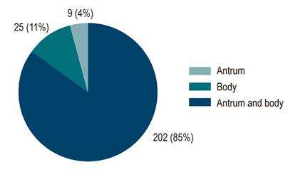
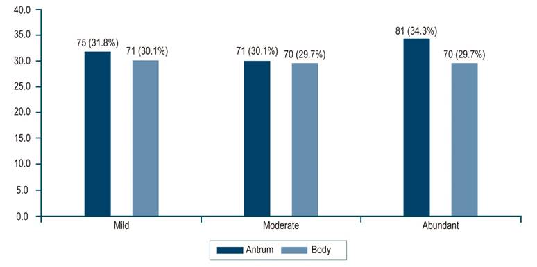
Table 2 shows the prevalence of H. pylori according to the EGD indication; the highest was observed in patients with an indication for H. pylori control. For the other indications, there were similar frequencies. Table 3 presents the association of specific clinical characteristics and endoscopic findings with H. pylori. Patients diagnosed with H. pylori were young, and PPI use in the last month was reported to be lower in the group with H. pylori. There was no significant difference in the frequency of a history of dyspepsia between patients with and without H. pylori. Of the total number of patients with dyspepsia as an EGD indication, 41% were diagnosed with H. pylori.
Table 2 Prevalence of H. pylori according to EGD indication
| EGD indication | Prevalence | 95% CI |
|---|---|---|
| H. pylori control | 50.0% | 31.4%-68.6% |
| Dyspepsia | 41.0% | 36.2%-45.9% |
| Other EGD indication | 35.2% | 26.1%-45.6% |
| Transplant protocol | 33.3% | 13.8%-60.9% |
| GERD | 30.6% | 20.6%-43.0% |
| Cancer screening | 25.0% | 13.3%-42.1% |
Source: The authors.
Table 3 Risk factors for H. pylori and related endoscopic findings
| Clinical feature | H. pylori | p-value | |
|---|---|---|---|
| No (n = 377) | Yes (n = 236) | ||
| Age | 54 (41-64) | 47 (34.5-58) | < 0.001 |
| Female | 242 (64.2) | 148 (62.7) | 0.711 |
| EGD indication | |||
| Dyspepsia | 233 (61.8) | 162 (68.6) | 0.085 |
| GERD | 43 (11.4) | 19 (8.1) | 0.180 |
| Cancer screening | 24 (6.4) | 8 (3.4) | 0.238 |
| H. pylori control | 12 (3.2) | 12 (5.1) | 0.107 |
| Transplant protocol | 8 (2.1) | 4 (1.7) | 0.775 |
| Other EGD indication | 57 (15.1) | 31 (13.1) | 0.495 |
| PPI use in the last month | 138 (36.6) | 59 (25.0) | 0.003 |
| ATB use in the last month | 41 (10.9) | 28 (11.9) | 0.706 |
| Endoscopic finding | |||
| Esophagus | |||
| Normal | 286 (75.9) | 203 (86.0) | 0.002 |
| Esophagitis | 66 (17.5) | 24 (10.2) | 0.013 |
| Barrett’s esophagus | 2 (0.5) | 0 | 0.526 |
| Ulcer | 4 (1.1) | 1 (0.4) | 0.654 |
| Other | 19 (5.0) | 8 (3.4) | 0.420 |
| Antrum | |||
| Normal | 5 (1.3) | 2 (0.8) | 0.712 |
| Possible atrophy | 26 (6.9) | 13 (5.5) | 0.473 |
| Metaplasia foci | 10 (2.7) | 3 (1.3) | 0.390 |
| Nodular-follicular appearance | 4 (1.1) | 23 (9.7) | < 0.001 |
| Erythematous gastropathy | 353 (93.6) | 219 (92.8) | 0.631 |
| Erosive gastritis | 63 (16.7) | 29 (12.3) | 0.128 |
| Ulcer | 5 (1.3) | 9 (3.8) | 0.048 |
| Neoplasm | 1 (0.3) | 0 | 1.000 |
| Polyps | 1 (0.3) | 0 | 1.000 |
| Other | 7 (1.9) | 3 (1.3) | 0.748 |
| Body | |||
| Normal | 270 (71.6) | 184 (78.0) | 0.019 |
| Possible atrophy | 28 (7.4) | 5 (2.1) | 0.006 |
| Metaplasia foci | 5 (1.3) | 1 (0.4) | 0.418 |
| Nodular-follicular appearance | 10 (2.7) | 7 (3.0) | 0.761 |
| Erythematous gastropathy | 54 (14.3) | 34 (14.4) | 0.838 |
| Erosive gastritis | 10 (2.7) | 5 (2.1) | 0.727 |
| Ulcer | 1 (0.3) | 0 | 1.000 |
| Neoplasm | 2 (0.5) | 0 | 0.530 |
| Polyps | 15 (4.0) | 2 (0.8) | 0.026 |
| Other | 4 (1.1) | 3 (1.3) | 1.000 |
| Duodenum | |||
| Normal | 355 (94.2) | 228 (96.6) | 0.172 |
| Duodenitis | 13 (3.4) | 6 (2.5) | 0.529 |
| Duodenal ulcer | 0 | 2 (0.8) | 0.148 |
| Other | 9 (2.4) | 0 | 0.015 |
Source: The authors.
A significant difference was observed for some endoscopic findings: a normal esophagus in patients with H. pylori was more frequent than esophagitis in patients without H. pylori. Mucosa with a nodular-follicular appearance in the antrum occurred in 4.4% of all patients, but it was highly specific for H. pylori infection (specificity: 98.9%; 95% CI: 97%-99%; likelihood ratio [LR+] = 9). An ulcer in the antrum was also frequent in patients diagnosed with H. pylori, although only 14 (2.2%) patients had it. Findings of possible atrophy and polyps in the body were more frequent in patients without H. pylori.
Table 4 introduces the histopathological findings related to H. pylori. Acute inflammation was noted in the antrum and body in 94.5% and 83.9% of patients infected with H. pylori; it was an infrequent finding in patients without H. pylori (5%-6%) and statistically significant. Atrophy and intestinal metaplasia in the antrum were not associated with H. pylori. The absence of this bacterium had a substantial relationship with atrophy and intestinal metaplasia in the body. The most frequent overall histopathology diagnosis was chronic non-atrophic gastritis (CNG).
Table 4 Histopathological findings
| H. pylori | Pathology | Total | ||||||
|---|---|---|---|---|---|---|---|---|
| Normal | CNG | MAG | MAG IM | Dysp | Ca | |||
| Negative | n | 23 | 279 | 4 | 68 | 1 | 2 | 377 |
| % | 6,1% | 74% | 1,1% | 18% | 0,3% | 0,5% | 100% | |
| Positive | n | 0 | 210 | 0 | 26 | 0 | 0 | 236 |
| % | 0% | 89% | 0% | 11% | 0% | 0% | 100% | |
| Total | n | 23 | 489 | 4 | 94 | 1 | 2 | 613 |
| % | 3,8% | 79,8% | 0,7% | 15,3% | 0,2% | 0,3% | 100% | |
Ca: cancer; Dysp: dysplasia; MAG: atrophic gastritis; MAG IM: atrophic gastritis with intestinal metaplasia; CNG: non-atrophic gastritis. Source: The authors.
Discussion
This study in adult patients undergoing gastric endoscopy for any indication reports a prevalence of H. pylori infection of 38.5% (95% CI: 34.7%-42.4%). This infection was negatively related to PPI use in the last month and identified in younger patients, probably due to the older age of patients with indications other than dyspepsia and lower infection frequencies; however, it was not associated with other clinical characteristics. No relationship with the indication for endoscopy was identified, highlighting the considerable prevalence of infection even in indications other than dyspepsia. The eradication of H. pylori is a form of prevention accepted worldwide to reduce the incidence of gastric cancer, a common pathology connected with high morbidity and mortality12-14.
The prevalence of H. pylori may vary widely depending on the study site and the inclusion of patients. In some studies, its manifestation has been linked to a low socioeconomic level. A higher prevalence has been reported in low- or middle-income countries with low levels of urbanization, sanitation, and access to drinking water1,2,15. These conditions are common in Colombia, where a high prevalence of H. pylori has been measured, such as that reported at 63% by Bravo et al. in 20033. Nonetheless, more recent studies have described lower prevalences, such as the one obtained in this study and by Correa et al.4) in Medellín, probably concerning a decrease in the risk factors for urban areas.
In studies in low- and middle-income countries, frequencies of H. pylori up to 50% mainly affect patients around ~10 years of age and adults around ~50 years of age, as shown in this study4,15,16. Reportedly, the male sex has been related to a higher prevalence of H. pylori17,18, unlike our study.
The negative association with PPI and ATB use in the last month for any medical indication is explained by the suppressive effect of H. pylori described for this treatment, in addition to the potential impact of reducing the sensitivity of the diagnostic method1,5. Therefore, it is advisable to suspend PPIs and ATBs before performing these tests19,20. Using these medications could be considered an exclusion criterion; however, this study wanted to assess the relationships mentioned and did not exclude patients for this reason.
H. pylori colonizes the antrum more often in patients with normal acid secretion having few acid-secreting parietal cells. In subjects with impaired secretion, for example, due to PPI use or vagotomy, H. pylori colonize the body more frequently21. Thus, finding H. pylori in the antrum and body simultaneously is expected, with greater abundance in the antrum, as revealed in this study.
Erythematous gastropathy in the antrum, considered endoscopic gastritis22, was present in almost all subjects. Like previous studies that report expected mucosa frequencies in the antrum that do not exceed 4% in the presence of H. pylori, only 0.8% of patients with H. pylori in our study had normal mucosa in the antrum3,23. A higher frequency of nodular-follicular appearance findings in the antral mucosa in the presence of H. pylori was observed, with results similar to those informed by other authors24,25. H. pylori has been identified in 95% of duodenal ulcers and 85% of gastric ulcers 26. In this study, the two patients with duodenal ulcers had H. pylori.
Regarding the histopathological findings, on the one hand, a significant relationship was found between acute inflammation and H. pylori in the antrum and the body, as reported by Garg and Mysorekar; this is a hallmark finding of infection, which is rare in the absence of H. pylori21,27. On the other hand, there was a negative association of H. pylori with gastric atrophy and intestinal metaplasia in the body. This association may be a consequence of previous treatment for H. pylori that eradicates the bacteria but does not reverse the histopathological changes in the mucosa7. According to the Sydney System, the most frequent global histopathological diagnosis was CNG (79.8%). Multifocal atrophic gastritis has diagnostic value for being a precursor lesion of malignancy5. Previous studies in the country found that the microorganism was associated with metaplasia, lymphoid follicles, and atrophy4. Concerning the endoscopic findings, this research has the limitation that, even though the six participating gastroenterologists have more than five years of experience, potential interobserver variability was not considered in this work.
Conclusion
The prevalence of H. pylori in patients undergoing EGD for all indications is 38.5% and has similar values in all EGD indication subgroups. The endoscopic search should be in the antrum and the body. Follicular nodular gastritis in the antrum and duodenal ulcer is associated with H. pylori. Studies are required to evaluate the epidemiological behavior in the general population.

