


 text in
  English (pdf)
 Article in xml format
 Article references
 Send this article by e-mail
 Cited by SciELO
  Cited by Google
  Similars in
SciELO
Similars in
SciELO  Similars in Google
 Permalink
Introduction
Acute pancreatitis is one of the main pathologies of the gastrointestinal tract treated in emergency services worldwide1. Colombia’s primary etiology is biliary (80%), followed by alcoholic (9%). Among the infectious causes, only 1.3% is due to ascariasis in some regions2. While usually mild and reversible, its manifestation can reach a mortality rate of up to 15%3. In severe cases, around 20-40% present with peripancreatic necrosis and organ dysfunction3. We present this case of severely compromised acute pancreatitis secondary to an uncommon infectious cause in our setting.
Case presentation
We present the case of a 35-year-old male patient, a public transport driver, from Bogotá, with a two-hour history of highly intense shooting retrosternal chest pain that radiated to the dorsal region and the epigastrium, with nausea, an emetic episode, and no other associated symptoms. He did not report any significant personal or family history or recent trips and did not smoke or drink alcohol.
The initial assessment found mild tachycardia (100 beats per minute), blood pressure and oximetry without alterations, and no additional findings upon cardiopulmonary auscultation or abdominal examination. An electrocardiogram was taken with sinus rhythm, finding no signs of myocardial injury or ischemia. Troponin I and D-dimer were taken in a chest pain study with typical values. The chest x-ray did not show consolidation or pleural effusion. Upon reassessment, he reported that the pain had migrated to the epigastrium with band irradiation, associated with multiple emetic episodes and a febrile peak, for which further studies were carried out to evaluate the acute biliopancreatic etiology and infectious involvement.
Markedly elevated amylase (2,902 U/L), a complete blood count with leukocytosis at the expense of neutrophilia (15,400 leukocytes/mL, 13,800 neutrophils/mL), and a slight elevation of alkaline phosphatase (254 U/L) with elevated transaminases ten times the upper limit (alanine transaminase [ALT]: 520 IU/L; aspartate aminotransferase [AST]: 441 IU/L) and no hyperbilirubinemia were documented. A diagnosis of mild acute pancreatitis is given by initial Atlanta classification, with an APACHE II score of 7 points. Management begins with the suspension of the oral route, analgesia, gastric protection, and fluid resuscitation.
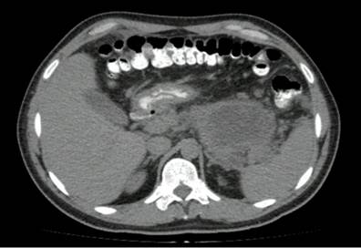
In search of the biliary etiology, an abdominal ultrasound was performed, showing a bile duct and common bile duct without alterations, the gallbladder without signs of cholecystitis or cholelithiasis, evidence of perisplenic fluid, and multiple collections in the paracolic gutters. The other etiology studies were without alterations, including a lipid profile and serum calcium levels. A contrast-enhanced abdominal scan was then performed, identifying a thickening of the body and tail of the pancreas with edema and diffuse peripancreatic fluid, consistent with pancreatitis in the edematous phase. The presence of endoluminal lesions near the gastric cavity with passage towards the intestinal loops and a filling defect of tubular morphology towards the duodenal papilla, with a length of approximately 76 mm, identified as a foreign body (Figure 1) were striking; therefore, there was a high suspicion of ascariasis.
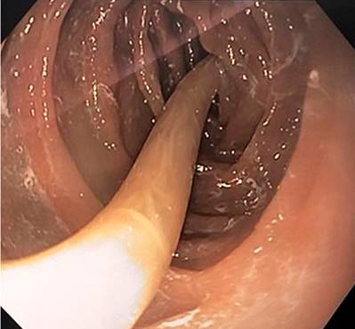
Antiparasitic management began with ivermectin, one drop per kilogram, and albendazole 400 mg daily for three days. An endoscopy of the digestive tract was carried out for viewing, noting an approximately 26 cm nematode at the second duodenal portion (Figure 2), whose proximal end is near the duodenal papilla and the distal end at the level of the duodenal lumen. It was removed with forceps, while the duodenum was thoroughly examined throughout its length with no evidence of another foreign body. Prophylactic antibiotic coverage with ampicillin/sulbactam was started, given the extent of the endoluminal compromise. Other possible causes related to pancreatitis were ruled out, with normal serum calcium (8.3 mg/dL) and a lipid profile with triglycerides less than 500 mg/dL. Therefore, it was considered that the directly related obstructive cause was pancreaticobiliary ascariasis.
On the fourth day of hospitalization, the patient presented with poor pain control, intolerance to the oral route, and signs of a systemic inflammatory response given by tachycardia, persistent fever, increased leukocytosis, and hyperlactatemia. Therefore, we decided to carry out an imaging follow-up to evaluate the complications, showing thickening of the body, uncinate process, and tail of the pancreas without enhancement associated with more collections than in the previous study. The most extensive collection was approximately 170 cm3 in volume, compatible with necrotic pancreatitis and associated intraparenchymal collections. A moderate-severe compromise was considered, with no signs of superinfection in collections. A procalcitonin test was performed with negative results. We decided to withdraw the oral route again, and the analgesic management was adjusted by the Pain Clinic group, with follow-up by the nutritional support group.
The patient exhibited good clinical evolution, hemodynamic stability, and adequate clearance of hyperlactatemia in the following days. The oral route was started with good tolerance, modulation of the signs of a systemic inflammatory response, and completion of antibiotic coverage for seven days. The patient completed 48 hours of tolerance with a soft diet. Progress was made in the diet, so we indicated discharge with outpatient follow-up by gastroenterology.
Discussion
Acute pancreatitis is a frequent reason for consultation in emergency services worldwide, and its incidence has increased in recent years4. The manifestation of this entity may vary in terms of clinical involvement and possible complications5. This report presents an atypical case of pancreatitis not only because of its unusual clinical manifestation (chest pain and dyspnea as initial clinical symptoms) but also because of its etiology.
Pancreatitis generally has common clinical manifestations, which are defined by their diagnostic criteria (two out of three): characteristic abdominal pain (epigastric with band irradiation), amylase or lipase levels above three times the upper limit of normal, or findings suggestive of inflammatory changes in the pancreas by imaging6. Upon reaching its diagnosis, initiating directed management and monitoring its possible complications is essential7. Although different drugs have been studied, such as immunomodulators or antisecretory agents, the cornerstone of its treatment is adequate hydration, pain management, and restriction to the oral route7-9.
After establishing its initial management, the next step is identifying its etiology. The initial approach must search for the most frequent causes in Colombia and the world. Biliary pathology is the leading cause of pancreatitis, followed by alcohol; thus, an upper abdominal ultrasound should be performed, and an appropriate history of alcohol use should be obtained from all patients. Although infrequent, the tumoral cause should be studied in patients older than 409. By excluding the most frequent causes, other serological and imaging studies should be performed serially to find its etiology, including serum levels of triglycerides and calcium1,10. In the case presented, there were no findings suggestive of biliary lithiasis etiology in the upper abdomen ultrasound, so we continued with serological tests and characterization by scan, which evidenced the cause.
About 10% of pancreatitis cases are related to infectious causes, with viral causes and tropical diseases being the most common. The parasitic cause is less frequent; however, it is an important entity, especially in developing countries11. Ascariasis is the parasitic cause most frequently related to pancreatitis, especially in children, occurring in approximately 23% of cases11.
Ascaris lumbricoides infection occurs initially by ingesting the infective egg of the larva, which migrates towards the intestinal loops and penetrates the colonic mucosa. Finally, it settles in the liver and lung tissue through hematogenous dissemination, penetrating the bronchial tissue and the oropharynx, where it is swallowed again and continues its maturation cycle near the intestinal lumen. Approximately ten days after primary infection, the larva matures in the small intestine, where it can reside and increase in size for up to 12 months. Therefore, the clinical manifestations can be masked by even asymptomatic pictures and self-resolving infections; however, cases as severe as intestinal obstruction requiring surgical management and high morbidity and mortality rates may occur12. In cases of pancreaticobiliary ascariasis, most of them manifest as biliary colic or acute cholangitis and, less frequently, due to pancreatitis (6%) or in the form of liver abscesses13.
Typically, cases of hepatobiliary pancreatitis go unnoticed or are little suspected, especially in adults in urban areas14, as in our patient. The initial suspicion was given by the incidental findings on the first tomography, whose purpose was to evaluate the involvement of the pancreatic parenchyma and, thus, to look for other etiologies, such as anatomical variants or tumors, as described in the diagnostic algorithms when ruling out the most common causes of this entity1,4,8. With this first approach, the patient underwent an upper GI endoscopy not only for diagnostic confirmation but also for possible therapeutic management. Since the entire helminth could be removed, with no evidence of remains or distal compromise, it was not necessary to perform an endoscopic retrograde cholangiopancreatography (ERCP) for its total removal, as described in cases related to this etiology13.
In addition to this management, based on removing the parasite from the gastrointestinal tract, the patient required intrahospital surveillance due to subsequent clinical deterioration. Therefore, as recommended in different management guidelines3,6,9, an imaging follow-up was performed with evidence of progression of the necroinflammatory involvement associated with necrotizing pancreatitis with collections; however, there were no imaging (gas) or clinical signs of infectious involvement. Since, at the time, he was under empirically established antibiotic management due to the high risk of endoluminal intestinal injury and bacterial translocation by the helminth, we considered not making any adjustments to the therapy and continuing with monitoring. The patient did not present with organic dysfunction or infectious deterioration; enteral nutrition was started with adequate tolerance. Finally, his discharge was indicated with imaging follow-up by gastroenterology.
This case is interesting for different reasons. First, as previously mentioned, it was an unusual manifestation due to the signal symptom of chest pain, even having ruled out potentially fatal causes such as acute coronary syndrome based on age and sex. Finding a “foreign body” near the duodenal papilla in the initial scan in looking for different etiologies of pancreatitis, having ruled out the most frequent ones, allowed its proper diagnosis and endoscopic therapeutic management. Furthermore, the presence of ascariasis in an adult residing in the urban area of Bogotá is noteworthy. Although ascariasis occurs more frequently in a tropical country such as Colombia than in others, it is odd to find it in a social and epidemiological context, such as our patient’s14,15.
Conclusion
Acute pancreatitis is a common condition worldwide. Its manifestation is usually classic; however, unusual symptoms such as chest pain may appear as the initial manifestation. The most frequent etiologies must be ruled out without neglecting infections as causal factors. It is unusual to find a parasitic compromise in general, especially in adults from urban areas; however, it should not be dismissed as a differential diagnosis. In addition to the initial management in the emergency room, consisting of adequate hydration, restriction of the oral route, and analgesic management, the treatment of pancreatitis due to Ascaris must be completed endoscopically with the extraction of the helminth. Close clinical follow-up of patients with pancreatitis and monitoring for possible complications, regardless of their cause, is vital.

