SciELO - Scientific Electronic Library Online

 
vol.37 número4Reservoritis secundaria a infección por citomegalovirus: reporte de caso y revisión de la literatura índice de autoresíndice de materiabúsqueda de artículos
Home Pagelista alfabética de revistas  

Servicios Personalizados

Revista

Articulo

Indicadores

Links relacionados

  • En proceso de indezaciónCitado por Google
  • No hay articulos similaresSimilares en SciELO
  • En proceso de indezaciónSimilares en Google

Compartir


Revista colombiana de Gastroenterología

versión impresa ISSN 0120-9957versión On-line ISSN 2500-7440

Rev. colomb. Gastroenterol. vol.37 no.4 Bogotá oct./dic. 2022  Epub 12-Jul-2023

https://doi.org/10.22516/25007440.815 

Case report

Peutz-Jeghers syndrome: Case report

Edgar Julián Ferreira-Bohórquez1  * 
http://orcid.org/0000-0003-0614-5932

Daniel Stiven Quintero-Rincón2 
http://orcid.org/0000-0002-2999-901X

Yulieth Vanessa Caro-Gamboa3 
http://orcid.org/0000-0003-4144-580X

María Camila Ayala-Forero4 
http://orcid.org/0000-0001-5016-9080

1Clinical-surgical gastroenterologist, Hospital Regional Manuela Beltrán del Socorro, Hospital Universitario de Bucaramanga, Clínica Los Comuneros, Gastrocal, Floridablanca. Bucaramanga, Colombia.

2Internist, Universidad Nacional de Colombia. Bogota, Colombia.

3Internist, Universidad de Santander. Bucaramanga, Colombia.

4Internist, Universidad de Santander. Bucaramanga, Colombia.


Abstract

Peutz-Jeghers syndrome is an autosomal dominant hereditary disease characterized by multiple hamartomatous-type gastrointestinal polyps associated with mucocutaneous hyperpigmentation.

A case of a 25-year-old male patient with a history of right hemicolectomy due to ileocolonic intussusception secondary to a giant polyp in the terminal ileum is reported. This patient consulted for rectal bleeding, with evidence on physical examination of dark brown hyperchromatic lesions on the buccal mucosa. A total colonoscopy was performed, noting multiple polyps. Endoscopic mucosectomy was conducted on some of them, being histopathologically compatible with hamartomatous polyps.

Keywords: Peutz-Jeghers syndrome; hamartomatous polyps; hyperchromatic lesions

Resumen

El síndrome de Peutz-Jeghers es una enfermedad hereditaria, autosómica dominante, caracterizada por la presencia de múltiples pólipos gastrointestinales de tipo hamartomatoso y se asocia con hiperpigmentación mucocutánea.

A continuación, se reporta un caso de un paciente de 25 años con historia de hemicolectomía derecha por una intususcepción ileocolónica secundaria a un pólipo gigante en el íleon terminal. Se trata de un paciente que consultó por rectorragia, con evidencia en el examen físico de lesiones hipercromáticas color café oscuro en la mucosa yugal. Se realizó una colonoscopia total, en la que se observaron múltiples pólipos. Se practicó una mucosectomía endoscópica a algunos de ellos, histopatológicamente compatibles con pólipos hamartomatosos.

Palabras clave: Síndrome de Peutz-Jeghers; pólipos hamartomatosos; lesiones hipercromáticas

Introduction

Peutz-Jeghers syndrome (PJS) is a rare, autosomal dominant, hereditary polyposis associated with a high frequency of STK11/LKB1 gene mutations located on the short arm of chromosome 191,4. It is characterized by mucocutaneous pigmentation and multiple hamartomatous polyps, with an increased risk of gastrointestinal malignancy. It has also been associated with an elevated risk of gynecological, testicular, and thyroid cancer1.

Mucocutaneous pigmentation is due to melanin-laden macrophages in the dermis and increased melanocytes at the dermal-epidermal junction, present in about 95% of patients. These usually appear in childhood around the mouth, nostrils, perianal region, fingers, and knuckles2,5.

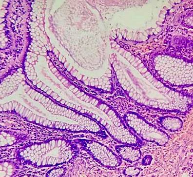
PJS polyps are usually located in the small intestine, mainly in the jejunum, followed by the colon, rectum, and stomach. They can also appear outside the gastrointestinal tract, including the renal pelvis, bladder, lungs, and nasopharynx5. Histological features include an elongated wave-shaped epithelial component, a cystic dilatation of the gland extending into the submucosa, and smooth muscle extending into the fronds of polyps2.

The clinical diagnosis should be considered in an individual meeting at least one of the following criteria1-3:

  1. Three or more histologically confirmed polyps

  2. Peutz-Jeghers polyps, regardless of number, in a subject with a family history of PJS

  3. Characteristic mucocutaneous pigmentation in an individual with a family history of PJS

  4. Peutz-Jeghers polyps associated with characteristic mucocutaneous pigmentation

Clinical case

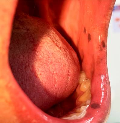
We present the case of a 25-year-old man from the urban area of ​​the municipality of Barbosa (Santander), who was admitted to the emergency department due to diffuse abdominal pain of three years of evolution and rectal bleeding. He had a history of right hemicolectomy for acute abdomen secondary to ileocolonic intussusception due to a giant polyp in the terminal ileum measuring 45 cm x 42 cm. Upon checking the surgical specimen, another giant polyp was found in the jejunum, 80 cm from the angle of Treitz, both with hamartomatous histopathology. The physical examination on admission revealed multiple hyperchromatic dark brown macular lesions on the buccal mucosa, with no other relevant findings (Figure 1).

Figure 1 Hyperchromatic spots on the buccal mucosa. Source: Authors’ archive. 

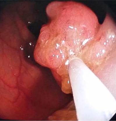
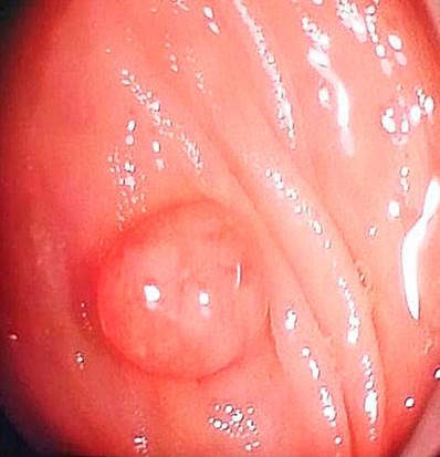
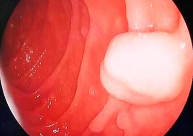
The patient was hospitalized and underwent a colonoscopy, noting two 7 mm polyps near the rectum and two pedunculated polyps in the proximal transverse muscle measuring 20 mm x 15 mm (Figure 2) and 20 mm x 20 mm (Figure 3), respectively. An upper digestive endoscopy was also performed, documenting multiple sessile polyps (around ten) between the first and second duodenal portions, with diameters ranging from 4 mm to 12 mm (Figures 4 and 5). All colon polyps were endoscopically resected, and one of the largest duodenal polyps was removed.

Figure 2 20 mm x 15 mm pedunculated polyp of the transverse colon. Source: Authors’ archive. 

Figure 3 20 mm x 20 mm transverse pedunculated polyp. Source: Authors’ archive. 

Figure 4 Hamartomatous-looking sessile polyp of the duodenum. Source: Authors’ archive. 

Figure 5 Hamartomatous-looking sub-pedunculated polyp of the duodenum. Source: Authors’ archive. 

The histopathological report of the colon polyps was of the hamartomatous type, and one was a tubular adenoma without dysplasia (Figures 6 and 7). The resected duodenal polyp was reported as a hamartomatous polyp.

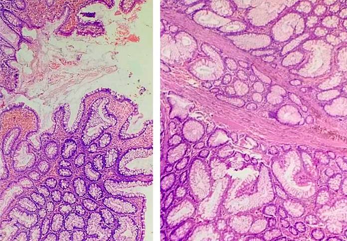
Figure 6 Polypoid lesions made up of glands of various shapes and sizes, supported by bands of smooth muscle fibers. Source: Authors’ archive. 

Figure 7 Different types of cells are present such as columnar, goblet, and endocrine cells. There are no signs of dysplasia. Source: Authors’ archive. 

Discussion

Peutz-Jeghers syndrome is a rare, autosomal dominant disease with few reports in our country. According to Wu et al., it has an estimated incidence of one case per 25,000-300,000 live births. It may occur in any ethnic group and affects men and women alike. Its average age of diagnosis is 23 years, and the average age for cancer development is 42 years6.

Moreover, according to Beggs et al., the first manifestation is often gastrointestinal obstruction due to intussusception by polyps, 69% of the time in the small intestine, and usually occurs between 6 and 18 years of age2,6.

The main goal in effectively treating patients with Peutz-Jeghers syndrome is based on surveillance, prevention, and treatment of complications. Recommendations include6-8:

  • Upper digestive tract endoscopy:

  • ○ It is initially detected from the age of 12.

  • ○ If polyps are found, repeat annually.

  • ○ Repeat every 2-3 years until adulthood in the absence of polyps

  • Colonoscopy:

  • ○ Initial screening starts at age 12 or earlier if symptoms are reported.

  • ○ If polyps are found, repeat annually.

  • ○ In the absence of polyps, repeat at intervals of 1-3 years

  • Pancreas:

  • ○ Magnetic resonance cholangiopancreatography (MRCP), biliopancreatic endoscopic ultrasound, or both: from 25-30 years of age; repeat every 1-2 years

  • Breast:

  • ○ Breast exam: clinical examination every six months from the age of 25.

  • ○ Mammography: from the age of 25

  • Gynecological:

  • ○ Papanicolaou smear: annually.

  • ○ Transvaginal ultrasound: annually from the age of 18

  • Testicles:

  • ○ Consider an annual exam and ultrasound starting at age 10

Finally, in terms of prevention, we advise providing genetic counseling to couples with a family history of Peutz-Jeghers syndrome who wish to have children6-10.

Conclusions

Peutz-Jeghers syndrome is a rare disease but with relevant clinical connotations. These patients have a 15-fold increased risk of gastrointestinal malignancy, although it has also been associated with extraintestinal neoplasms.

It is crucial to know its initial clinical manifestation: hyperpigmented mucocutaneous macules (present in more than 95% of cases), a positive family history of Peutz-Jeghers syndrome, and hamartomatous-type gastrointestinal polyposis.

Thus, a prompt diagnosis would be achieved to implement adequate surveillance, as previously recommended, to avoid complications such as intestinal obstruction secondary to intussusception and emergency surgical interventions and monitor the appearance of premalignant and malignant lesions.

Referencias

1. Daniell J, Plazzer JP, Perera A, Macrae F. An exploration of genotype-phenotype link between Peutz-Jeghers syndrome and STK11: a review. Fam Cancer. 2018;17(3):421-27. https://doi.org/10.1007/s10689-017-0037-3Links ]

2. Beggs A, Latchford A, Vasen H, Moslein G, Alonso A, Aretz S, et al. Peutz-Jeghers syndrome: a systematic review and recommendations for management. Gut. 2010;59(7):975-86. https://doi.org/10.1136/gut.2009.198499Links ]

3. Aguilera Matos I, Díaz Oliva S, Velazco Villaurrutia Yd, García Bacallao E, Labrada Moreno LM. Síndrome de Peutz-Jeghers, experiencia de casos en el Instituto de Gastroenterología. Arch.cuba.gastroenterol. 2019;1(1). Disponible en: http://revgastro.sld.cu/index.php/gast/article/view/18Links ]

4. Parga J, Otero Regino W, Gómez Zuleta M. Prevalencia y características histológicas de los pólipos diminutos del recto y del sigmoides en una población colombiana. Rev. colomb. Gastroenterol. 2020;35(1):25-32. https://doi.org/10.22516/25007440.363Links ]

5. Ospina Nieto J, Pío Quintero Á. Síndrome de Peutz-Jeghers. Presentación de casos y revisión de la literatura. Rev. colomb. Gastroenterol. 2009;24(2):188-99. Disponible en: https://www.redalyc.org/articulo.oa?id=337731592013Links ]

6. Wu M, Krishnamurthy K. Peutz-Jeghers Syndrome. [Actualizado el 21 de agosto de 2022]. En: StatPearls [Internet]. Treasure Island (FL): StatPearls Publishing; 2022. Disponible en: https://www.ncbi.nlm.nih.gov/books/NBK535357/ [ Links ]

7. Signoretti M, Bruno MJ, Zerboni G, Poley JW, Delle Fave G, Capurso G. Results of surveillance in individuals at high-risk of pancreatic cancer: A systematic review and meta-analysis. United European Gastroenterol J. 2018 May;6(4):489-499. https://doi.org/10.1177/2050640617752182Links ]

8. Spoto CPE, Gullo I, Carneiro F, Montgomery EA, Brosens LAA. Hereditary gastrointestinal carcinomas and their precursors: An algorithm for genetic testing. Semin Diagn Pathol. 2018;35(3):170-83. https://doi.org/10.1053/j.semdp.2018.01.004Links ]

9. Grez I, M., Prado A, R., Lahsen H, J. and Hernández M, J. Síndrome de Peutz-Jeghers complicado: Reporte de un caso. Revista chilena de cirugía, vol 60, núm. 3, junio, 2008, pp. 249-254. https://doi.org/10.4067/S0718-40262008000300015Links ]

10. Rodríguez Lagos F, Sorlí Guerola J, Romero Martínez I, Codoñer Franch P. Registro y seguimiento clínico de pacientes con síndrome de Peutz Jeghers en Valencia. Revista de Gastroenterología de México. 2020;85(2):123-139. https://doi.org/10.1016/j.rgmx.2019.02.005Links ]

Citation: Ferreira-Bohórquez EJ, Quintero-Rincón DS, Caro-Gamboa YV, Ayala-Forero MC. Peutz-Jeghers syndrome: Case report. Revista. colomb. Gastroenterol. 2022;37(4):502-506. https://doi.org/10.22516/25007440.815

Received: August 17, 2021; Accepted: November 05, 2021

*Correspondence: Edgar Julián Ferreira-Bohórquez. ejulferbo@yahoo.com

Creative Commons License This is an open-access article distributed under the terms of the Creative Commons Attribution License