SciELO - Scientific Electronic Library Online

 
vol.38 issue1Endoscopic Findings in Patients with Moderate to Severe COVID-19: A Cross-sectional StudyFatty Liver (part 2): Clinical Approach and Treatment author indexsubject indexarticles search
Home Pagealphabetic serial listing  

Services on Demand

Journal

Article

Indicators

Related links

  • On index processCited by Google
  • Have no similar articlesSimilars in SciELO
  • On index processSimilars in Google

Share


Revista colombiana de Gastroenterología

Print version ISSN 0120-9957On-line version ISSN 2500-7440

Rev. colomb. Gastroenterol. vol.38 no.1 Bogotá Jan./Mar. 2023  Epub June 06, 2023

https://doi.org/10.22516/25007440.966 

Original article

Whipple’s Disease: A Systematic Review of the Literature

Ledmar Jovanny Vargas-Rodríguez1  * 
http://orcid.org/0000-0001-6001-5720

Jeinny Lucero Ruiz-Muñoz2 
http://orcid.org/0000-0001-5107-7364

Paola Andrea Bolívar-Córdoba2 
http://orcid.org/0000-0001-8432-4472

Mónica Dayana Romero-Cely2 
http://orcid.org/0000-0001-5801-7479

Ervirson Jair Cañón-Abril2 
http://orcid.org/0000-0003-3937-4942

Zulma Marisol Suárez-Correa2 
http://orcid.org/0000-0002-3535-8446

María Angélica Mendoza-Cáceres3 
http://orcid.org/0000-0002-4479-6851

1MD, Epidemiology, Universidad de Boyacá. Tunja, Colombia.

2Medical Student, Universidad de Boyacá. Tunja, Colombia.

3Physician and Surgeon, Universidad Industrial de Santander. Emergency Physician, Clínica Roma, Colsubsidio. Bogotá, Colombia.


Abstract

Introduction:

Whipple’s disease is a chronic systemic disease with a predilection for the digestive system, especially the small intestine. It was first described in 1907 by George H. Whipple, who named it intestinal lipodystrophy. It is caused by a gram-positive bacterium belonging to the Actinomycetaceae family called Tropheryma whipplei.

Objective:

To characterize patients with Whipple’s disease.

Materials and methods:

A systematic literature review was carried out using the DeCS terms enfermedad de Whipple (Whipple’s disease) or (Tropheryma whipplei) in the Pubmed/Medline, Scopus, Scielo, Science Direct, Embase, Cochrane Library, BIREME, Proquest, and Redalyc databases; 123 articles were analyzed.

Results:

123 published articles corresponding to case reports and series were examined, noting a higher prevalence in males (70.6%). The most frequent manifestations were joint symptoms (61%), followed by weight loss (47.1%) and diarrhea (43.4%). The most used diagnostic method was polymerase chain reaction (PCR) (63.2%), followed by biopsy (50.7%) and pathological examination with PAS (periodic acid Schiff) granules (47.8%). The management most used was antibiotic therapy with a predominance of trimethoprim/sulfamethoxazole and ceftriaxone.

Conclusions:

Whipple’s disease has a low prevalence, occurs more frequently in white people, mainly affects the elderly, has a predilection for the male sex, and is characterized as a chronic systemic disease with a predilection for the digestive system, especially the small intestine.

Keywords: Whipple’s disease; Tropheryma; systematic review.

Resumen

Introducción:

la enfermedad de Whipple es una enfermedad crónica sistémica con predilección por el aparato digestivo, especialmente el intestino delgado. Fue descrita por vez primera en 1907 por George H. Whipple, quien la denominó lipodistrofia intestinal. Es causada por una bacteria grampositiva perteneciente a la familia de los Actinomycetaceae denominada Tropheryma whipplei.

Objetivo:

caracterizar a los pacientes con enfermedad de Whipple.

Materiales y métodos:

se realizó una revisión sistemática de la literatura, de los términos DeCS enfermedad de Whipple (whipple Disease) o (Tropheryma whipplei) en las bases de datos Pubmed/Medline, Scopus, Scielo y Science Direct, Embase, Cochrane Library, BIREME, Proquest y Redalyc; se analizaron 123 artículos.

Resultados:

se analizaron 123 artículos publicados que correspondían a reportes y series de casos en los cuales se evidenció una mayor prevalencia en varones (70,6%). Las manifestaciones más frecuentes fueron los síntomas articulares (61%), seguidos de pérdida de peso (47,1%) y diarrea (43,4%). El método diagnóstico más usado fue la reacción en cadena de la polimerasa (PCR) (63,2%), seguido por la biopsia (50,7%) y, por último, examen anatomopatológico con gránulos PAS (ácido peryódico de Schiff) (47,8%). El manejo más empleado fue la antibioticoterapia con predominio de trimetoprima/sulfametoxazol y ceftriaxona.

Conclusiones:

la enfermedad de Whipple tiene una baja prevalencia, se presenta con mayor frecuencia en personas de raza blanca, afecta principalmente a los adultos mayores, tiene predilección por el sexo masculino y se caracteriza por ser una enfermedad crónica sistémica con predilección por el aparato digestivo, especialmente el intestino delgado.

Palabras clave: Enfermedad de Whipple; Tropheryma; revisión sistemática

Introduction

Whipple’s disease (WD) is a chronic systemic disease likely to manifest in the digestive system, particularly in the small intestine. It was first reported in 1907 by George H. Whipple, who named it intestinal lipodystrophy.1,2 The disease is caused by a gram-positive bacterium, Tropheryma whipplei (TW), which belongs to the Actinomycetaceae family, and causes an infection with nonspecific symptoms. However, the typical symptoms usually start with arthralgia and then diarrhea, weight loss, fever, adenopathy, and, in some cases, neurological, cardiac, or ocular manifestations. Although atypical, the disease may also present as a febrile syndrome of unknown origin.1-4

This study aims to provide a systematic literature review that characterizes patients with WD.

Materials and methods

Type of Study

The systematic literature search for this study was carried out in accordance with the 2020 “Preferred Reporting Items for Systematic Reviews and Meta-Analyses” (PRISMA) statement and the Cochrane Handbook for Systematic Reviews of Interventions, version 5.1.022.

Population

The study included all individuals who had been diagnosed with WD and had received treatment, as well as articles published from 2015 to 2021. The latest developments related to clinical presentations, diagnosis, laboratory tests, treatment, and prognosis were also taken into consideration.

Inclusion Criteria

We included case reports and case series articles published in English or Spanish within the last six years.

Exclusion Criteria

Studies, topic reviews, and editorial letters were excluded from our analysis.

Variables

The variables analyzed in this study were sex (male/female), country of origin, year of publication, medical history, clinical manifestations, laboratory tests, and medical management approaches used.

Search Strategies

Between September 1 and September 15, 2021, the researchers conducted a search using the following databases: PubMed/Medline, Scopus, Scielo, Science Direct, Embase, Cochrane Library, BIREME, Proquest, and Redalyc. The search terms used were “Whipple Disease” (Enfermedad de Whipple) or “Tropheryma”, based on the DeCS [MeSH] system.

Search Restrictions

The search was restricted to literature related to humans and published in English and Spanish.

Data Extraction

The articles found in the literature search were recorded in an MS Excel database, and duplicates were removed. We discussed doubts and reasons for possible exclusions as a team and made consensus-based decisions. The statistical software SPSS version 22 was used to analyze the data. Descriptive statistics were used to conduct a univariate analysis and determine the absolute and relative frequencies of the qualitative variables, as well as measures of central tendency and dispersion for the quantitative variables. Finally, we reviewed the full articles to select those to be included in this study.

Biases

In this study design, it is important to control several biases. The first is related to poor population selection, which is why we created inclusion and exclusion criteria. The second one is measurement bias, which we addressed using a data collection sheet to be applied by the researchers. Finally, we had to deal with potential information gaps since the data come from case reports and case series records that may have incomplete information.

Results

Selection of Studies

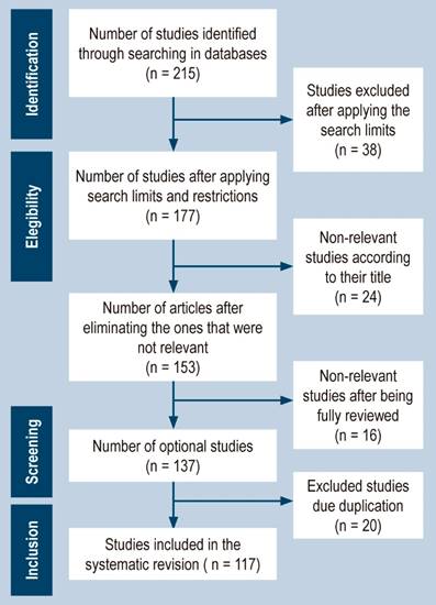
After implementing the search strategy as described, we initially identified 215 articles from the selected databases. However, we excluded 38 articles due to not meeting our search criteria. Following this, we screened the titles and abstracts of the remaining 177 articles and eliminated 24 articles. This resulted in 153 articles that were eligible for full-text review. After reviewing the full-text articles, we excluded 16 articles that did not meet the study’s inclusion criteria, resulting in 137 articles. We then removed 20 duplicate articles, resulting in 117 published articles consisting of case reports and case series. We adhered to the PRISMA statement guidelines for the selection process of the included studies, and a flowchart outlining the study selection process can be found in Figure 1.

Figure 1 PRISMA flowchart. Source: Authors’ own research. 

Sociodemographic Characteristics of Patients

The data interpretation regarding the most common age of onset for WD reveals that it can occur at any stage of life, with documented cases ranging from a 4-year-old girl to an 82-year-old man. However, the disease’s highest prevalence falls within the approximate age range of 50 to 65 years, with 67 reported cases. Furthermore, a greater incidence was observed in the 50-58 (2.9%) and 63 (6.6%) age groups. Males showed a higher incidence rate, with 96 cases and a percentage of 70.6% out of the total cases.1-117

Clinical Characteristics

The researchers conducted an analysis of the clinical case series and identified the most common manifestations of WD. Arthralgia was found in 83 patients (61%), weight loss in 64 (47.1%), diarrhea in 59 (43.4%), fever in 45 (33.1%), abdominal pain in 31 (22.8%), and neurological symptoms in 30 cases (22.1%), as shown in Table 1.1-117

Table 1 Clinical characteristics 

Prevalence n % Prevalence n %
Clinical manifestations Studies
Diarrhea 59 43.4 Hemoglobin (anemia) 40 29.4
Bloating 14 10.3 Leukocytosis 21 15.4
Rectal bleeding 3 2.2 Neutrophilia 11 8.1
Abdominal pain 31 22.8 Increased ESR 114 83.8
Dyspnea 19 14.4 PCR for T. Whipple positive 86 63.2
Headache 4 2.9 Human rheumatoid factor 1 0.7
Fever 45 33.1 Anatomopathological examination with PAS granules 65 47.8
Hyporexia 11 8.1 Fecal occult blood 3 2.2
Anorexia 7 5.1 Imaging findings 90 66.2
Weight loss 64 47.1 Colonoscopy 9 6.6
Asthenia 10 7.4 Endoscopic findings 25 18.4
Polyarthritis 25 18.4 Biopsy findings 69 50.7
Myalgias 7 5.1 Autopsy 1 0.7
Arthralgias 83 61.0
Adynamia 3 2.2
Fatigue 14 10.3
Weakness 10 7.4
Vomit 8 5.9
Neurological symptoms 30 22.1
Ophthalmoplegia 3 2.2
Cardiac symptoms 10 7.4
Tachycardia 3 2.2
Hyperpigmentation of the skin 9 6.6
Apathy 2 1.5
Irritability 2 1.5
Lymphadenopathy 24 17.6
Pericarditis 1 0.7
Endocarditis 2 1.5
Pleural effusion 3 2.2
Personality changes 4 2.9
Uveitis 5 3.7
Insomnia 2 1.5
Night sweats 12 8.8
Hypotension 4 2.9
Ocular symptoms 18 13.2
Depression and anxiety 5 3.7
Edema in lower limbs 18 13.2
Swelling of limbs 1 0.7
Paresthesia in lower limbs 3 2.2
Hypoesthesia and hyporeflexia 2 1.5

PAS: Periodic acid-Schiff. Source: Authors’ own research.

Based on the clinical cases analyzed by the research group, it was found that the three most commonly used diagnostic tools for generating a possible diagnosis were PCR for T. Whipple in 86 patients (63.2%), biopsy in 69 patients (50.7%), and pathological examination with positive staining for Periodic acid-Schiff (PAS) in 65 cases (47.8%). In addition, imaging findings from tests such as radiography, magnetic resonance imaging (MRI), and computed tomography (CT) were essential for the diagnosis, as they were helpful in 90 patients (66.2%). The erythrocyte sedimentation rate (ESR), high in 114 patients (83.8%), was also significant in aiding diagnosis.

Therapeutic characteristics

The results of the study showed that antibiotic therapy was the preferred approach for treating WD, with a high tendency towards the use of trimethoprim/sulfamethoxazole (61.0%), ceftriaxone (57.4%), and doxycycline (31.6%). Hydroxychloroquine (26.5%) and corticosteroids/prednisone (15.4%) were also used. Other antibiotic-based treatments, such as cefotaxime, meropenem, vancomycin, imipenem, tigecycline, and cefixime, were used less frequently. A detailed description of the results can be found in Table 2.1-117

Table 2 Therapeutic features1-117  

Prevalence n %
Therapeutic characteristics
Trimethoprim/sulfamethoxazole 83 61
Ceftriaxone 78 57.4
Metronidazole 1 0.7
Vancomycin 3 2.2
Penicillin 6 4.4
Meropenem 7 5.1
Tigecycline 2 1.5
Doxycycline 43 31.6
Cefotaxime 1 0.7
Cefixime 2 1.5
Imipenem 3 2.2
Ciprofloxacin 1 0.7
Hydroxychloroquine 36 26.5
Corticosteroids or prednisone 21 15.4

Source: Authors’ own research.

In addition, other treatment options were employed to manage specific associated symptoms. These included methotrexate (1.5%), non-specified antibiotics (1.5%), nonsteroidal anti-inflammatory drugs (NSAIDs) (1.5%), ofloxacin/prednisone drops (0.7%), palliative stereotactic surgery (0.7%), caspofungin (0.7%), and gentamicin plus noradrenaline plus dopamine (0.7%), as reported in Table 3.

Table 3 Other treatment options 

Prevalence n %
Therapeutic characteristics
NSAIDs 2 1.5
Amoxicillin 1 0.7
Gentamicin + Amoxicillin 1 0.7
Caspofungin 1 0.7
Ceftazidime 1 0.7
Cyclophosphamide 1 0.7
Co-trimoxazole 4 2.9
Streptomycin 1 0.7
Gentamicin + noradrenaline + dopamine 1 0.7
Leflunomide 1 0.7
Methotrexate 2 1.5
Ofloxacin drops + prednisone drops 1 0.7
Piperazine 1 0.7
Sulfadiazine 1 0.7
Non-specified antibiotic 2 1.5
Stereotactic palliative surgery 1 0.7

Source: Authors’ own research.

Discussion

WD is a systemic condition caused by Tropheryma whipplei, a gram-positive bacterium. The disease is named after the Greek words trophe (food) and eryma (barrier) due to its association with poor absorption of nutrients. It was first described in 1907 by George Hoyt Whipple as a chronic and systemic disease linked to intestinal lipodystrophy. It was not until 1952 that its etiology was postulated to be bacterial, based on clinical improvement after antibiotic treatment. Electron microscopy findings in 1960 provided further evidence for this hypothesis, and its ribonucleic acid (RNA) was amplified using PCR in 1990. Cultivation of the bacterium was achieved in 1997.42 The disease can occur at any age, as there are reports of cases from individuals aged 4 to 82.1-117 While the highest prevalence is found among individuals between 50 to 65 years old, the disease onset can occur at any age. Males have a higher predisposition to the disease, with 96 cases reported and a percentage of 70.6.1-117

The clinical presentation of WD is variable and not specific, with different organs affected and requiring diverse treatments. The disease may manifest as gastrointestinal histological lesions or isolated neurological cases. Typically, joint involvement is the first clinical sign, as observed in 83 patients (61%), consistent with a study by Moreno García (60% of cases). Weight loss is present in 64 patients (47.1%), diarrhea in 59 (43.4%), fever in 45 (33.1%), abdominal pain in 31 (22.8%), and neurological symptoms in 30 cases (22.1%). Joint involvement typically appears as intermittent migratory arthralgia, arthritis, or both, without deformity. It usually affects multiple joints, although it can be oligoarticular in large joints.9

Ocular involvement is not common in WD and typically presents at a later stage. It should be considered when bilateral choroidal granulomatosis is observed in the context of an atypical systemic disease. A limited number of cases of ocular involvement in WD have been reported in recent scientific literature, with eighteen cases identified in the review, representing a prevalence of 13.2%.29

According to Gundling F et al., central nervous system (CNS) manifestations occur in up to 15% of cases. These may occur rarely with little or no gastrointestinal damage, as isolated involvement, or associated with gastrointestinal disease. They may also occur as neurological relapse in previously treated disease,58 which is consistent with reports of cases that found CNS involvement in 30 patients (22.1%). Histological alterations in the brain have been found in autopsies. This review found alterations in one case (0.7%) where WD organisms significantly affected several organ systems, confirmed by histochemical and molecular evaluation.

The frequency of cardiac involvement in WD varies widely among different studies. Our research found involvement in ten cases (7.4%), while other authors, such as Parodi R., reported a range of 17% to 55% of cases. Pericarditis is a common manifestation, while myocarditis is less frequent but can be the initial presentation of cardiac failure or sudden death. Endocarditis can also occur but is difficult to diagnose due to negative blood cultures, lack of fever, and valve destruction. Its diagnosis is confirmed through PCR analysis of surgically obtained valvular material.9

In terms of diagnosing Wilson’s disease (WD), the three most commonly utilized diagnostic tools are the PCR for TW, biopsy, and anatomopathological examination with PAS granules. Fernández-Mondelo et al. propose two diagnostic criteria for WD: positive PAS staining in a small intestine biopsy and positive results in two tests (PAS, PCR, or immunohistochemistry) of TW from gastrointestinal or extraintestinal samples. Gastroscopy with biopsy remains the most frequently used technique for diagnosing WD. Macroscopically, a woolly, yellow, and pale mucosa alternating with an erythematous, erosive, or slightly friable mucosa in the post-bulbar region of the duodenum or jejunum is a characteristic finding. Biopsies taken at this level reveal positive foamy macrophages for PAS. Identifying the bacillus through PCR in the same duodenal tissue can confirm the diagnosis. In patients with suspected WD but without gastrointestinal symptoms, samples should be collected from relevant anatomical sites such as synovial fluid and lymph nodes, among others.91

Differential Diagnoses

When joint symptoms are predominant, other rheumatic diseases should be ruled out before considering WD. In cases where joint disease does not improve or worsens with biological therapy, WD should be suspected. HIV infection can also present with similar symptoms, including enteropathy and wasting syndrome. If endocarditis is present, the most common pathogens should be ruled out first. The presence of systemic granulomas can be mistaken for sarcoidosis. In cases of abdominal involvement, other causes of malabsorptive syndrome should be investigated, such as celiac disease and other infections. A diagnosis of abdominal angina9 should also be considered for patients with vascular disease.

The recommended treatment for WD includes antibiotics that can penetrate the blood-brain barrier, such as trimethoprim/sulfamethoxazole or ceftriaxone, to prevent neurological relapses.8 In our research, we observed a preference for trimethoprim/sulfamethoxazole (83 cases; 61.0%) followed by ceftriaxone (78 cases; 57.4%). Currently, the recommended treatment involves ceftriaxone (2 g/day) or meropenem (3 g/day) administered parenterally, followed by trimethoprim/sulfamethoxazole (800-160 mg/ times daily) administered orally for at least one to two years. In cases of isolated neurological forms, ceftriaxone (2-4 g/day) is administered intravenously for 15-30 days, followed by trimethoprim/sulfamethoxazole (1600-320 mg/day) or doxycycline (200 mg/day) or cefixime (400 mg/day) until cerebrospinal fluid (CSF) PCR results are negative.8

The treatment goals for Whipple’s disease are to minimize morbidity, prevent complications, and eliminate the infection. Without appropriate treatment, the mortality rate is almost 100%. However, the prognosis is favorable with complete antibiotic therapy.

Referencias

1. Valdés Álvarez K, Nievas Sánchez M. Enfermedad de Whipple en paciente con fiebre de origen desconocido. Rev Cubana Med. 2018;57(1):33-7. [ Links ]

2. Dolmans RA, Boel CH, Lacle MM, Kusters JG. Clinical Manifestations, Treatment, and Diagnosis of Tropheryma whipplei Infections. Clin Microbiol Rev. 2017;30(2):529-555. https://doi.org/10.1128/CMR.00033-16Links ]

3. Cardoso J, Gomes L, Santos S, Moreira H, Gomes P, Rua J, et al. Whipple’s disease: A rare cause of malabsorption syndrome. GE Port J Gastroenterol. 2020;27(4):283-9. https://doi.org/10.1159/000504760Links ]

4. Pankl S, Baez M, Young P, Bruetman J, Rausch A, Zubiarurre I, et al. Enfermedad de Whipple e hipertensión pulmonar reversible. Medicina. 2021;81(1):91-95. [ Links ]

5. Kadian R, Wang J, Freitas E. Could CT abdomen and PET/CT be helpful in early diagnosis of Whipple’s disease? A case report. IDCases. 2021;26(e01286):e01286. https://doi.org/10.1016/j.idcr.2021.e01286Links ]

6. Brönnimann D, Vareil M-O, Sibon I, Lagier J-C, Lepidi H, Puges M, et al. Limbic encephalitis as a relapse of Whipple’s disease with digestive involvement and spondylodiscitis. Infection. 2019;47(4):637-41. https://doi.org/10.1007/s15010-018-1173-xLinks ]

7. Lopes A, Santos AF, Alvarenga MJ, Mello E, Silva A. Whipple’s disease: a rare case of malabsorption. BMJ Case Rep. 2018; 2018:bcr2017222955. http://dx.doi.org/10.1136/bcr-2017-222955Links ]

8. Crespo E, Lemus A, Gonzáles S. Enfermedad de Whipple, una causa poco frecuente de diarrea, Rev Ciencias Médicas. 2017. 21(3):133-137. [ Links ]

9. Parodi R, Ibarzábal J, Román R, Alasino M, Varela M, Díaz S. Enfermedad de Whipple. Comunicación de un caso y revisión de la literatura. Acta Gastroenterológica Latinoamericana. 2019;49(3):229-240. [ Links ]

10. Kukull B, Mahlow J, Hale G, Perry LJ. Whipple’s disease: a fatal mimic. Autops Case Rep. 2021;11:e2020237. https://doi.org/10.4322/acr.2020.237Links ]

11. Melas N, Amin R, Gyllemark P, Younes AH, Almer S. Whipple’s disease: the great masquerader-a high level of suspicion is the key to diagnosis. BMC Gastroenterol. 2021;21(1):128. https://doi.org/10.1186/s12876-021-01664-1Links ]

12. da Silva GAR, Neto JSP. Whipple’s disease manifested as difficult-to-diagnose polyarthralgia: a case report and literature review. Rev Bras Reumatol Engl Ed. 2017;57(5):483-6. https://doi.org/10.1016/j.rbre.2015.05.003Links ]

13. Turcan S, Tofan-Scutaru L, Istrate V, Tirbu V. Whipple’s disease? A case report and discussion. Med Pharm Rep. 2021;94(Suppl 1):S76-8. https://doi.org/10.15386/mpr-2237Links ]

14. Alsarhani WK, Alkhalifah MI, Alkatan HM, Alsolami AL, Maktabi AMY, Alsuhaibani AH. Whipple’s disease scleral nodules: a novel presentation in 2 consecutive patients. BMC Ophthalmol. 2020;20(1):413. https://doi.org/10.1186/s12886-020-01695-4Links ]

15. Brevet P, Rottenberg P, Viacroze C, Schleifer D, Lequerre T, Vittecoq O. A case of Whipple’s disease mimicking auto-inflammatory disease and revealed by severe right cardiac failure under anakinra. Joint Bone Spine. 2020;87(4):365-6. https://doi.org/10.1016/j.jbspin.2019.11.004Links ]

16. Gruber JR, Sarro R, Delaloye J, Surmely J-F, Siniscalchi G, Tozzi P, et al. Tropheryma whipplei bivalvular endocarditis and polyarthralgia: a case report. J Med Case Rep. 2015;9(1):259. https://doi.org/10.1186/s13256-015-0746-xLinks ]

17. Ramos JM, Pasquau F, Galipienso N, Valero B, Navarro A, Martinez A, et al. Whipple’s disease diagnosed during anti-tumor necrosis factor alpha treatment: two case reports and review of the literature. J Med Case Rep. 2015;9(1):165. https://doi.org/10.1186/s13256-015-0632-6Links ]

18. Scheurwater MA, Verduin CM, van Dantzig J-M. Whipple’s endocarditis: a case report of a blood culture-negative endocarditis. Eur Heart J Case Rep. 2019;3(4):1-6. https://doi.org/10.1093/ehjcr/ytz222Links ]

19. Alozie A, Zimpfer A, Köller K, Westphal B, Obliers A, Erbersdobler A, et al. Arthralgia and blood culture-negative endocarditis in middle Age Men suggest tropheryma whipplei infection: report of two cases and review of the literature. BMC Infect Dis. 2015;15(1):339. https://doi.org/10.1186/s12879-015-1078-6Links ]

20. Li W, Zhang Q, Xu Y, Zhang X, Huang Q, Su Z. Severe pneumonia in adults caused by Tropheryma whipplei and Candida sp. infection: a 2019 case series. BMC Pulm Med. 2021;21(1):29. https://doi.org/10.1186/s12890-020-01384-4Links ]

21. McGee M, Brienesse S, Chong B, Levendel A, Lai K. Tropheryma whipplei Endocarditis: Case Presentation and Review of the Literature. Open Forum Infect Dis. 2019;6(1):ofy330. https://doi.org/10.1093/ofid/ofy330Links ]

22. Jos S-L, Angelakis E, Caus T, Raoult D. Positron emission tomography in the diagnosis of Whipple’s endocarditis: a case report. BMC Res Notes. 2015;8(1):56. https://doi.org/10.1186/s13104-015-1022-2Links ]

23. Testi I, Tognon MS, Gupta V. Ocular Whipple disease: Report of three cases. Ocul Immunol Inflamm. 2019;27(7):1117-20. https://doi.org/10.1080/09273948.2018.1518461Links ]

24. Dick J, Krauß P, Hillenkamp J, Kohlmorgen B, Schoen C. Postoperative Tropheryma whipplei endophthalmitis - a case report highlighting the additive value of molecular testing. JMM Case Rep. 2017;4(10):e005124. https://doi.org/10.1099/jmmcr.0.005124Links ]

25. Liersch J, Carlotti A, Theunis A, Leonard A, Barrett M, Carlson JA, et al. Erythema nodosum leprosum-like lesions are a histopathologic pattern in Whipple’s disease and a sign of the immune reconstitution inflammatory syndrome: A case series and review of the literature. Am J Dermatopathol. 2017;39(4):259-66. https://doi.org/10.1097/DAD.0000000000000641Links ]

26. Vural A, Acar NP, Soylemezoglu F, Oguz KK, Dericioğlu N, Saka E. Isolated central nervous system Whipple’s disease: Two cases. Clin Neurol Neurosurg. 2015;139:91-4. https://doi.org/10.1016/j.clineuro.2015.08.028Links ]

27. Peregrin J, Malikova H. Primary Whipple disease of the brain: case report with long-term clinical and MRI follow-up. Neuropsychiatr Dis Treat. 2015;11:2461-9. https://doi.org/10.2147/NDT.S92066Links ]

28. Karlowee V, Kolakshyapati M, Amatya VJ, Takayasu T, Nosaka R, Sugiyama K, et al. Diffuse leptomeningeal glioneuronal tumor (DLGNT) mimicking Whipple’s disease: a case report and literature review. Childs Nerv Syst. 2017;33(8):1411-4. https://doi.org/10.1007/s00381-017-3405-2Links ]

29. de Saint-Martin G, Urbanski G, Beucher A-B, Ebran J-M. An ophthalmologic complication of Whipple’s disease: Case report. J Fr Ophtalmol. 2018;41(8):e387-9. https://doi.org/10.1016/j.jfo.2017.11.036Links ]

30. Citerne Q, Honstettre S, Ouichka R, Loeuille D, Gillet P, Chary-Valckenaere I. Atypical response of spondyloarthritis to biologics revealing Whipple’s disease: A case-report. Therapie. 2018;73(5):437-9. https://doi.org/doi:10.1016/j.therap.2018.02.008Links ]

31. Sampaio F, Moreira J, Jordão S, Vieira B, Pereira S, Carvalho R. Whipple’s disease orbitopathy: case report and review of literature. Orbit. 2022;41(1):112-117. https://doi.org/10.1080/01676830.2020.1820044Links ]

32. Saraci G. Enfoque diagnóstico de una condición rara: la enfermedad de Whipple. Rev Gastroenterol Méx (Engl Ed). 2020;85(4):477-8. https://doi.org/10.1016/j.rgmx.2020.07.006Links ]

33. Santos Seoane SM, Martínez Gutiérrez R, Venta Menéndez VI. Whipple’s disease: when diarrhea is absent. Rev Esp Enferm Dig. 2019;111(6):492-3. https://doi.org/10.17235/reed.2019.6015/2018Links ]

34. Chou J-W, Hsu B-C, Chang C-H. A rare cause of chronic diarrhea and fever. Gastroenterology. 2020;158(8):e5-6. https://doi.org/10.1053/j.gastro.2020.01.018Links ]

35. Saito H, Shiode J, Ohya S, Yao A, Saito S, Fujii M, et al. Whipple’s disease with long-term endoscopic follow-up. Intern Med. 2018;57(12):1707-13. https://doi.org/10.2169/internalmedicine.9631-17Links ]

36. Damaraju D, Steiner T, Wade J, Gin K, FitzGerald JM. Clinical problem-solving. A surprising cause of chronic cough. N Engl J Med. 2015;373(6):561-6. https://doi.org/10.1056/NEJMcps1303787Links ]

37. Ruggiero E, Zurlo A, Giantin V, Galeazzi F, Mescoli C, Nante G, et al. Short article: Relapsing Whipple’s disease: a case report and literature review. Eur J Gastroenterol Hepatol. 2016;28(3):267-70. https://doi.org/10.1097/MEG.0000000000000539Links ]

38. Guiomar V, Pinto MJ, Gomes C, Correia C, Tavares S, Chaves V, et al. Whipple’s disease as the first manifestation of chronic Lymphocytic leukaemia. Eur J Case Rep Intern Med. 2019;6(10):001270. https://doi.org/10.12890/2019_001270Links ]

39. Fontana M, Cerri S, Bernardelli G, Brugioni L, Clini E, Tonelli R. Unusual effectiveness of systemic steroids in Whipple disease. Pulmonology. 2020;26(6):415-7. https://doi.org/10.1016/j.pulmoe.2020.02.007Links ]

40. Sullivan A, Shrestha P, Basnet S, Herb R, Zagorski E. A rare case of Whipple’s disease with endocarditis in a patient with dextrocardia. SAGE Open Med Case Rep. 2020;8:2050313X20936952. https://doi.org/10.1177/2050313X20936952Links ]

41. Balducci C, Foresti S, Ciervo A, Mancini F, Nastasi G, Marzorati L, et al. Primary Whipple disease of the Central Nervous System presenting with rhombencephalitis. Int J Infect Dis. 2019;88:149-51. https://doi.org/10.1016/j.ijid.2019.08.019Links ]

42. Sid’Amar S, Puppa G. Whipple’s disease affecting ileal Peyer’s patches: The first case report. Case Rep Pathol. 2019;2019:1509745. https://doi.org/10.1155/2019/1509745Links ]

43. Juanmartiñena Fernández JF, Oyón Lara D, Rázquin Lizarraga S, Fernández Urien I. Whipple’s disease under the vision of capsule endoscopy. Rev Esp Enferm Dig. 2016;108(9):606. https://doi.org/10.17235/reed.2016.4362/2016Links ]

44. Bally JF, Méneret A, Roze E, Anderson M, Grabli D, Lang AE. Systematic review of movement disorders and oculomotor abnormalities in Whipple’s disease: Movement Disorders in Whipple’s Disease. Mov Disord. 2018;33(11):1700-11. https://doi.org/10.1002/mds.27419Links ]

45. Priest DH, Grote TH, Staley SL, Berger WS, Norman ES, Smith BS. Secondary immune thrombocytopenia (ITP) as an initial presentation of Whipple’s disease. IDCases. 2018;12:e4-6. https://doi.org/10.1016/j.idcr.2017.05.010Links ]

46. Gaudé M, Tébib J, Puéchal X. Atypical focal forms of Whipple’s disease seen by rheumatologists. Joint Bone Spine. 2015;82(1):56-9. https://doi.org/10.1016/j.jbspin.2014.08.005Links ]

47. Ankli B, Khanlari B, Pegios V, Zettl A, Daikeler T. Whipple’s disease mimicking an auto-inflammatory disease with myositis and soft tissue inflammation. Joint Bone Spine 2018;85(5):645-646. https://doi.org/10.1016/j.jbspin.2018.01.003Links ]

48. Seddon O, Hettiarachchi I. Whipple’s endocarditis presenting as ulnar artery aneurysm; if you don’t look, you won’t find. BMJ Case Rep. 2017;bcr2017221327. https://doi.org/10.1136/bcr-2017-221327Links ]

49. Olano C, Dorelo R, Oricchio M, Mendez D, Canavesi A, Pitetta C. Capsule endoscopy aiding diagnosis of a rare condition - Whipple’s disease. Endoscopy. 2019;51(9):E272-3. https://doi.org/10.1055/a-0896-2269Links ]

50. Totschnig D, Seitz T, Zoufaly A, Hagenauer-Drektraan S, Wenisch C. Whipple’s disease diagnosed in a patient with suspected sarcoidosis. Int J Infect Dis. 2021;106:41-2. https://doi.org/10.1016/j.ijid.2021.03.053Links ]

51. Hamza Bin Waqar S, Diks J, Zaman U, Sharif S, Sheikh T, Kolla S, et al. Refractory effusions, crumbly bones, mystifying cachexia and an absent mind: An unusual presentation of Whipple’s disease with review of literature. Am J Med Case Rep. 2021;9(7):348-53. https://doi.org/10.12691/ajmcr-9-7-2Links ]

52. Loiodice A, Losurdo G, Iannone A, Rossi R, Fiore MG, Piscitelli D. Transmission electron microscopy helpfulness in Whipple’s disease masked by immunosuppressant therapy for arthritis. APMIS. 2018;126(1):92-6. https://doi.org/10.1111/apm.12782Links ]

53. Kono M, Yamamoto K, Nagamatsu M, Kutsuna S. Use of polymerase chain reaction in the diagnosis of Whipple’s disease. J Infect Chemother. 2015;21(12):885-8. https://doi.org/10.1016/j.jiac.2015.08.010Links ]

54. Boban M, Gjadrov-Kuveždić K, Jakić-Razumović J. Cytology of cerebrospinal fluid in CNS Whipple disease. Acta Neurol Belg. 2017;117(4):935-936. https://doi.org/10.1007/s13760-017-0824-5Links ]

55. Wartique L, Lagier JC, Raoult D, Jamilloux Y, Sève P. Mesenteric lymphadenitis as a presenting feature of Whipple’s disease: Value of PCR analysis. Int J Infect Dis. 2018;75:15-17. https://doi.org/10.1016/j.ijid.2018.08.003Links ]

56. Giunchi D, Marcoli N, Deabate L, Delcogliano M, Testa E, Candrian C, et al. Isolated Knee Arthritis as Early and Only Symptom of Whipple’s Disease. Case Rep Med. 2018;2018:3417934. https://doi.org/10.1155/2018/3417934Links ]

57. Kundu A, Sen P, Khurana S. Isolated CNS Whipple’s disease: a diagnostic dilemma. BMJ Case Rep. 2015;2015:bcr2015211784. https://doi.org/10.1136/bcr-2015-211784Links ]

58. Gundling F, Wittenburg H, Tannapfel A, Mossner J. Neurological presentation of Whipple’s disease after long-term antibiotic treatment: a case report. J Med Case Rep. 2008;2:191. https://doi.org/10.1186/1752-1947-2-191Links ]

59. Zhang WM, Xu L. Pulmonary parenchymal involvement caused by Tropheryma whipplei. Open Med (Warsz). 2021;16(1):843-6. https://doi.org/10.1515/med-2021-0297Links ]

60. Zhu B, Tang J, Fang R, Fei X, Wang Q, Wang W, et al. Pulmonary coinfection of Mycobacterium tuberculosis and Tropheryma whipplei: a case report. J Med Case Rep. 2021;15(1):359. https://doi.org/10.1186/s13256-021-02899-yLinks ]

61. Obma KL, Marx GE, Mauchley D, Seres T, Babu A, Saveli CC, et al. CASE 12--2015: Tropheryma Whipplei Endocarditis. J Cardiothorac Vasc Anesth. 2015;29(6):1712-6. https://doi.org/10.1053/j.jvca.2014.11.011Links ]

62. Vindigni SM, Taylor J, Quilter LAS, Hyun TS, Liu C, Rosinski SL, et al. Tropheryma whipplei infection (Whipple’s disease) in a patient after liver transplantation. Transpl Infect Dis. 2016;18(4):617-24. https://doi.org/10.1111/tid.12562Links ]

63. Muretti M, Keiralla A, Jeffery K, Krasopoulos G. Tropheryma whipplei endocarditis: An uncommon infection with potentially fatal consequences. J Card Surg. 2020;35(4):923-5. https://doi.org/10.1111/jocs.14467Links ]

64. Heavener T, Thompson M, Patel C, Forrester L, Rawls D. An unusual presentation of Tropheryma whipplei infection. Proc (Bayl Univ Med Cent). 2017;30(4):429-30. https://doi.org/10.1080/08998280.2017.11930215Links ]

65. Lagier JC, Cammilleri S, Raoult D. Classic Whipple’s disease diagnosed by (18)F-fluorodeoxyglucose PET. Lancet Infect Dis. 2016;16(1):130. https://doi.org/10.1016/S1473-3099(15)00503-4Links ]

66. He YT, Peterson K, Crothers J, Dejace J, Hale AJ. Endocarditis and systemic embolization from Whipple’s disease. IDCases. 2021;24(e01105):e01105. https://doi.org/10.1016/j.idcr.2021.e01105Links ]

67. Tatsuki M, Ishige T, Igarashi Y, Hatori R, Hokama A, Hirato J, et al. Whipple disease mimicking inflammatory bowel disease. Intest Res. 2021;19(1):119-25. https://doi.org/10.5217/ir.2019.09177Links ]

68. Sanchez A, Del Giudice P, Mantion C, Mazellier S, Boukari F, Roger P-M, et al. Erythematous skin nodules during treatment of Whipple’s disease. Med Mal Infect. 2021;51(4):397-9. https://doi.org/10.1016/j.medmal.2020.10.006Links ]

69. Yan J, Zhang B, Zhang Z, Shi J, Liu S, Qi J, et al. Case report: Tropheryma whipplei hide in an AIDS patient with Pneumocystis pneumonia. Front Public Health. 2021;9:663093. https://doi.org/10.3389/fpubh.2021.663093Links ]

70. Branquinho DF, Pinto-Gouveia M, Mendes S, Sofia C. From past sailors’ eras to the present day: scurvy as a surprising manifestation of an uncommon gastrointestinal disease. BMJ Case Rep. 2015;2015:bcr2015210744. https://doi.org/10.1136/bcr-2015-210744Links ]

71. Rezk A, Gunnerson AC, Komar M. A disease that is often missed without gastrointestinal symptoms. Gastroenterology. 2016;150(5):1096-7. https://doi.org/10.1053/j.gastro.2015.11.054Links ]

72. Chizinga M, Schiliro D, Mullin B, Barrie RL. Mesenteric lymphadenitis as a presenting feature of Whipple’s disease. IDCases. 2017;9:50-2. https://doi.org/10.1016/j.idcr.2017.06.002Links ]

73. De Francesco V, Corsi F, Pennella A, Bellesia A, Fiorini G, Vaira D, et al. Whipple’s disease: case report and review of the literature. J Gastrointestin Liver Dis. 2018;27(3):331-6. https://doi.org/10.15403/jgld.2014.1121.273.fraLinks ]

74. Henriques MS de M, da Paz AR, Gaertner ABP, Melo CI, Filgueiras PL, Jerome RA. Deep vein thrombosis as initial manifestation of Whipple disease. Case Rep Gastroenterol. 2016;10(3):640-5. https://doi.org/10.1159/000452206Links ]

75. Parkash V, Mudhar HS, Wagner BE, Raoult D, Batty R, Lepidi H, et al. Bilateral ocular myositis associated with Whipple’s disease. Ocul Oncol Pathol. 2017;3(1):17-21. https://doi.org/10.1159/000448622Links ]

76. Kilani M, Njim L, Nsir AB, Hattab MN. Whipple disease presenting as cystic brain tumor: Case report and review of the literature. Turk Neurosurg. 2018;28(3):495-9. https://doi.org/10.5137/1019-5149.JTN.17111-16.2Links ]

77. Puéchal X, London J. Clinical image: Whipple’s destructive septic arthritis. Arthritis rheumatol. 2017;69(3):559. https://doi.org/10.1002/art.39999Links ]

78. Elfanagely Y, Jamot S, Dapaah-Afriyie K, Fine S. Whipple’s disease mimicking common digestive disorders. R I Med J (2013). 2021;104(4):43-5. [ Links ]

79. Papakonstantinou D, Riste MJ, Langman G, Moran E. Misdiagnosing Whipple’s disease in the young. BMJ Case Rep. 2017;2017:bcr2016218866. https://doi.org/10.1136/bcr-2016-218866Links ]

80. Dubost J-J, Couderc M, Mathieu S, Tournadre A, Soubrier M. Chronic bursitis and tenosynovitis revealing Whipple’s disease. Joint Bone Spine. 2020;87(5):481-2. https://doi.org/10.1016/j.jbspin.2020.01.010Links ]

81. Sarvananthan S, Velissaris T, Miskolczi S, Yam T, Shah BN. Tropheryma whipplei endocarditis. Echocardiography. 2021;38(4):697-700. https://doi.org/10.1111/echo.15007Links ]

82. Vayssade M, Tournadre A, D’Incan M, Soubrier M, Dubost J-J. Immune reconstitution inflammatory syndrome during treatment of Whipple’s disease. Joint Bone Spine. 2015;82(2):122-4. https://doi.org/10.1016/j.jbspin.2014.09.002Links ]

83. Quartuccio L, Giovannini I, Pizzolitto S, Scarpa M, De Vita S. Seronegative arthritis and Whipple disease: Risk of misdiagnosis in the era of biologic agents. Case Rep Rheumatol. 2019;2019:3410468. https://doi.org/10.1155/2019/3410468Links ]

84. Emonet S, Wuillemin T, Harbarth S, Wassilew N, Cikirikcioglu M, Schrenzel J, et al. Relapse of Tropheryma whipplei endocarditis treated by trimethoprim/sulfamethoxazole, cured by hydroxychloroquine plus doxycycline. Int J Infect Dis. 2015;30:17-9. https://doi.org/10.1016/j.ijid.2014.11.003Links ]

85. Moreno García MS, Casorrán Berges M, Del Río-Martínez PS, Bosque Peralta MT. The great unknown, Whipple’s disease. Reumatol Clin. 2017;13(4):243-4. https://doi.org/10.1016/j.reuma.2016.08.002Links ]

86. Van der Bent S, van Vugt M, Amir A, van der Wal A, Mekkes J. Cutaneous manifestations in treated Whipple’s disease. Int J Dermatol. 2017;56(4):e82-e84. https://doi.org/10.1111/ijd.13479Links ]

87. Chiu M, Moore S. Bilateral optic disc swelling in Whipple’s disease: Letter to the Editor. Clin Experiment Ophthalmol. 2017;45(6):641-3. https://doi.org/10.1111/ceo.12933Links ]

88. Van Bockstal M, Hoorens A, Van den Bosch F, Creytens D, Verbeke S, Van Dorpe J. Whipple’s disease in granulomatous disguise: a challenging diagnosis with many histopathological pitfalls. Virchows Arch. 2017;470(4):465-8. https://doi.org/10.1007/s00428-017-2084-4Links ]

89. Aamar A, Madhani K, Anwar MS, Singh P, Garsten J. Whipple’s disease manifested as recurrent ascites. Cureus. 2017;9(3):e1108. https://doi.org/10.7759/cureus.1108Links ]

90. Chandra SR, Raj P, Pai AR, Reddy N. A case of Whipple’s disease: A very rare cause for rapidly progressive dementia. Indian J Psychol Med. 2018;40(3):280-3. https://doi.org/10.4103/IJPSYM.IJPSYM_149_17Links ]

91. Fernández-Mondelo J, Cervero-Jiménez M. Forma atípica de presentación de la enfermedad de Whipple. Reporte de un caso. Rev Esp Casos Clin Med Intern (RECCMI). 2018;3(2):56-58. [ Links ]

92. Brönnimann D, Vandenhende M-A, Viallard J-F. Gamma delta T cell expansion in Whipple’s disease with muscular granulomatous vasculitis. Infection. 2018;46(4):573-6. https://doi.org/10.1007/s15010-018-1143-3Links ]

93. Herráez A, Valmaseda M, Chao M, Reinoso M. Manifestaciones neuropsiquiátricas de la enfermedad de Whipple en una adolescente: a propósito de un caso. Dig Liver Dis. 2018;27(2):92-5. [ Links ]

94. Methotrexate. Masking and worsening of Whipple’s disease: case report. React Wkly. 2018;1692(1):224. https://doi.org/10.1007/s40278-018-42751-4Links ]

95. Kutlu O, Erhan SS, Gökden Y, Kandemir O, Tükekc T. Whipple’s Disease: A Case Report. Med Princ Pract. 2020; 29(1):90-93. Disponible en: https://www.ncbi.nlm.nih.gov/pmc/articles/PMC7024871/Links ]

96. Loiodice A. The conundrum of arthritis and chronic diarrhea could be unveiled as Whipple’s disease. Dig Liver Dis. 2017;2:3-10. https://doi.org/10.1016/S1590-8658(17)30459-0Links ]

97. Bosa L. Tropheryma whipplei infection in a pediatric kidney transplanted patient: A case report. Dig Liver Dis. 2017;49(4):E257. https://doi.org/10.1016/j.dld.2017.09.041Links ]

98. Herbette M, Cren JB, Joffres L, Lucas C, Ricard E, Salliot C, et al. Usefulness of polymerase chain reaction for diagnosing Whipple’s disease in rheumatology. PLoS ONE. 2018;13(7):e0200645. https://doi.org/10.1371/journal.pone.0200645Links ]

99. Maciorkowska M, Ustymowicz, Zakrzewski M, Pryczynicz A, Markowski A, Romatowski J, et al. Whipple’s disease as a systemic infectious disease - a case presentation. Prog Health Sci. 2018;8(1):212-218. https://doi.org/10.5604/01.3001.0012.1332Links ]

100. Delarbre D, Gan L, Antoine C, Poisnel E, Cambon A, Dutasta F, et al. Difficultés diagnostiques de la maladie de Whipple au cours des rhumatismes inflammatoires chroniques: trois observations. La Revue de Médecine Interne. 2021;42(11):801-804. [ Links ]

101. Lenfant M, Callemeyn J, Alerts H, Meersseman W, Van Moerkercke W. Whipple’s disease in a man of north african descent: Case report and brief review of the literature. Acta Gastroenterol Belg. 2019;82(1):83-86. [ Links ]

102. Thornton S, Wang Y, Köebel M, Bernard K, Burdz T, Maitland A, et al. Another Whipple’s triad? Pericardial, myocardial and valvular disease in an unusual case presentation from a Canadian perspective. BMC Cardiovascular Disorders. 2019;19(1):312. https://doi.org/10.1186/s12872-019-1257-2Links ]

103. Spoerl D, Bär D, Cooper J, Vogt T, Tyndall A, Walker UA. Multisegmental spondylitis due to Tropheryma whipplei: case report. Orphanet J Rare Dis. 2019;4:13. https://doi.org/10.1186/1750-1172-4-13Links ]

104. Le Blay P, Rakotonirain H, Lagier JC, Raoult D, Puechal X, Pers YM. A severe Whipple disease with an immune reconstitution inflammatory syndrome: an additional case of thalidomide efficiency. Joint Bone Spine. 2014;81(3):260-2. https://doi.org/10.1016/j.jbspin.2013.10.007Links ]

105. Fenollar F, Nicoli F, Paquet C, Lepidi H, Cozzone P, Antoine J-C, et al. Progressive dementia associated with ataxia or obesity in patients with Tropheryma whipplei encephalitis. BMC Infect Dis. 2018;11:171. [ Links ]

106. Erak P, Murillo D. Enfermedad de Whipple: presentación clínica sin afección articular. Rev Clin Esc Med. 2019;9(4):64-68. [ Links ]

107. Hujoel IA, Johnson DH, Lebwohl B, Leffler D, Kupfer S, Wu TT, et al. Tropheryma whipplei Infection (Whipple Disease) in the USA. Dig Dis Sci. 2019;64(1):213-223. https://doi.org/10.1007/s10620-018-5033-4Links ]

108. Lenfant M, Callemeyn J, Alaerts H, Meersseman W, Van Moerkercke W. Whipple’s disease in a man of North African descent: case report and brief review of the literature. Acta Gastroenterol Belg. 2019;82(1):83-86. [ Links ]

109. Kutlua O, Şengiz Erhanb S, Gökdena Y, Kandemira O, Tükekc T. Whipple’s Disease: A Case Report. Med Princ Pract 2020;29(1):90-93. https://doi.org/10.1159/000498909Links ]

110. Aguiar A, Gomes P, Curvo-Semedo L, Donato P. Whipple’s disease: imaging contribution for a challenging case. BMJ Case Rep. 2020;13(2):e233071. https://doi.org/10.1136/bcr-2019-233071Links ]

111. Al-Hamoudi W, Habbab F, Nudo C, Nahal A, Flegel K. Eosinophilic vasculitis: a rare presentation of Whipple’s disease. Can J Gastroenterol. 2017;21(3):189-91. [ Links ]

112. Verbrugghe L, Verraes K, Vanderschueren S, Vermeire S, Pollet S, De Leyn P, et al. Mesenteric panniculitis as a presentation of Whipple›s disease: case report and review of the literature. Acta Gastroenterol Belg. 2020;83(4):666-668. [ Links ]

113. Olivier M, Licitra C, Field Z, Ge L, Hill D, Madruga M, et al. Thrombocytopenia and endocarditis in a patient with Whipple’s disease: case report. BMC Infect Dis. 2020;20(1):71. https://doi.org/10.1186/s12879-020-4799-0Links ]

114. Rey R, Orozco L, Marrugo K, Lopez R, Perez E, De la Hoz J, et al. Whipple disease diagnosed by enteroscopy: first case report in Colombia of an underdiagnosed disease and literature review. BMC Gastroenterol. 2020;20(1):197. https://doi.org/10.1186/s12876-020-01302-2Links ]

115. Foteinogiannopoulou K, Mouzas I, Tzardi M, Orfanoudaki E, Theodoraki E, Kofteridis D. First case of Whipple’s disease successfully treated with tigecycline. Germs. 2021;11(1):105-110. https://doi.org/10.18683/germs.2021.1246Links ]

116. Hofmann P, Durisch N, Buetikofer C, Helmchen B. Granulomatous lung disease and immune reconstitution inflammatory syndrome in Whipple’s disease. BMJ Case Rep. 2021;14(6):e243633. https://doi.org/10.1136/bcr-2021-243633Links ]

117. Tandon P, Huang V, Jaffer N, Kirsch R, Croitoru K. A rare presentation of hypovolemic shock secondary to Whipple’s disease. Eur J Gastroenterol Hepatol. 2019;31(5):642-5. https://doi.org/10.1097/MEG.0000000000001363Links ]

Citation:

1Vargas-Rodríguez LJ, Ruiz-Muñoz JL, Bolívar-Córdoba PA, Romero-Cely MD, Cañón-Abril EJ, Suárez-Correa ZM, Mendoza-Cáceres MA. Whipple’s Disease: A Systematic Review of the Literature. Revista. colomb. Gastroenterol. 2023;38(1):35-45. https://doi.org/10.22516/25007440.966

Received: September 15, 2022; Accepted: September 23, 2022

*Correspondence: Ledmar Jovanny Vargas-Rodríguez. lejovaro@gmail.com

The authors declare that they have no conflicts of interest in conducting this study. Additionally, all authors were involved in the design, data collection, analysis, interpretation, and drafting of the manuscript.

Creative Commons License This is an open-access article distributed under the terms of the Creative Commons Attribution License