


 texto en
  Inglés (pdf)
 Articulo en XML
 Referencias del artículo
 Enviar articulo por email
 Citado por SciELO
  Citado por Google
  Similares en
SciELO
Similares en
SciELO  Similares en Google
 Permalink
Introduction
The ingestion of foreign bodies is a common medical problem, especially in the emergency department, which has greater relevance in three populations: pediatric patients, patients with some cognitive disability, and patients with psychiatric pathologies and a history of psychoactive substance use given the higher risk of complications and difficulties in extracting them1. Although these elements generally manage to pass spontaneously through the GI tract, in 20% of cases, endoscopic intervention will be required for their removal, and at least 1% of cases will require some surgical intervention2,3.
International guidelines such as those proposed by the European Society of Gastrointestinal Endoscopy (ESGE) recommend emergent upper gastrointestinal endoscopy (EGD) (in the first 2 to 6 hours after ingestion) in three main scenarios: cases of complete esophageal obstruction, sharp-pointed foreign bodies given the high risk of perforation, and the ingestion of cells or batteries due to the risk of liquefactive necrosis and perforation, especially in sites of stricture (upper esophageal sphincter, aortic arch, lower esophageal sphincter, pylorus, ileocecal valve, and anus). If these parameters are not met, it has been said that the endoscopic study can be performed urgently in the first 24 hours4.
In Colombia, ingesting foreign bodies is a frequent reason for consultation; however, there is no data to establish the incidence of this problem in the country, nor to establish the type of foreign body ingested and its most common complications. This work aims to conduct a retrospective analysis of the experience of Clínica Universitaria Colombia gastroenterology and digestive endoscopy group in ingesting foreign bodies.
Materials and methods
This descriptive, retrospective study included patients with suspected ingestion of foreign bodies admitted to the gastroenterology and digestive endoscopy department of the Clínica Universitaria Colombia between January 2007 and August 2020. The cases were identified by reviewing the medical record, the reason for consultation, the reported symptoms, the reason for requesting EGD, and the report of endoscopic procedures. After excluding those patients with incomplete data and those under 18 years of age, 2,307 patients were included in the analysis.
The following demographic, clinical, and endoscopic data were collected: age, sex, time of onset of symptoms before consultation, primary symptoms, findings on neck X-ray, type of foreign body, location of the foreign body, extraction method, and associated complications.
Results
Population characteristics
The age of occurrence was 18 to 95 years; the average age was 45 years, and the age range in which the event occurred the most was between 27 to 59 years. The female sex was predominant (62%).
Most patients consulted early within the first 24 hours of ingesting the foreign body (n = 1,786, 77.5%). The most common symptoms were feelings of discomfort (53.32%), foreign body sensation (17.21%), and dysphagia (13.4%). To a lesser extent, patients reported pain (12.8%), hypersalivation (2.56%), and dyspnea (0.5%).
Endoscopic features
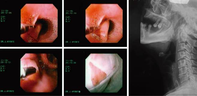
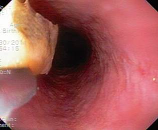
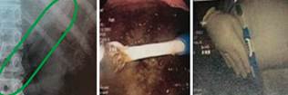
The foreign bodies ingested and found were variable; the most frequent were fishbones, representing 64.11% of the cases, followed by the ingestion of chicken bones and food impaction with 17.43% and 9.19%, respectively. Table 1 summarizes reported ingested foreign bodies. Figures 1 to 4 correspond to some examples of ingested foreign bodies found during the endoscopic study.
Table 1 Reported ingested foreign bodies
| Strange body | Number | Percentage |
|---|---|---|
| Fishbone | 1479 | 64.11% |
| Chicken bone | 402 | 17.43% |
| Food impaction | 212 | 9.19% |
| Incomplete MR data | 47 | 2.04% |
| Dental prosthesis | 36 | 1.56% |
| Orthodontic wire | 24 | 1.04% |
| Glass | 22 | 0.95% |
| Plastic element (straw, piece of package) | 19 | 0.82% |
| Fruit seed | 16 | 0.69% |
| Tablets | 15 | 0.65% |
| Toothpick | 15 | 0.65% |
| Worm | 7 | 0.30% |
| Pin | 5 | 0.22% |
| Coin | 3 | 0.13% |
| Eggshell | 3 | 0.13% |
| Bezoar - hair | 1 | 0.04% |
| Battery | 1 | 0.04% |
| Total | 2307 | 100% |
MR: medical record.

Taken from: Personal atlas of Dr. Diego Aponte.
Figure 1 Plastic ring located in the hypopharynx; 2007.
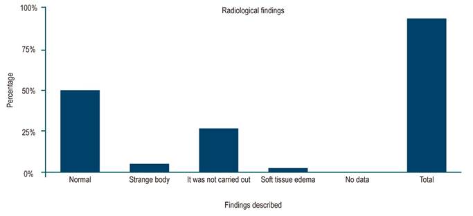
Imaging studies were performed in 66% of the patients before endoscopy and neck X-ray, obtaining normal results in 55.5% of the patients. It was possible to detect the foreign body in only 10.6% of the cases, and, as an additional finding, there was soft tissue edema in 48% of the cases. Figure 5 records the radiological findings.
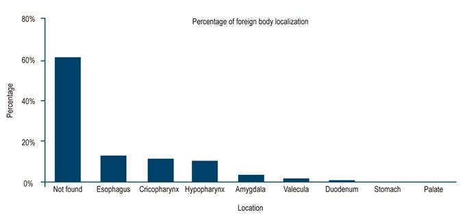
Of note is that in more than half of the cases, the foreign body was not identified for extraction (62.3%), only 38% of the patients required foreign body extraction, and the most frequently used tool was the foreign body clamp in 34.89% of the cases. The primary location was the esophagus in 12.53% of cases, followed by the cricopharynx and hypopharynx in 11.18% and 10%, respectively. Only a small percentage of patients had to undergo surgery due to suspected complications associated with the ingestion of the foreign body (n = 20, 0-87%). Figure 6 shows the location distribution of ingested foreign bodies.
In the endoscopic studies, the most frequent findings were lacerations at the pharynx level in 35%, followed by a typical endoscopic study in 31% of cases, and lacerations in the esophagus in 10.23% of patients. These findings are shown in Table 2.
Table 2 Endoscopic findings
| Endoscopic findings | Number | Percentage |
|---|---|---|
| Laceration in the pharynx | 809 | 35.07% |
| Normal | 717 | 31.08% |
| Laceration in the esophagus | 236 | 10.23% |
| Hematoma in the pharynx | 215 | 9.32% |
| Laceration, tear, or edema in the tonsil | 81 | 3.51% |
| Hematoma in the esophagus | 46 | 1.99% |
| Not classified | 44 | 1.91% |
| Laceration, tear in the vallecula | 39 | 1.69% |
| Ulcer in the esophagus | 37 | 1.60% |
| Suspected perforation in the esophagus | 29 | 1.26% |
| Suspected perforation in the pharynx | 23 | 1.00% |
| Ulcer in the pharynx | 23 | 1.00% |
| Perforation in the palate | 4 | 0.17% |
| Suspected perforation in the stomach | 2 | 0.09% |
| Suspected perforation in the duodenum | 2 | 0.09% |
We know that the ingested objects with the highest risk of perforation are those with sharp points, mainly fishbones and chicken bones, the most commonly ingested in the population studied. Of the 1,479 patients who ingested fishbones, 2% had findings suggestive of perforation, while of the 402 patients who ingested chicken bones, 3% showed findings suggestive of perforation (Table 3).
The predominant symptoms associated with foreign bodies identified in the endoscopic study were the sensation of discomfort in 28.9% and dysphagia in 25%, which occurred in the case of ingestion of fishbones in 64%, chicken bones in 16%, and food impaction in 10.9%.
Discussion
The ingestion of foreign bodies continues to be a frequent reason for consultation and, in some circumstances, is still considered an emergency in gastroenterology. EGD is the method of choice as it is diagnostic and therapeutic3,5. It has been described that the vast majority of ingested foreign bodies, approximately 80%, manage to reach the stomach and, once there, cross the digestive tract smoothly1,2. However, we see how, in our series, it was only possible to find the ingested foreign body in 62.3% of the cases, possibly due to the early performance of endoscopic studies in our department.
Complications from foreign body ingestion are usually mild and include erosions, superficial lacerations, edema, and hematomas. However, serious complications such as perforation, mediastinitis, cardiac tamponade, and the development of fistulas may occur6. Among the risk factors for these complications, the presence of foreign bodies visible on cervical X-rays, impaction in the cricopharyngeus, and evolution of the impaction greater than 24 hours have been described6,7. The risk and severity of these complications go hand in hand with the ingested object’s characteristics and impaction site5,6. Among the most frequent sites of foreign body impaction are the esophagus, in places of anatomical narrowness (at the level of the cricopharyngeal muscle, aortic arch, and gastroesophageal junction)1, the stomach, the pharynx, and the duodenum5,7. Although imaging studies, such as cervical X-rays, are often used as part of the initial evaluation of these patients, it is known that they have a limited sensitivity in the scenario found, between 25% and 55%8.
In this series, fishbones, which are recognized for the difficult visualization both in imaging studies and in endoscopy7,8, were the most commonly ingested element and represented 64% of the cases, followed by chicken bones and food impaction, findings similar to those described in other series and reviews3,9,10. Still, radiological identification was achieved in only 10% of the cases, which speaks of the limited use of this diagnostic tool in this scenario, as described in other reports, and is of greater importance if there is suspicion of perforation11,12.
Regarding location, these foreign bodies were found most frequently at the esophagus level, followed by the cricopharynx and hypopharynx, results consistent with previous studies13-15. The clinical manifestations associated with ingesting foreign bodies are related to the site of impaction and the duration of the condition2. In this series, it is clear that the most common symptoms were foreign body sensation and dysphagia concerning foreign body impaction in the hypopharynx and esophagus.
EGD continues to be the diagnostic and therapeutic tool of choice in these cases2, which additionally has multiple tools with which the endoscopist must be familiar and achieves success rates of up to 95% for managing these patients14,15. In this series, in the patients with a foreign body, endoscopic removal was achieved, the impaction was resolved favorably in all cases, and foreign body forceps was the most used tool. Only a small percentage of patients required surgical management due to suspicion of perforation or other serious complications (0.84%), possibly related to the patients’ early intervention, which could justify emergent EGD in this scenario.
Conclusions
The ingestion of foreign bodies continues to be a frequent reason for consultation and is considered one of the emergencies in gastroenterology. While simple X-ray studies are recommended to locate the ingested foreign body, this diagnostic aid has limited use in this scenario. EGD remains the procedure of choice for visualization and removal and is safe and highly effective.
This work is probably the most extensive series published worldwide, with findings very similar to those published in other series regarding the type of foreign body ingested, location, and associated complications.

